Diffusion | Convection | Osmosis | Three-Pore Model | Dwell Time | Residual Kidney Function
CHAPTER MISSION Build a bedside transport model of peritoneal dialysis: know what crosses, through which pathway, in which direction, and how time, osmotic force, membrane characteristics and residual kidney function change the clinical result. The goal is to predict what a prescription will do before changing it.
MASTER PRINCIPLE Peritoneal dialysis is not a bag-exchange ritual. It is a time-dependent transport system. Every prescription change should name the clinical goal, the transport mechanism being altered, the expected trade-off and the reassessment endpoint.
0. One-page chapter map
Table 1.1 - The eight decisions that govern PD physiology.
| Decision | Core question | Bedside output |
|---|---|---|
| 1. Barrier | What structure actually separates capillary blood from dialysate? | Microvascular transport model |
| 2. Gradient | Which concentration or osmotic gradient is driving movement? | Direction of solute/water transfer |
| 3. Pathway | Diffusion, small-pore convection, AQP1 free water or large-pore transport? | Mechanism named |
| 4. Time | Where is the patient within the dwell: early, intermediate or late? | Expected equilibration/UF state |
| 5. Water + sodium | Is the problem water removal, sodium removal, or both? | Volume strategy matched to physiology |
| 6. Kidney | How much residual kidney function remains? | Total therapy interpreted correctly |
| 7. Lever | Which change alters the failing mechanism with least burden? | Mechanism-based prescription change |
| 8. Verify | Did symptoms, volume, biochemistry and delivered therapy improve? | Clinical success, not a number alone |
Learning outcomes
Explain why the peritoneal microvascular wall, rather than the mesothelium alone, is the dominant early transport barrier.
Distinguish diffusion, convection, crystalloid osmosis, free-water transport and intraperitoneal fluid absorption.
Use the three-pore model to explain small-solute transfer, protein loss, aquaporin-mediated water movement and sodium sieving.
Predict how urea/creatinine, glucose and intraperitoneal volume change during a dwell without treating the exchange as static.
Explain why rapid solute transport can coexist with poor long-dwell glucose ultrafiltration.
Distinguish water removal from sodium removal and recognise why short glucose dwells can accentuate free-water removal.
Integrate residual kidney function into every assessment of clearance and volume rather than adding peritoneal dose reflexively.
Choose prescription levers by mechanism and define a reassessment endpoint before changing treatment.
EVIDENCE POSTURE ISPD goal-directed prescribing recommendations (2020) and membrane-dysfunction recommendations (2021) anchor the clinical framework. Contemporary CJASN and Frontiers reviews (2024) update transport biology. Foundational three-pore, AQP1, CANUSA and ADEMEX studies remain essential because they established mechanisms and challenged clearance-only definitions of adequacy. PET thresholds and formal membrane classification are intentionally deferred to Chapter 3. [1–13]
1. Core concept: a biological dialyzer, not a bag exchange
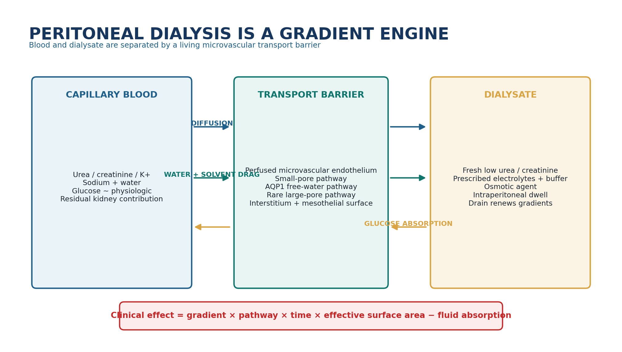
Peritoneal dialysis works because dialysate in the peritoneal cavity is brought into close functional contact with a perfused microvascular bed. Solutes move because concentration gradients exist; water moves because hydrostatic and osmotic forces act across the capillary endothelium; solutes may also accompany water by convection. The exchange is dynamic: the strongest gradients are usually present just after fresh dialysate is instilled and then weaken as equilibration proceeds. [3–5,14,15]
The drained bag is therefore the final read-out of several simultaneous processes: solute diffusion into or out of the cavity, transcapillary water entry, solute-coupled convection, osmotic-agent absorption and ongoing fluid absorption away from the cavity. If one of these components changes, the same nominal prescription can produce a different clinical result.
Table 1.2 - A PD exchange in one minute.
| Phase | What is happening | Clinical meaning |
|---|---|---|
| Fill | Fresh dialysate establishes solute and osmotic gradients. | Maximum opportunity for gradient-driven transport begins. |
| Early dwell | Small-solute diffusion and glucose-driven water transport are brisk; free-water transport is most visible. | Strongest sodium sieving and early UF physiology. |
| Intermediate dwell | Dialysate solute concentrations rise; glucose falls; gradients narrow. | Useful clearance continues but marginal transfer slows. |
| Late dwell | Small-solute equilibration is advanced; glucose osmotic force may be substantially weaker. | Longer is not automatically better for glucose-driven UF. |
| Drain | Dialysate carrying solute and net fluid is removed. | New fill renews gradients; incomplete drainage distorts apparent UF. |
BEDSIDE TRANSLATION When a PD prescription “stops working,” ask which part of the exchange changed: the gradient, the membrane pathway, the dwell time, the residual kidney contribution, or the mechanical delivery. Do not start with a larger glucose bag.
2. The transport barrier: the microvascular wall is central
The phrase “peritoneal membrane” is clinically convenient but anatomically oversimplified. Transport occurs across a composite system that includes capillary blood, the microvascular endothelium and its transport pathways, basement membrane, interstitial tissue and the mesothelial surface. Contemporary physiology places the microvascular wall of peritoneal tissues at the centre of initial solute and water transfer. The mesothelium matters biologically and in long-term membrane health, but it is not the main semipermeable barrier determining routine early exchange kinetics. [2–4]
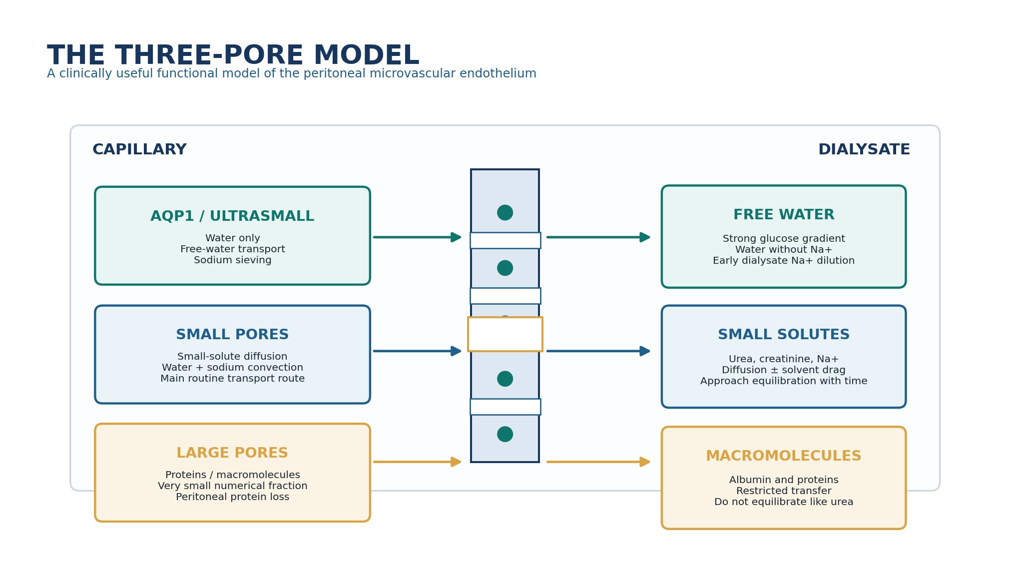
Table 1.3 - Layer of the system -> physiologic role -> bedside relevance.
| Layer / element | Physiologic role | Why it matters clinically |
|---|---|---|
| Perfused capillaries | Provide blood-side solute and water source. | Effective perfused surface area influences transport capacity. |
| Endothelial small-pore pathway | Diffusion of small solutes plus solute-coupled water flow. | Main route for routine urea/creatinine and sodium transport. |
| AQP1 | Transcellular water movement without accompanying solute. | Explains free-water transport and sodium sieving. |
| Large-pore pathway | Restricted macromolecule transfer. | Explains peritoneal protein loss; negligible role in small-solute equilibration. |
| Interstitium | Distance/matrix between capillaries and cavity; important in long-term structural change. | Fibrosis and vascular/interstitial changes can alter transport over time. |
| Mesothelium | Biologically active surface and host-defense interface. | Important for membrane health, inflammation and long-term remodeling; not the main early transport barrier. |
MECHANISM RULE Do not teach “the peritoneum” as one uniform sheet. What the clinician measures is the integrated behavior of perfused capillaries, endothelial pathways, interstitium, contact area and time.
3. The three-pore model turns anatomy into clinical prediction
The classic three-pore model describes three functional transport pathways across the microvascular endothelium. Abundant small pores, with a functional radius of roughly 4-5 nm, carry most small-solute diffusion and a large share of solute-coupled water transport. Rare large pores, roughly 25 nm in radius, permit protein and macromolecule transfer. Ultrasmall pores, later identified with endothelial AQP1 water channels, provide a water-selective pathway with an effective radius below about 0.3 nm. [2,3,5–7]
Table 1.4 - Three-pore model: what to remember.
| Pathway | Main cargo | Dominant force | Clinical signature |
|---|---|---|---|
| AQP1 / ultrasmall | Water without meaningful solute | Crystalloid osmotic gradient, especially glucose | Free-water transport; early sodium sieving |
| Small pores | Urea, creatinine, electrolytes + water | Diffusion + hydrostatic/osmotic convection | Most routine solute clearance and sodium-coupled UF |
| Large pores | Albumin and larger macromolecules | Pressure/concentration dependent restricted transfer | Protein loss; slow, incomplete macromolecule equilibration |
DO NOT OVER-LITERALISE The model is a functional explanation of measured transport. The exact anatomic counterparts of small and large pores are less directly defined than AQP1, whose role as the ultrasmall water pathway is experimentally established.
4. Diffusion: the main engine of small-solute clearance
Diffusion is net solute movement down a concentration gradient. For urea and creatinine, blood concentration is usually higher than fresh dialysate concentration, so net movement is from capillary blood into dialysate. For glucose the direction is reversed: dialysate glucose greatly exceeds plasma glucose, so glucose diffuses into the patient while simultaneously acting as the osmotic agent that drives water movement. [3,14,15]
A useful bedside approximation is that diffusive transfer rises with the concentration gradient and effective mass-transfer capacity, then slows as dialysate approaches equilibrium. The mass transfer area coefficient (MTAC) is an integrated expression of permeability and effective transport area for a particular solute; it is not a fixed “membrane quality” number independent of solute, perfusion and clinical state. [14,15]
Table 1.5 - What determines diffusive transfer?
| Determinant | If it increases | Bedside implication |
|---|---|---|
| Concentration gradient | Diffusive driving force increases. | Fresh dialysate is most efficient at renewing the gradient. |
| Molecular size | Larger molecules diffuse more slowly. | Urea kinetics cannot represent every retained solute. |
| Effective vascular/contact area | Transfer capacity rises until recruitment plateaus. | Fill volume can influence clearance but is not limitless. |
| Peritoneal solute transfer rate | Dialysate approaches plasma faster. | High PSTR improves rapid equilibration but changes long-dwell UF behavior. |
| Dwell time | More time allows equilibration. | Marginal gain falls as the gradient disappears. |
| Repeated exchanges | Fresh gradients are recreated. | More cycles can improve small-solute clearance but increase burden/exposure. |
THRESHOLD DISCIPLINE There is no single “best dwell time” from diffusion alone. Short dwells preserve gradients but permit less equilibration per exchange; long dwells permit more equilibration but progressively lose marginal efficiency. Prescription requires the clinical goal and the patient’s transport physiology.
5. Convection: solute travels with moving water
Convection is solute transport coupled to bulk water movement, often called solvent drag. Water traversing small pores carries dissolved solutes according to their permeability and sieving characteristics. Thus, diffusion and convection are not competing explanations; they occur together during a PD dwell. [5,14,15]
Table 1.6 - Diffusion versus convection.
| Feature | Diffusion | Convection |
|---|---|---|
| Driving force | Solute concentration gradient | Bulk water movement |
| What moves | Solute | Water plus permeable solutes |
| Important pathway | Mostly small pores for routine small solutes | Small-pore water flow; not AQP1 free water |
| Clinical dependence | Gradient, MTAC, time | Transcapillary water flow and solute sieving |
| Key trap | Assuming longer dwell always gives proportionally more clearance | Assuming all UF removes the same amount of sodium |
BEDSIDE TRANSLATION A litre of net UF is not a chemically identical litre from one prescription to another. The fraction moving through AQP1 versus small pores changes how much sodium accompanies the water.
6. Osmosis and ultrafiltration: net fluid removal is a balance
Glucose-based PD creates a crystalloid osmotic gradient across the peritoneal microvasculature. Water moves from capillary blood toward the dialysate through both AQP1 and small-pore pathways. At the same time, fluid is absorbed away from the peritoneal cavity through tissues and lymphatic pathways. The drained net ultrafiltration is therefore the result of opposing fluxes, not simply the amount of water that crossed from blood. [3,4,7,15]
PHYSIOLOGY EQUATION Net UF = transcapillary water entry - intraperitoneal fluid absorption. A low drain volume can therefore arise from weak osmotic water entry, excessive absorption, or a mechanical failure to recover the fluid.
Table 1.7 - Forces acting on water movement.
| Force / process | Direction / effect | Clinical relevance |
|---|---|---|
| Glucose crystalloid osmosis | Capillary -> cavity | Major early water-removal force in glucose dwells. |
| Hydrostatic pressure | Generally favors capillary filtration early in the microcirculation | Contributes to baseline transcapillary flow. |
| Oncotic forces | Oppose movement according to protein gradients | Part of net Starling balance; altered by protein concentration and interstitium. |
| AQP1 free-water pathway | Capillary -> cavity without sodium | Creates sodium sieving. |
| Small-pore water flow | Capillary -> cavity with sodium/solutes | Important for sodium removal and convection. |
| Fluid absorption | Cavity -> tissue/lymphatic return | Reduces final drained net UF, especially as osmotic force wanes. |
7. Aquaporin-1 and sodium sieving: free water made visible
AQP1 is the molecular counterpart of the ultrasmall water pathway predicted by the three-pore model. Experimental deletion of AQP1 abolishes sodium sieving and markedly reduces early solute-free water transport despite preserved small-solute transport, providing direct mechanistic evidence that this channel is central to glucose-driven free-water movement. [6]
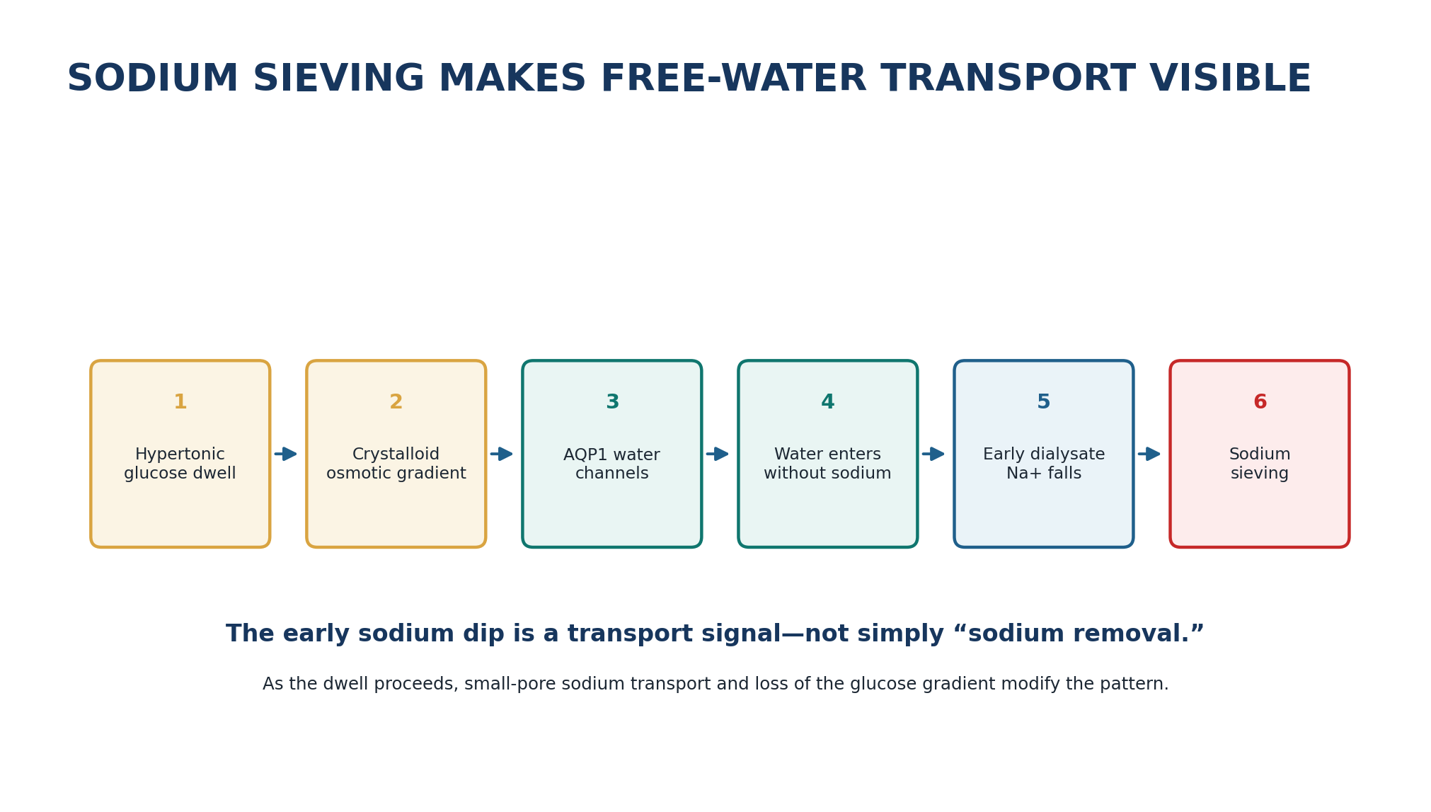
Table 1.8 - Sodium sieving: correct interpretation.
| Observation | Mechanism | Do not misread it as |
|---|---|---|
| Early dialysate sodium falls | AQP1-mediated solute-free water dilutes dialysate | Proof of large sodium removal |
| Free-water fraction is prominent early | Glucose gradient is strongest early | A fixed proportion throughout the dwell |
| Sodium later rises toward equilibrium | Small-pore sodium diffusion/convection + declining free-water effect | Loss of all water transport |
| Reduced sodium dip in an abnormal UF phenotype | May reflect impaired free-water/osmotic conductance | Diagnosis without standardized testing |
CLINICAL RULE Water balance and sodium balance must be assessed separately. A short glucose dwell can remove water efficiently yet remove less sodium than the same UF volume generated predominantly through small-pore transport.
8. Dwell time: the exchange is continuously changing
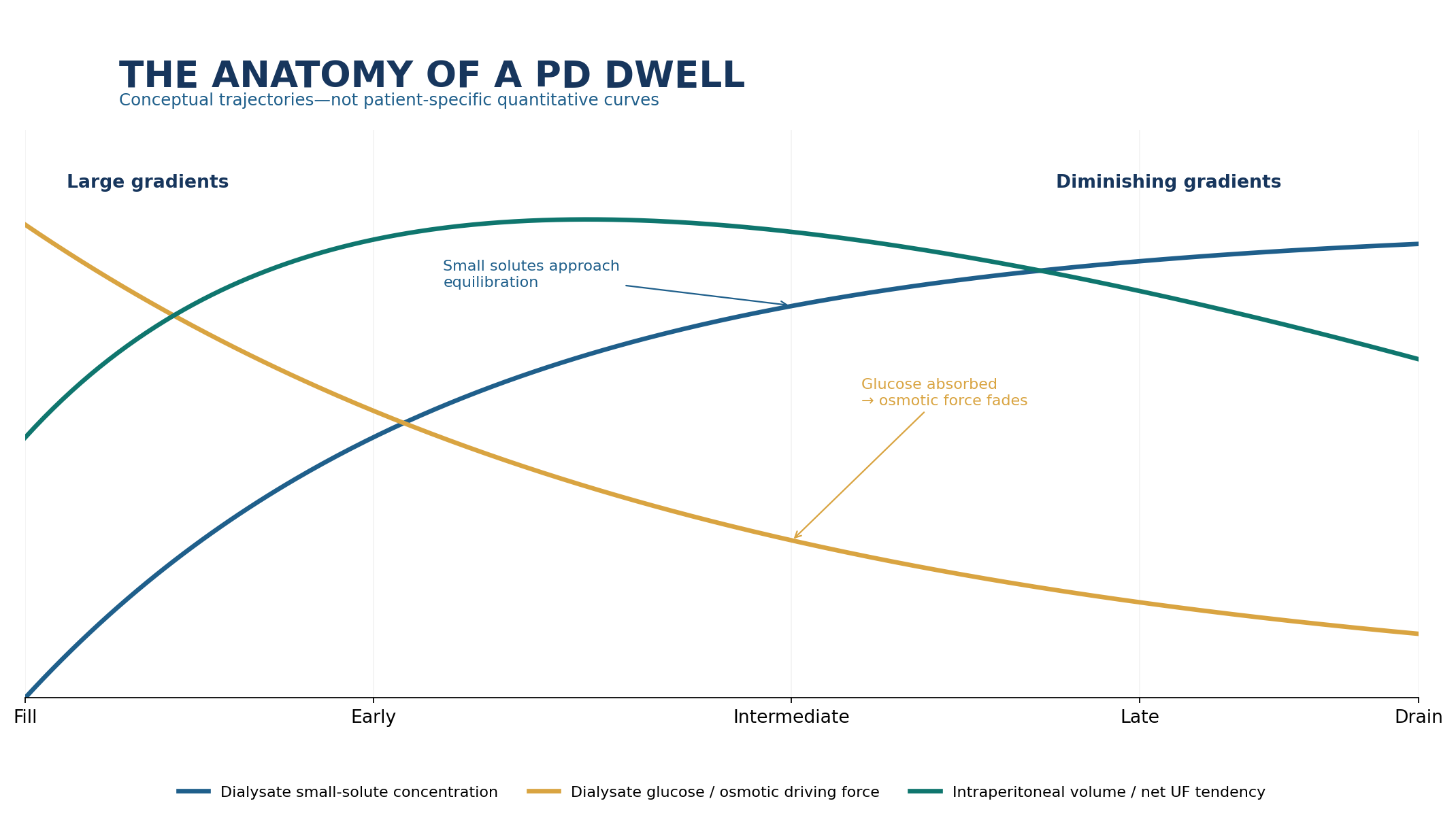
The central time-dependent conflict in PD is simple: longer time favors solute equilibration, whereas prolonged glucose dwell time can erode the osmotic gradient needed for net UF. Therefore the dwell that best serves small-solute clearance may not be the dwell that best serves fluid removal. This tension is most clinically visible in patients with rapid peritoneal solute transfer, in whom glucose can be absorbed quickly. [2–4]
Table 1.9 - Early, intermediate and late dwell physiology.
| Dwell stage | Small-solute gradient | Glucose osmotic force | Likely clinical emphasis |
|---|---|---|---|
| Early | Large | Strong | Rapid diffusion + prominent free-water transport |
| Intermediate | Narrowing | Declining | Continued clearance and useful UF |
| Late | Small for rapidly equilibrating solutes | May be weak | Marginal clearance gain; UF may plateau |
| Overlong glucose dwell in susceptible physiology | Near equilibration | Substantially dissipated | Fluid absorption may offset ongoing water entry |
BEDSIDE TRANSLATION If a long glucose dwell produces poor UF in a fast transporter, “stronger glucose” is not the only physiologic answer. The dwell may be outlasting its useful crystalloid osmotic gradient.
9. Peritoneal solute transfer rate: fast is not simply good
Peritoneal solute transfer rate (PSTR) describes how rapidly small solutes equilibrate between plasma and dialysate. A higher PSTR can be advantageous for short-dwell small-solute transfer because dialysate concentrations rise quickly. The same physiology can be unfavorable for a prolonged glucose dwell because glucose also leaves the cavity quickly, shortening effective osmotic time. [2,3,13]
Table 1.10 - Solute transport and UF are different axes.
| Pattern | Small-solute behavior | Glucose-dwell UF behavior | Prescription implication |
|---|---|---|---|
| Faster PSTR | Rapid approach to equilibration | Osmotic gradient may dissipate earlier | Shorter glucose dwells may be advantageous; long dwell often needs different strategy |
| Slower PSTR | Slower equilibration | Glucose gradient may persist longer | Longer dwell may be needed for clearance; avoid assuming poor early D/P means poor membrane |
| Low UF with expected PSTR | Solute transport alone does not explain problem | Consider osmotic conductance, absorption or mechanics | Formal membrane assessment if persistent |
| Abrupt change from baseline | Could reflect acute inflammation or measurement conditions | UF may change simultaneously | Repeat under standardized conditions and assess clinical context |
DEFERRED DETAIL The standardized peritoneal equilibration test, transport categories, serial PET interpretation and formal UF-failure phenotypes are developed in Chapter 3. Here, remember the physiologic relationship: solute transfer rate and water-removal efficiency are linked but not interchangeable.
10. Fill volume, contact area and intraperitoneal pressure
Increasing fill volume can recruit additional effective contact area and improve solute transfer up to the point at which further recruitment becomes limited. At the same time, a larger intraperitoneal volume increases intraperitoneal pressure, which may impair comfort, breathing, abdominal-wall tolerance and mechanical safety in susceptible patients. Physiologic benefit therefore plateaus before “the largest tolerable fill” becomes a universal target. [3,14]
Table 1.11 - Fill volume is a lever with two sides.
| Change | Potential gain | Potential cost | How to reassess |
|---|---|---|---|
| Increase fill volume | More effective contact area; possible clearance gain | Higher intraperitoneal pressure, discomfort, leak/hernia risk | Symptoms, mechanical tolerance, delivered clearance |
| Reduce fill volume | Lower pressure; useful in leaks, early starts or intolerance | Less contact area and clearance per exchange | Compensate only when clinically needed with time/exchanges |
| Supine versus upright dwell | Contact distribution and pressure differ | Patient-specific comfort and mechanics | Interpret symptoms in actual treatment position |
THRESHOLD DISCIPLINE Do not import a single fill-volume or intraperitoneal-pressure cutoff into every adult. Body size, posture, abdominal mechanics, catheter function, leak risk and treatment goal determine what is usable. Exact prescription construction belongs to Chapter 6.
11. Glucose: an effective osmotic agent with a biologic price
Glucose works because its dialysate concentration is far above plasma concentration, creating a powerful crystalloid osmotic gradient. Yet glucose is small enough to be progressively absorbed into the systemic circulation. The intervention that creates UF therefore simultaneously erodes its own driving force and adds metabolic and membrane glucose exposure. [3,4,7,14]
This explains why glucose concentration should be thought of as an osmotic dose, not simply a “strength.” More hypertonic glucose can produce greater early osmotic water movement, but the trade-off includes greater systemic carbohydrate exposure and cumulative local glucose burden. Long-term glucose exposure is implicated in structural and functional membrane changes; exact solution composition and biocompatibility are developed in Chapter 2 and long-term membrane failure in Chapter 14. [4]
Table 1.12 - When glucose is increased, what else changes?
| Immediate effect | Downstream consequence | Clinical question before escalation |
|---|---|---|
| Crystalloid osmotic gradient rises | Early water movement tends to increase | Is low UF truly due to inadequate osmotic force? |
| More dialysate glucose available for absorption | Systemic carbohydrate exposure rises | Could dwell redesign or icodextrin solve the problem with less glucose? |
| Greater local glucose exposure | Potential long-term membrane/metabolic burden | Is this a temporary rescue or chronic strategy? |
| More water may move through AQP1 early | Water removal may exceed sodium removal proportionally | Is the clinical goal water, sodium or both? |
PRESCRIPTION RULE Never write “increase glucose” without completing the sentence: increase glucose to create a stronger crystalloid osmotic gradient, because ___ is the suspected mechanism; accept ___ as the trade-off; reassess ___ after the change.
12. Icodextrin: different osmotic physics for the long dwell
Icodextrin is a glucose polymer that generates sustained osmotic water movement during a long dwell by colloid-like osmotic mechanisms. Its larger fractions diffuse across the small-pore pathway far less readily than glucose, so the osmotic effect is sustained and is largely independent of AQP1. This is physiologically different from simply using “stronger glucose.” [7,8]
Table 1.13 - Glucose versus icodextrin: mechanism, not brand.
| Feature | Glucose | Icodextrin |
|---|---|---|
| Osmotic principle | Crystalloid osmosis | Sustained macromolecule-associated / colloid osmosis |
| Key water pathways | AQP1 + small pores | Predominantly small-pore pathway; AQP1 not required |
| Time profile | Strong early effect that fades as glucose is absorbed | Designed for sustained long-dwell UF |
| Typical physiologic problem solved | Need for glucose-driven UF across short/intermediate dwell | Long dwell in which glucose osmotic force would dissipate |
| Metabolic glucose burden | Adds absorbed glucose | Can reduce glucose exposure versus glucose long-dwell alternative |
Randomized-trial meta-analysis supports greater long-dwell UF and fewer fluid-overload episodes with icodextrin compared with glucose-only long-dwell strategies in appropriate patients, while patient selection and product-specific safety remain essential. [8]
CLINICAL RULE Think “long-dwell osmotic strategy,” not “icodextrin is stronger glucose.” The two agents exploit different transport physics.
13. Sodium removal is not the same as water removal
Net volume removal is clinically important, but extracellular volume is fundamentally a sodium-and-water problem. Sodium can leave through small-pore diffusion/convection and through residual kidney excretion. Free water moving through AQP1 does not carry sodium. Therefore the sodium concentration of removed fluid changes across the dwell and across prescriptions. [2,3]
Table 1.14 - Why water and sodium must be tracked separately.
| Scenario | Water behavior | Sodium behavior | Bedside risk |
|---|---|---|---|
| Very short hypertonic glucose dwell | Prominent early free-water removal | Relatively less sodium accompanies AQP1 water | Good UF number can overstate sodium removal |
| Longer small-pore-dominant phase | Free-water fraction falls | More sodium can accompany convective/diffusive transport | May improve sodium removal but glucose UF may later fade |
| High dietary sodium | PD may remove fluid but total sodium balance remains positive | Thirst and extracellular volume expand | Escalating glucose alone treats consequence, not intake |
| Falling residual urine | Loss of continuous water excretion | Loss of renal sodium excretion | Same PD prescription can become insufficient for volume |
BEDSIDE TRANSLATION For edema or hypertension, ask “What is total sodium balance?” before asking only “What was yesterday’s UF?” The bag drain and the urine bottle belong in the same fluid assessment.
14. Residual kidney function: part of the prescription even though it is not in the bag
Residual kidney function (RKF) contributes continuous water and sodium excretion plus clearance of urea, creatinine, phosphate, potassium and other retained solutes. Observational evidence consistently links RKF with better outcomes. The CANUSA reanalysis showed that renal clearance carried a stronger association with survival than equivalent increments in peritoneal clearance, undermining the assumption that renal and peritoneal clearances are biologically interchangeable. [9,12,16]
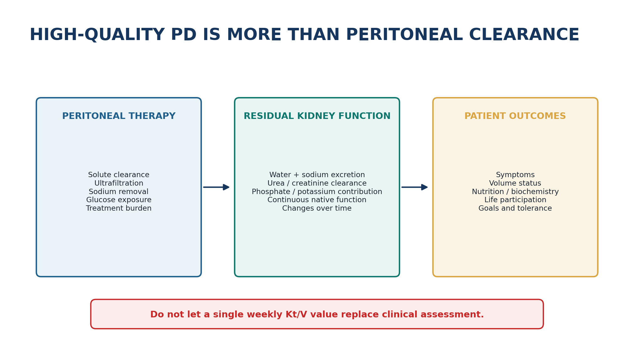
Table 1.15 - What RKF contributes beyond weekly Kt/V arithmetic.
| Contribution | Why the patient feels it | What happens when it falls |
|---|---|---|
| Water excretion | More flexible fluid intake and less glucose dependence | Volume control becomes harder |
| Sodium excretion | Reduces chronic positive sodium balance | Thirst, hypertension and edema risk rise |
| Small-solute clearance | Adds continuous clearance | Total clearance falls even if PD prescription is unchanged |
| Phosphate/potassium handling | Helps biochemical control | Diet and PD requirements may intensify |
| Treatment flexibility | Allows incremental/less burdensome prescriptions in selected patients | Treatment burden may need to increase |
PRESERVATION PRINCIPLE Avoiding preventable RKF loss is a dialysis intervention. Contemporary practice emphasizes individualized volume management, avoidance of unnecessary nephrotoxicity and hypotension, infection prevention and glucose-aware prescribing. Evidence for several specific preservation strategies varies; Chapter 7 treats RKF formally. [12,16]
15. Clearance and adequacy: why a good Kt/V is not a good patient
Small-solute clearance remains measurable and clinically useful, but randomized and observational evidence does not support using it as the sole definition of adequate PD. ADEMEX showed no survival benefit from increasing peritoneal small-solute clearance above conventional therapy, while CANUSA reanalysis emphasized the disproportionate importance of RKF. The 2020 ISPD evidence update concluded that evidence for exact small-solute targets is weak and that increasing weekly Kt/V above approximately 1.7 has not shown a survival advantage. [9–11]
Table 1.16 - What weekly Kt/V can and cannot tell you.
| Kt/V can help describe | Kt/V cannot independently establish |
|---|---|
| Delivered urea clearance over time | Euvolemia or adequate sodium removal |
| Change in total small-solute clearance as RKF falls | Freedom from uremic symptoms |
| Whether a major prescription delivery problem may exist | Adequate phosphate/middle-molecule removal |
| A conventional small-solute adequacy benchmark | Nutrition, life participation or treatment burden |
| Peritoneal + renal urea clearance arithmetic | That renal and peritoneal clearance are biologically equivalent |
EVIDENCE INTERPRETATION For anuric adults, weekly Kt/V around 1.7 remains a conventional small-solute reference supported by low-certainty evidence; it is not a universal definition of “adequate dialysis.” High-quality PD is goal-directed and integrates symptoms, volume status, nutrition/biochemistry, RKF, membrane function, treatment burden and patient priorities. [1,11]
16. Prescription levers: every change should name its mechanism
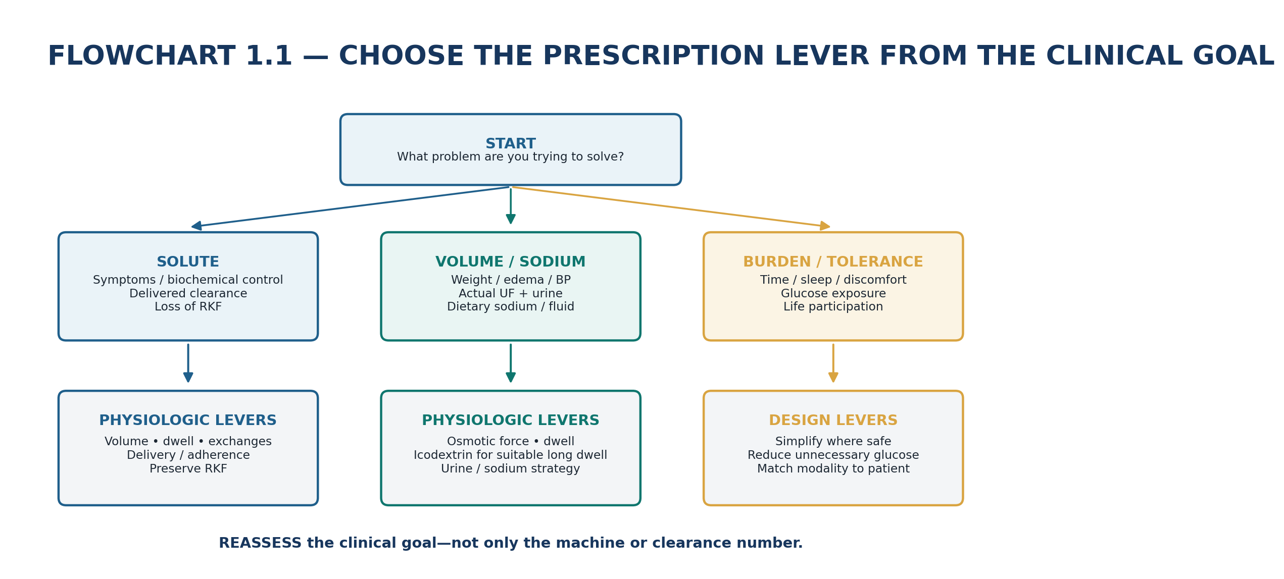
Table 1.17 - Prescription lever -> mechanism -> trade-off -> reassessment.
| Lever | Main mechanism | Trade-off | Reassess |
|---|---|---|---|
| Fill volume up | More effective contact area | Pressure, discomfort, leak risk | Tolerance + delivered clearance |
| Dwell longer | More time for equilibration | Glucose gradient may fade; burden | Solute control + net UF |
| Dwell shorter | Renew gradient before glucose dissipation | Less equilibration per dwell; more cycles | UF + sodium + clearance |
| More exchanges | More frequent gradient renewal | Treatment time, glucose exposure | Delivered clearance + burden |
| Higher glucose exposure | Stronger crystalloid osmotic force | Metabolic/membrane glucose load | Volume endpoint; minimize chronic use |
| Icodextrin long dwell | Sustained long-dwell osmotic force | Agent-specific safety and technique | Long-dwell UF + volume status |
| Preserve RKF | Maintains continuous native excretion/clearance | Requires careful volume/drug strategy | Urine volume + renal clearances |
PRESCRIPTION LANGUAGE Avoid “increase exchanges” or “increase glucose.” Write the full physiologic sentence: problem -> mechanism -> lever -> expected effect -> trade-off -> reassessment.
17. Reduced net ultrafiltration: first decide whether it is real
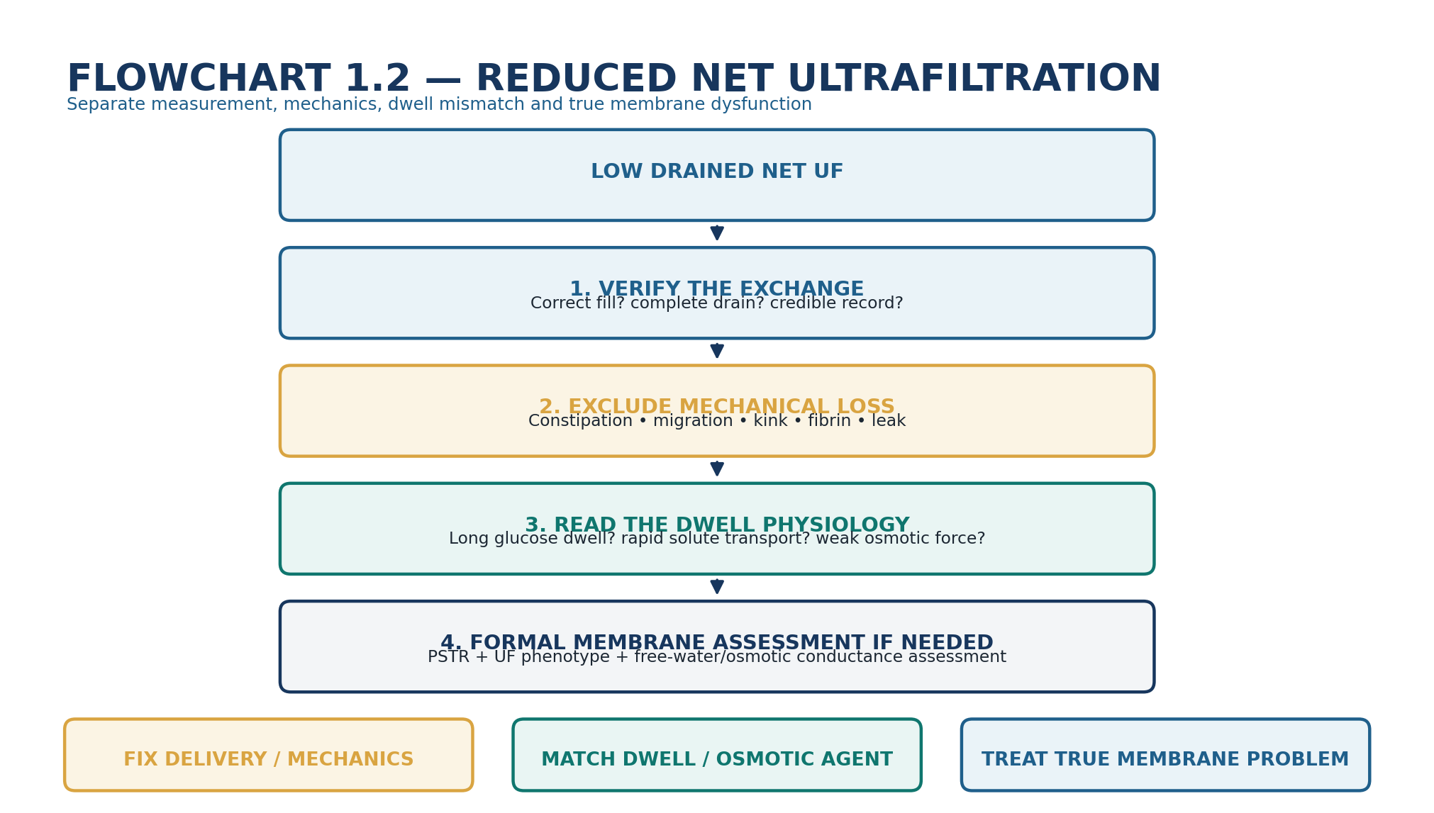
Table 1.18 - Low net UF: rapid differential.
| Cause | Clue | Mechanism | Next step |
|---|---|---|---|
| Incomplete drain / constipation | Fullness, variable drains, positional pattern | Fluid remains in cavity | Correct mechanics and re-measure |
| Leak | Unexpected low recovery, edema at leak site or other mechanical clues | Dialysate leaves intended cavity | Leak pathway evaluation |
| Fast PSTR + long glucose dwell | Good solute equilibration but poor long-dwell UF | Glucose gradient dissipates early | Match dwell/agent to transport |
| Insufficient osmotic force | Low UF despite credible drain and suitable dwell | Water-entry force inadequate | Review osmotic prescription and intake |
| Low osmotic conductance / free-water problem | Persistent abnormal UF phenotype on standardized testing | Reduced effective crystalloid water transport | Formal membrane evaluation |
| High absorption / long dwell | Volume peaks then declines | Absorption offsets transcapillary UF | Redesign dwell; investigate context |
DIAGNOSTIC RULE Low drained volume is a phenotype. “Ultrafiltration failure” should not be assigned until fill accuracy, drainage, leaks and standardized membrane behavior have been considered.
18. Apparent underdialysis: do not add dose until the missing component is named
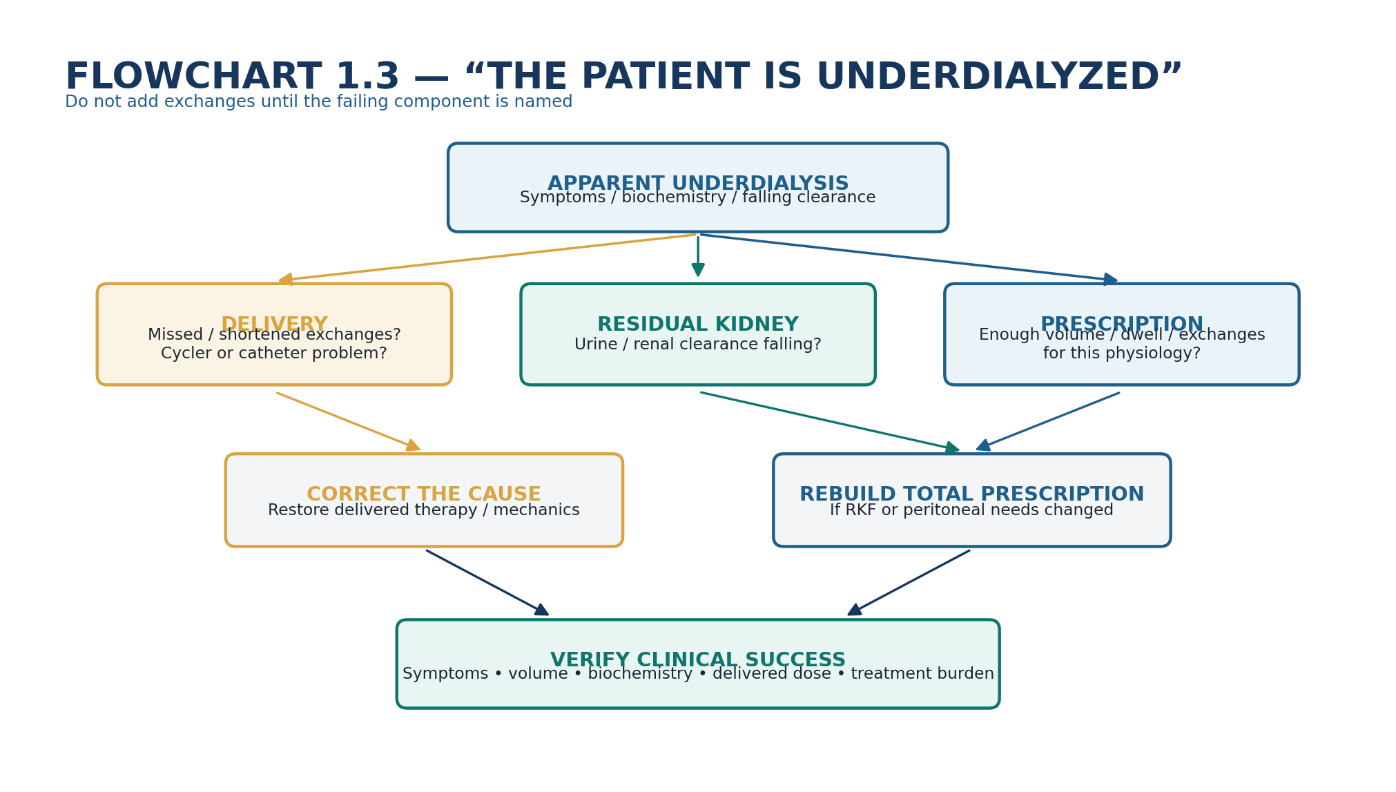
Table 1.19 - Falling clinical control: mechanism before dose.
| Finding | First hypothesis | Important alternative | Action direction |
|---|---|---|---|
| Uremic symptoms + falling urine | Loss of RKF | Missed exchanges or intercurrent illness | Reassess total delivered therapy |
| Low measured clearance | Under-delivery / collection error | True prescription insufficiency | Verify technique and collections before escalation |
| Edema with acceptable Kt/V | Sodium/water problem | Cardiac/liver disease | Volume-focused assessment |
| Good UF but hyperphosphatemia | Solute-specific problem + diet/RKF | Adherence or binder issue | Do not assume more glucose solves clearance |
| Cycler alarms + low clearance | Mechanical/delivery problem | Access dysfunction or poor prescription | Fix delivery before increasing prescribed dose |
GOAL-DIRECTED RULE The endpoint of a prescription change is not “the machine completed.” It is improvement in the clinical target with acceptable burden and no new volume, metabolic or mechanical harm.
19. Mini-cases: decisions, not trivia
Case 1 - Good clearance, worsening edema
A 52-year-old on CAPD has acceptable small-solute clearance but progressive edema and higher blood pressure. The longest glucose dwell gives little net UF; shorter exchanges drain well.
REASONING Separate solute from volume physiology. A rapid PSTR can yield good urea/creatinine equilibration while glucose is absorbed quickly during the long dwell, erasing osmotic force. Check sodium intake, urine, mechanics and transport phenotype; redesign the long dwell rather than chasing a higher Kt/V.
Case 2 - The same prescription stopped working
A patient has used the same PD prescription for two years. Urine volume falls substantially over several months and she develops fatigue, rising phosphate and edema.
REASONING The peritoneal prescription is unchanged, but total kidney replacement is not. Loss of RKF removes continuous solute, sodium and water clearance. Reassess renal clearances, urine, volume and delivered PD before increasing treatment burden.
Case 3 - More glucose is not the first answer
An APD patient has poor daytime UF during a prolonged glucose dwell. The proposed response is chronic escalation to a more hypertonic glucose solution.
REASONING Ask whether the dwell outlasts the useful crystalloid osmotic gradient. If transport is rapid, a sustained long-dwell strategy such as icodextrin may address the physiology with less glucose exposure. Confirm mechanics and clinical volume need before changing therapy.
Case 4 - A low drain that is not membrane failure
A previously stable patient develops several low drain volumes after three days of constipation and reports abdominal fullness.
REASONING Low drained net UF does not prove low transcapillary UF. Treat the mechanical problem, confirm complete drainage and remeasure before ordering a membrane-failure work-up or escalating osmotic therapy.
Case 5 - Short cycles, high UF, still hypertensive
An APD prescription produces impressive overnight UF, but the patient remains thirsty, hypertensive and edematous with high sodium intake.
REASONING Water removal is not identical to sodium removal. Short glucose cycles may accentuate free-water removal while dietary sodium maintains extracellular volume and thirst. Reframe the problem around total sodium balance, urine and dwell architecture.
Case 6 - The Kt/V is “on target” but the patient is not
An anuric patient has a weekly Kt/V around the conventional target but persistent nausea, poor appetite and difficult volume control.
REASONING Do not certify adequacy from Kt/V alone. Reassess symptoms, volume/sodium, nutrition, biochemical control, membrane function, delivered treatment and competing diagnoses. A small-solute benchmark is one dimension of high-quality PD, not the endpoint.
20. Common pitfalls - and the correction
Table 1.20 - High-frequency errors in PD physiology.
| Pitfall | Why it fails | Correction |
|---|---|---|
| “The peritoneum is the mesothelium.” | Misses the microvascular barrier that drives transport. | Think capillary endothelium + pathways + interstitium. |
| “Longer dwell means more dialysis.” | Marginal diffusion falls and glucose osmotic force can dissipate. | Match dwell to solute and UF goals. |
| “High transporter means good membrane.” | Fast solute equilibration may impair long-glucose UF. | Interpret PSTR and UF together. |
| “One litre UF = one litre sodium-rich fluid.” | AQP1 free water carries little sodium. | Assess sodium removal and total sodium balance. |
| “Poor drain = UF failure.” | Mechanics/leak can reduce recovered volume. | Verify fill-drain mechanics first. |
| “More glucose solves edema.” | May ignore sodium intake, RKF loss, dwell mismatch or mechanics. | Name mechanism before osmotic escalation. |
| “Kt/V on target = adequate patient.” | Kt/V does not measure symptoms, volume or burden. | Use goal-directed multidimensional assessment. |
| “Renal and peritoneal clearance are interchangeable.” | Outcome relationships differ; kidney function is continuous and broader. | Protect and measure RKF separately. |
| “Icodextrin is stronger glucose.” | They use different osmotic mechanisms. | Use icodextrin as a sustained long-dwell strategy when appropriate. |
| “Cycler settings are the physiology.” | The cycler only manipulates volume, time and exchanges. | Translate every setting into a transport effect. |
21. Flashcards and active recall
1. Q: What is the dominant early transport barrier in PD? A: The peritoneal microvascular wall, especially capillary endothelium, with the interstitium contributing to the composite barrier.
2. Q: What drives most small-solute transfer? A: Diffusion down a concentration gradient through the small-pore pathway.
3. Q: What is MTAC conceptually? A: An integrated measure of solute permeability multiplied by effective transport area.
4. Q: Why does diffusion slow during a dwell? A: Dialysate approaches plasma concentration, so the driving gradient shrinks.
5. Q: What is convection? A: Solute transport coupled to bulk water flow - solvent drag.
6. Q: Net UF equals what? A: Transcapillary water entry minus intraperitoneal fluid absorption.
7. Q: Which channel is the molecular correlate of the ultrasmall pore? A: Aquaporin-1.
8. Q: What is sodium sieving? A: Early dialysate sodium dilution caused by solute-free AQP1 water movement during a glucose dwell.
9. Q: Why can rapid solute transport impair long glucose UF? A: Glucose is absorbed rapidly, so the crystalloid osmotic gradient disappears earlier.
10. Q: Why can shortening a dwell improve UF in a fast transporter? A: Drain occurs before osmotic force has dissipated and before absorption dominates.
11. Q: What may be lost when the dwell is shortened? A: Solute equilibration per exchange; more cycles may be needed for clearance.
12. Q: Why is icodextrin suited to a long dwell? A: Its larger glucose polymers generate more sustained small-pore/colloid osmotic water movement and are absorbed more slowly than glucose.
13. Q: Does good net UF prove good sodium removal? A: No; the free-water fraction may be high and dietary/renal sodium balance still matters.
14. Q: Why is RKF disproportionately valuable? A: It provides continuous water, sodium and solute handling and is more strongly linked to outcomes than equivalent peritoneal clearance increments.
15. Q: What did ADEMEX teach? A: Increasing peritoneal small-solute clearance above conventional therapy did not improve survival.
16. Q: What does weekly Kt/V not measure? A: Volume status, sodium balance, symptoms, quality of life, treatment burden and many non-urea solutes.
17. Q: First step in low drained UF? A: Verify fill/drain measurement and exclude mechanical problems or leak.
18. Q: What makes a prescription change complete? A: A named mechanism, lever, expected trade-off and clinical reassessment endpoint.
22. Final revision sheet
TEN TAKE-HOME RULES 1) PD is a gradient engine. 2) The microvascular wall is the key transport barrier. 3) Small pores handle most routine solute transfer. 4) AQP1 carries free water and explains sodium sieving. 5) Glucose creates osmotic force and simultaneously diffuses away. 6) Dwell time changes clearance and UF in different directions. 7) Fast solute transport is not automatically good long-dwell UF. 8) Water removal is not sodium removal. 9) RKF is part of the prescription even though it is not in the bag. 10) High-quality PD is goal-directed; Kt/V is a component, not the patient.
Table 1.21 - One-minute bedside synthesis.
| If you see... | Think... | Do now... |
|---|---|---|
| Good clearance + edema | Solute and volume physiology are discordant | Assess sodium, urine, actual UF, dwell and PSTR |
| Low long-dwell glucose UF | Gradient dissipation, mechanics or membrane dysfunction | Verify drain; assess transport; match dwell/agent |
| Falling urine + same PD | Total therapy has fallen | Reassess RKF, volume and total clearance |
| Short-cycle high UF + hypertension | Free-water removal may exceed sodium removal | Reassess sodium balance and dwell strategy |
| Low drain after constipation | Mechanical retention | Correct constipation/catheter mechanics and re-measure |
| Kt/V acceptable + symptomatic | Adequacy is multidimensional | Assess symptoms, volume, nutrition, biochemistry and delivery |
| Need more clearance | Gradient/time/volume/exchange problem | Choose the least-burdensome effective lever |
| Need more fluid removal | Osmotic/dwell/sodium/RKF problem | Treat the mechanism, not the bag strength alone |
Table 1.22 - Before and after any physiology-driven prescription change.
| Check | Pass criterion |
|---|---|
| Clinical goal | Specific: solute symptoms, volume/sodium, biochemical control or burden. |
| Mechanism | Named and plausible from delivery + membrane + RKF data. |
| Lever | Chosen because it changes that mechanism. |
| Trade-off | Glucose exposure, dwell burden, pressure, sodium removal or clearance consequence anticipated. |
| Safety | Mechanical tolerance and product/device-specific precautions addressed. |
| Reassessment | A date/endpoint is defined: symptoms, weight/edema/BP, UF, urine, labs, delivered dose, burden. |
| Escalation | If response is absent, re-open the mechanism rather than simply intensifying the same intervention. |
FINAL MENTAL MODEL Clinical problem -> verify delivery -> identify gradient -> identify pathway -> place the exchange in time -> add residual kidney contribution -> choose the smallest effective prescription lever -> anticipate the trade-off -> reassess the patient.
Rapid oral viva
Explain why the mesothelium alone is not the functional “dialysis membrane.”
Trace urea, glucose, sodium and water during the first hour of a glucose dwell and name the pathway for each.
Explain sodium sieving without using the phrase “because sodium is removed.”
A fast transporter has poor long-dwell UF: give the mechanism and two physiologic prescription directions.
Explain why a patient can have an acceptable Kt/V and still require a major prescription review.
Give a cause-based differential for low drained UF before diagnosing membrane failure.
SCOPE BOUNDARY This chapter teaches the transport physics and clinical reasoning that underlie PD. Solution composition and cycler systems are developed in Chapter 2; standardized PET and membrane dysfunction in Chapter 3; initial prescription in Chapter 6; adequacy/RKF in Chapter 7; volume and UF failure in Chapter 8; and CAPD/APD optimization in Chapter 9.
23. Selected authoritative references
1. Brown EA, Blake PG, Boudville N, et al. International Society for Peritoneal Dialysis practice recommendations: Prescribing high-quality goal-directed peritoneal dialysis. Perit Dial Int. 2020;40(3):244–253. https://doi.org/10.1177/0896860819895364. PMID: 32063219.
2. Morelle J, Stachowska-Pietka J, Oberg C, et al. ISPD recommendations for the evaluation of peritoneal membrane dysfunction in adults: classification, measurement, interpretation and rationale for intervention. Perit Dial Int. 2021;41(4):352–372. https://doi.org/10.1177/0896860820982218. PMID: 33563110.
3. Morelle J, Lambie M, Oberg CM, Davies S. The Peritoneal Membrane and Its Role in Peritoneal Dialysis. Clin J Am Soc Nephrol. 2024;19(2):244–253. https://doi.org/10.2215/CJN.0000000000000282. PMID: 37616463; PMCID:PMC10861113.
4. Krediet RT. Physiology of peritoneal dialysis; pathophysiology in long-term patients. Front Physiol. 2024;15:1322493. https://doi.org/10.3389/fphys.2024.1322493. PMID: 39193440; PMCID:PMC11347314.
5. Rippe B. A three-pore model of peritoneal transport. Perit Dial Int. 1993;13 Suppl 2:S35–S38. https://doi.org/10.1177/089686089301302S09. PMID: 8399608.
6. Ni J, Verbavatz JM, Rippe A, et al. Aquaporin-1 plays an essential role in water permeability and ultrafiltration during peritoneal dialysis. Kidney Int. 2006;69(9):1518–1525. https://doi.org/10.1038/sj.ki.5000285. PMID: 16508653.
7. Morelle J, Sow A, Fustin CA, et al. Mechanisms of Crystalloid versus Colloid Osmosis across the Peritoneal Membrane. J Am Soc Nephrol. 2018;29(7):1875–1886. https://doi.org/10.1681/ASN.2017080828. PMID: 29844208; PMCID:PMC6050940.
8. Goossen K, Becker M, Marshall MR, et al. Icodextrin Versus Glucose Solutions for the Once-Daily Long Dwell in Peritoneal Dialysis: An Enriched Systematic Review and Meta-analysis of Randomized Controlled Trials. Am J Kidney Dis. 2020;75(6):830–846. https://doi.org/10.1053/j.ajkd.2019.10.004.
9. Bargman JM, Thorpe KE, Churchill DN. Relative contribution of residual renal function and peritoneal clearance to adequacy of dialysis: a reanalysis of the CANUSA study. J Am Soc Nephrol. 2001;12(10):2158–2162. https://doi.org/10.1681/ASN.V12102158. PMID: 11562415.
10. Paniagua R, Amato D, Vonesh E, et al. Effects of increased peritoneal clearances on mortality rates in peritoneal dialysis: ADEMEX, a prospective, randomized, controlled trial. J Am Soc Nephrol. 2002;13(5):1307–1320. https://doi.org/10.1681/ASN.V1351307. PMID: 11961019.
11. Boudville N, de Moraes TP. 2005 Guidelines on targets for solute and fluid removal in adults being treated with chronic peritoneal dialysis: 2019 update of the literature and revision of recommendations. Perit Dial Int. 2020;40(3):254–260. https://doi.org/10.1177/0896860819898307. PMID: 32048566.
12. Chen CH, Perl J, Teitelbaum I. Prescribing high-quality peritoneal dialysis: The role of preserving residual kidney function. Perit Dial Int. 2020;40(3):274–281. https://doi.org/10.1177/0896860819893821. PMID: 32063188.
13. Twardowski ZJ, Nolph KD, Khanna R, et al. Peritoneal equilibration test. Perit Dial Int. 1987;7(3):138–148. https://doi.org/10.1177/089686088700700306.
14. Khanna R. Solute and Water Transport in Peritoneal Dialysis: A Case-Based Primer. Am J Kidney Dis. 2017;69(3):461–472. https://doi.org/10.1053/j.ajkd.2016.11.007. PMID: 28111028.
15. Leypoldt JK. Solute transport across the peritoneal membrane. J Am Soc Nephrol. 2002;13 Suppl 1:S84–S91. https://doi.org/10.1681/ASN.V13suppl_1s84. PMID: 11792767.
16. Yeter HH. Preserving residual kidney function in peritoneal dialysis: from conventional approaches to contemporary practice. Clin Kidney J. 2026;19(5):sfag131. https://doi.org/10.1093/ckj/sfag131. PMID: 42137551; PMCID:PMC13170695.
SOURCE NOTE ISPD recommendations, PubMed-indexed source metadata and contemporary open-access reviews were checked 1 September 2026. This chapter intentionally avoids treating local solution brands, cycler settings, PET thresholds or prescription volumes as universal; current manufacturer information, local PD protocols and later chapter-specific guidance govern those details.