Solutions | Connectology | CAPD | APD | Cycler Logic | Alarms | Delivered Therapy
CHAPTER MISSION Build a bedside system map from sterile bag to peritoneal cavity and back again: know what is in the solution, why the bag is constructed the way it is, how transfer sets and disconnect systems prevent contamination, how CAPD and APD move fluid, what a cycler alarm actually means, and how to verify that the prescription was truly delivered.
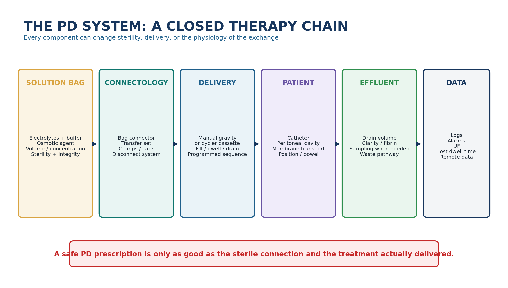
MASTER PRINCIPLE The PD system is not “bag plus catheter.” It is a sterile fluid-delivery platform. Solution choice determines physiology; connectology determines contamination risk; cycler logic determines what is actually delivered. The safest clinician reads all three together.
0. One-page chapter map
Table 2.1 - The eight decisions that govern the PD system.
| Decision | Core question | Bedside output |
|---|---|---|
| 1. Solution | What fluid is prescribed - electrolyte frame, buffer, osmotic agent, concentration and volume? | Correct solution matched to the clinical goal |
| 2. Bag | Is the product intact, in date, correctly mixed when compartmentalised, clear and safely warmed? | Released bag safe to connect |
| 3. Connect | Is the transfer set, cap and disconnect pathway closed and aseptic? | Sterile fluid path maintained |
| 4. Exchange | Can drain -> flush -> fill -> dwell occur in the correct direction without contamination or air? | Successful manual exchange |
| 5. Cycler | Are solution sources, disposable set and program aligned with the prescription? | Correct APD setup |
| 6. Flow | Do fill and drain behavior fit the catheter, position, bowel state and programmed cycle? | Mechanism-based response to alarms |
| 7. Delivery | Did the patient actually receive prescribed fills, dwell time, cycles and last fill? | Prescribed-versus-delivered comparison |
| 8. Verify | Is the clinical target met without excess glucose exposure, contamination risk or treatment burden? | Safe continuation or focused redesign |
Learning outcomes
Name the functional components of the PD system from unopened solution bag to effluent disposal and treatment data.
Read a PD solution label as a clinical prescription: osmotic agent, glucose concentration convention, buffer, calcium/magnesium formulation, volume and bag architecture.
Explain why neutral-pH, low-glucose-degradation-product (GDP) solutions are usually compartmentalised and what the evidence does - and does not - show about clinical benefit.
Distinguish glucose, icodextrin and amino-acid solutions by mechanism, intended role, limitations and safety signals.
Perform a system-level CAPD exchange model using drain, flush-before-fill, fill and disconnect logic without relying on one brand-specific connector.
Distinguish dry from wet contamination and state why wet contamination requires immediate PD-team assessment.
Trace APD fluid movement through solution source, disposable set/cassette, patient line, peritoneal cavity and drain pathway.
Interpret initial drain, fill, dwell, drain, cycles, therapy time, last fill and tidal therapy as program variables rather than machine jargon.
Localise common cycler alarms to setup, connection, flow/mechanics or programming before repeatedly resetting the device.
Verify delivered treatment using actual volumes, dwell time, interruptions, ultrafiltration and patient symptoms rather than assuming completion from a green screen alone.
EVIDENCE POSTURE ISPD goal-directed prescribing (2020), peritonitis prevention/treatment guidance (2022) and the 2025 ISPD training position paper anchor solution choice, connectology and home-technique safety. Evidence for neutral-pH/low-GDP fluids and icodextrin comes from randomized trials and Cochrane/meta-analytic synthesis; disconnect systems have randomized evidence for lower peritonitis risk than old spike systems. APD device behavior is manufacturer-specific, so the chapter teaches conserved clinical logic and uses contemporary lost-dwell-time and remote-monitoring literature without turning one cycler interface into a universal standard. [1–12]
1. Core concept: PD is a sterile fluid-delivery system
Chapter 1 established the transport physics. Chapter 2 adds the hardware and solution architecture that make those gradients possible at home. A PD prescription only becomes therapy when a specific fluid is selected, a sterile connection is made, the intended volume reaches the cavity, the dwell actually occurs, the effluent drains, and the system records or communicates what happened.
This distinction matters because the same clinical phenotype can arise from very different system failures. Low ultrafiltration may reflect an inappropriate osmotic strategy, a long glucose dwell, an incomplete drain, a cycler interruption, an incorrect bag concentration, or a leak. “The prescription is adequate” is therefore not a valid conclusion until delivery is verified.
Table 2.2 - The PD system in one minute.
| Component | Primary job | Failure phenotype |
|---|---|---|
| Solution bag | Creates the prescribed chemical and osmotic environment | Wrong concentration, wrong calcium/buffer, under- or over-ultrafiltration |
| Bag seals / chambers | Preserve sterility and separate components until use | Leak, incomplete mixing, wrong final pH/composition |
| Transfer set | Reusable patient-side interface between catheter and exchange system | Touch contamination, damage, leak, disconnection |
| Disconnect/Y-set/twin-bag system | Routes drain, flush and fill while reducing open manipulation | Contamination or wrong-path flow if sequence is breached |
| Cycler disposable set/cassette | Routes and meters APD fluid | Setup alarms, occlusion, incorrect source connection |
| Patient line + catheter | Carries fluid to and from the cavity | Slow fill/drain, pain, obstruction, migration |
| Drain path | Recovers effluent and permits volume assessment | Backpressure, incomplete drain, lost treatment time |
| Program/data layer | Defines and records sequence, time and volumes | Wrong prescription loaded, hidden lost dwell time, misread UF |
BEDSIDE TRANSLATION When PD “does not work,” first decide whether the failing domain is solution, sterility/connectology, flow mechanics, program delivery or membrane physiology. That classification prevents a machine problem from being treated with stronger glucose - and a true volume problem from being dismissed as an alarm issue.
2. The PD solution is a prescribed drug
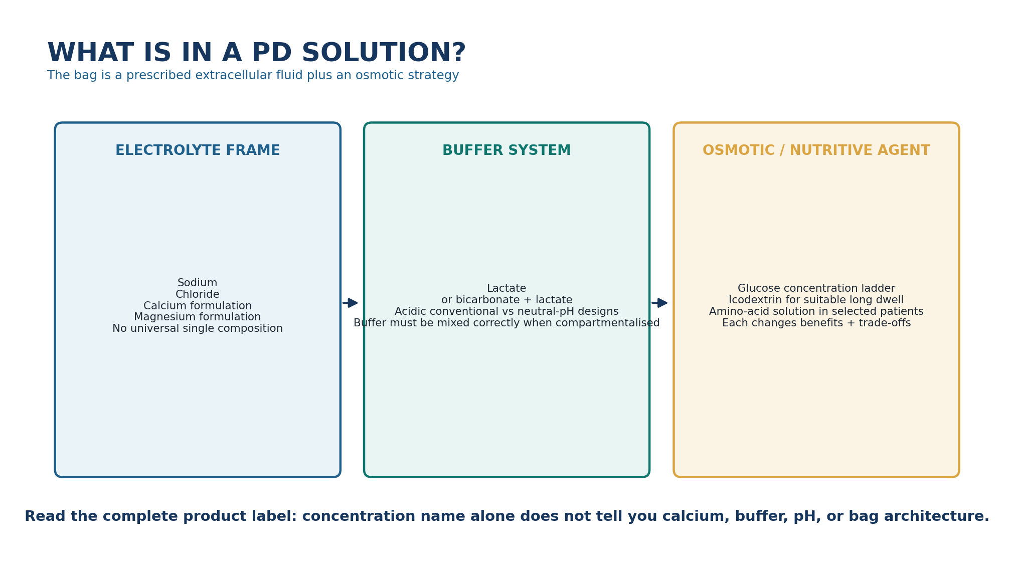
PD fluid is often described casually as “1.5%” or “2.5%,” but the glucose label is only one dimension of the prescription. Commercial solutions vary in buffer composition, calcium concentration, magnesium concentration, glucose labeling convention, pH/GDP profile and bag design. The clinician should therefore prescribe and document the complete product formulation rather than assume that two bags with the same apparent glucose strength are chemically interchangeable. [1,15,16]
Table 2.3 - Read the label in this order.
| Label element | What it controls | Why it changes care |
|---|---|---|
| Osmotic agent | Water-removal strategy and metabolic exposure | Glucose, icodextrin and amino acids are not interchangeable |
| Glucose strength / convention | Crystalloid osmotic force | Anhydrous and monohydrate labels can look numerically different |
| Volume | Intraperitoneal dose and pressure burden | Wrong bag volume can alter fill tolerance and delivered clearance |
| Sodium / chloride | Dialysate electrolyte frame | Influences sodium gradient and chloride load; exact product values vary |
| Calcium | Dialysate calcium balance | Interacts with CKD-MBD therapy and serum calcium |
| Magnesium | Dialysate magnesium balance | May influence serum magnesium; formulations differ |
| Buffer | Acid-base support and inflow tolerance | Lactate vs bicarbonate/lactate designs differ |
| Bag architecture | Whether mixing is required before use | Failure to mix compartmentalised fluid can make the solution unsafe |
PRESCRIPTION LANGUAGE Do not write only “2.5% bag.” Record the locally used product or formulation, glucose convention, volume, calcium formulation when clinically relevant, osmotic agent and whether a last-fill/long-dwell solution is intended.
3. Glucose solutions: understand the concentration convention before prescribing
Glucose remains the most widely used osmotic agent in PD. A higher dialysate glucose concentration creates a larger crystalloid osmotic gradient and generally greater early ultrafiltration, at the cost of greater glucose exposure and absorption. The concentration ladder is familiar, but the numbers printed on bags vary by market and whether glucose is expressed as anhydrous glucose or glucose monohydrate. [15,16]
Table 2.4 - A common glucose-labeling equivalence: check the actual product label.
| Anhydrous glucose label | Approximate glucose-monohydrate label | Clinical interpretation |
|---|---|---|
| 1.36% w/v | 1.5% w/v | Lower glucose strength within the common commercial ladder |
| 2.27% w/v | 2.5% w/v | Intermediate glucose strength |
| 3.86% w/v | 4.25% w/v | Higher glucose strength; repeated use should trigger review of the underlying volume problem |
The pairs above are not separate physiologic prescriptions; they are alternative labeling conventions used in commercially available fluids. This is exactly why copying a percentage from a foreign guideline, teaching slide or discharge letter without checking the local product can create error. [15,16]
THRESHOLD DISCIPLINE Use the lowest osmotic burden that achieves the required clinical fluid goal. KDIGO volume guidance and product labeling both support avoiding reflex dependence on the most hypertonic glucose solution; recurrent need for very hypertonic exchanges should prompt review of sodium/fluid intake, residual urine, dwell design, mechanical function and membrane physiology. [13,16]
4. Buffer, pH and glucose degradation products: why modern bags may have two chambers
Traditional glucose-based PD fluids were commonly lactate buffered and acidic to reduce glucose degradation during heat sterilization and storage. More biocompatible designs separate glucose from buffer/electrolytes during sterilization, then mix the compartments immediately before use to generate a neutral or near-physiologic pH solution with lower GDP exposure. The bag design is therefore part of the chemistry. [5,6,14,16]
Table 2.5 - Conventional versus neutral-pH/low-GDP solution design.
| Feature | Conventional single-compartment concept | Neutral-pH/low-GDP concept |
|---|---|---|
| Storage / sterilization | Glucose and buffer stored together in acidic environment | Components separated to reduce heat-generated GDP formation |
| Final pH | Typically acidic | Closer to physiologic after mixing |
| Buffer | Often lactate only | May use lactate or bicarbonate/lactate depending product |
| Bag preparation | No compartment mixing step | Internal seals must be opened and contents completely mixed |
| Biologic rationale | Greater local acidic/GDP exposure | Reduced GDP and improved mesothelial biocompatibility markers |
| Hard outcomes | Standard comparator | Patient/technique survival advantage remains uncertain |
Randomized studies, including balANZ, and systematic reviews suggest better preservation of residual kidney function or urine volume with neutral-pH/low-GDP solutions, while effects on peritonitis, technique survival and mortality are less certain. The clinical signal is therefore real but not a licence to promise broad outcome superiority. [5,6,14]
MIXING RULE A multi-compartment solution is not ready because it is warm or hanging on the pole. Release the internal seals exactly as instructed, visually confirm complete mixing, and only then connect. A partially mixed bag can deliver the wrong pH and composition.
5. Electrolytes: similar does not mean identical
Most PD solutions use an extracellular-fluid-like sodium/chloride framework and contain calcium, magnesium and buffer. However, exact values vary by manufacturer and formulation. For example, contemporary product families may offer more than one calcium concentration, while neutral-pH bicarbonate/lactate solutions have a different buffer profile from lactate-only fluids. [15,16]
Table 2.6 - Electrolyte component -> clinical question.
| Component | Clinical question | Common error |
|---|---|---|
| Sodium | Is the patient sodium overloaded, and what is the total sodium balance across PD + urine + intake? | Assuming bag sodium alone determines volume control |
| Calcium | Does dialysate calcium fit serum calcium, PTH/bone strategy and calcium-based therapies? | Treating all glucose bags as the same calcium formulation |
| Magnesium | Could dialysate concentration contribute to hypo- or hypermagnesemia? | Never checking formulation when magnesium changes |
| Potassium | Most standard chronic PD fluids contain no potassium; does the patient have a clinical reason for prescribed supplementation? | Adding electrolytes without compatibility/sterility protocol |
| Buffer | Is acid-base correction adequate and is inflow pain linked to solution characteristics? | Assuming buffer type is clinically irrelevant |
SAFETY BOUNDARY Electrolyte or drug additives to PD bags require a validated sterile process and compatibility information. This chapter does not provide a generic recipe for potassium, insulin, heparin or antibiotic admixture; later clinical chapters and local pharmacy/PD protocols govern exact preparation.
6. Icodextrin: a long-dwell osmotic strategy with a unique safety issue
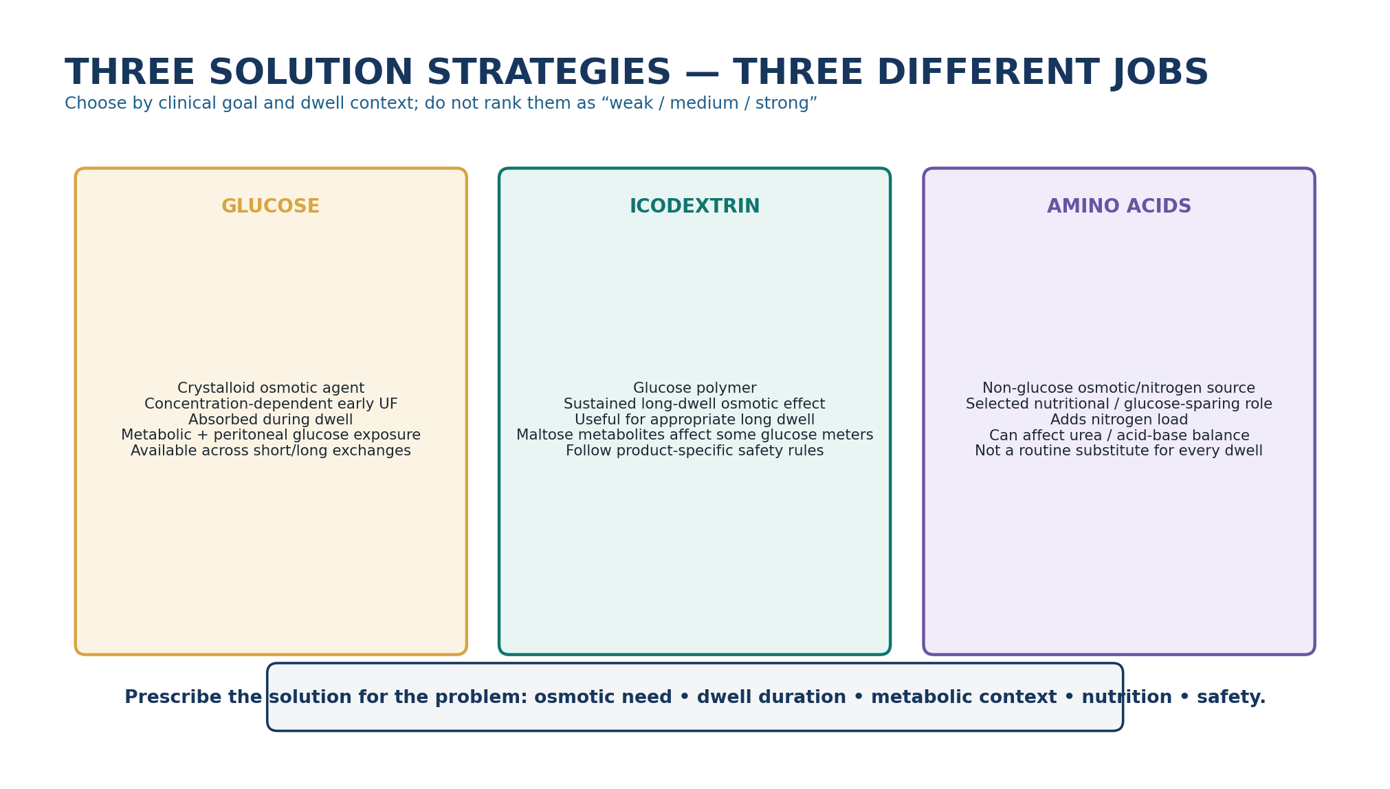
Icodextrin is a starch-derived glucose polymer used as an alternative osmotic agent for a suitable long dwell. Its macromolecular osmotic effect is sustained more effectively than the crystalloid glucose gradient during a long dwell, and randomized-trial syntheses support improved long-dwell ultrafiltration and reduced episodes of uncontrolled fluid overload in selected patients. [5,7,13]
Table 2.7 - Icodextrin: bedside use and safety.
| Domain | Clinical meaning | What must be verified |
|---|---|---|
| Role | Long-dwell osmotic alternative to glucose | The intended long dwell and local licensed use |
| Fluid effect | Often improves long-dwell UF compared with glucose | Volume status; avoid excessive volume depletion |
| Glucose sparing | Reduces glucose exposure for the replaced dwell | Total daily prescription, not one bag in isolation |
| Metabolites | Maltose and related metabolites circulate | Compatible glucose-measurement method |
| Hypersensitivity / sterile inflammation | Uncommon but clinically important | Rash, abdominal symptoms, cloudy effluent require assessment |
| Hospital transition | Safety risk if staff do not know the patient uses icodextrin | Medication reconciliation + glucose-meter compatibility alert |
HIGH-ALERT SAFETY Icodextrin metabolites can falsely elevate readings with some non-glucose-specific blood glucose methods, potentially causing dangerous insulin administration and missed hypoglycaemia. The product warning extends beyond the last dwell because interference can persist after discontinuation. Every admission should explicitly flag icodextrin use and verify the glucose meter/test strip method. [17]
7. Amino-acid solutions: selected nutritional and glucose-sparing use
Amino-acid PD solutions use amino acids rather than glucose as the principal osmotic/nitrogen source. Commercial 1.1% formulations are intended for selected patients, particularly when nutritional support or glucose-sparing is desired, and are generally used as part of a broader PD prescription rather than as a replacement for every exchange. [18,19]
Table 2.8 - Amino-acid solution: what it can and cannot do.
| Potential role | Potential advantage | Limitation / monitoring |
|---|---|---|
| Nutritional support | Provides absorbable amino acids during a dwell | Benefit depends on overall energy intake and catabolic state |
| Glucose sparing | Replaces one glucose exposure in selected regimens | Does not remove the need for adequate osmotic/volume strategy |
| Solute clearance | Can support dialysis exchange similarly to low-strength glucose in selected contexts | Not prescribed for clearance alone |
| Nitrogen load | May support protein balance | Can raise urea generation and requires metabolic follow-up |
| Acid-base / electrolytes | Product-specific formulation | Monitor bicarbonate and potassium; exact risks depend on patient and formulation |
EVIDENCE CALIBRATION The evidence base for amino-acid PD solution is much smaller and older than for glucose or icodextrin, and clinically important long-term outcomes remain uncertain. Use it as a targeted tool within a nutritional plan, not as a universal “biocompatible” replacement. [18,19]
8. Bag release: inspection, mixing and warming are clinical safety steps
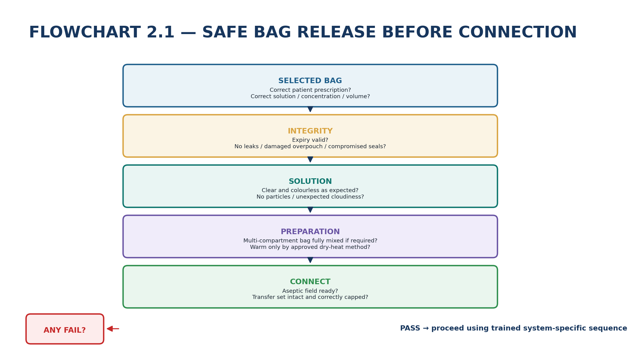
Table 2.9 - Pre-connection bag check.
| Check | Pass criterion | If it fails |
|---|---|---|
| Prescription | Correct solution type, strength, calcium/buffer formulation and volume | Do not connect; reconcile order |
| Expiry / storage | Within expiry and stored according to product requirements | Quarantine bag |
| Overpouch / seams | No leak, puncture or compromised seal | Discard/quarantine |
| Clarity | Solution is clear and expected colour; no visible particles | Do not infuse; investigate |
| Compartment mixing | Internal seals fully opened; solution completely mixed | Repeat approved mixing steps or replace bag |
| Temperature | Comfortably warmed by approved method if warming used | Do not microwave or improvise unsafe heating |
| Additives | Only prescribed, compatible, aseptically prepared additives present | Do not use if identity/compatibility uncertain |
Current product information for modern PD fluids commonly permits warming in the overpouch using dry heat and warns against microwave heating; some labels also warn against water-bath warming because of contamination risk. The exact warming device and procedure remain governed by the product and local PD program. [16,18]
VISUAL RULE Cloudiness belongs to the clinical pathway, not the warming tray. Never “warm and see if it clears” or infuse a visibly abnormal bag. Likewise, after the exchange, inspect effluent: new cloudiness is peritonitis until appropriately evaluated.
9. Transfer sets and disconnect systems: the sterile interface
The transfer set is the patient-side extension connected to the PD catheter and repeatedly used to connect fresh exchange systems. It is a high-frequency contamination interface. Modern disconnect systems - Y-set and twin/double-bag designs - reduce the need for direct spiking/manipulation and permit a drain/flush/fill sequence within a closed fluid path. Randomized evidence supports lower peritonitis risk with Y-set or twin-bag systems compared with older conventional spike systems. [8,9]
Table 2.10 - Connectology: component -> purpose -> failure clue.
| Component | Purpose | Failure clue |
|---|---|---|
| Transfer set | Reusable patient-side connector and clamp interface | Crack, leak, loose connection, damaged threads, contamination |
| Disposable connector / Y-set | Links fresh solution and drain path | Wrong-path clamp sequence, touch contamination |
| Drain bag / line | Receives prior effluent and flush volume | Backpressure, leak, accidental elevation or occlusion |
| Fresh bag line | Delivers prescribed solution | Wrong bag connected, clamp closed, air or incomplete priming |
| Sterile cap / disconnect shield | Protects patient-side connector between exchanges | Cap loss, wetness, touched critical surface |
| Clamps | Control direction and maintain closure | Wrong clamp opened or left open during contamination |
INFECTION-PREVENTION PRINCIPLE Disconnect technology reduces risk; it does not replace technique. The strongest system still fails if critical sterile surfaces are touched, a contaminated open pathway is used, or a damaged transfer set is ignored. [2,3,8]
10. CAPD exchange logic: drain, flush-before-fill, fill, dwell
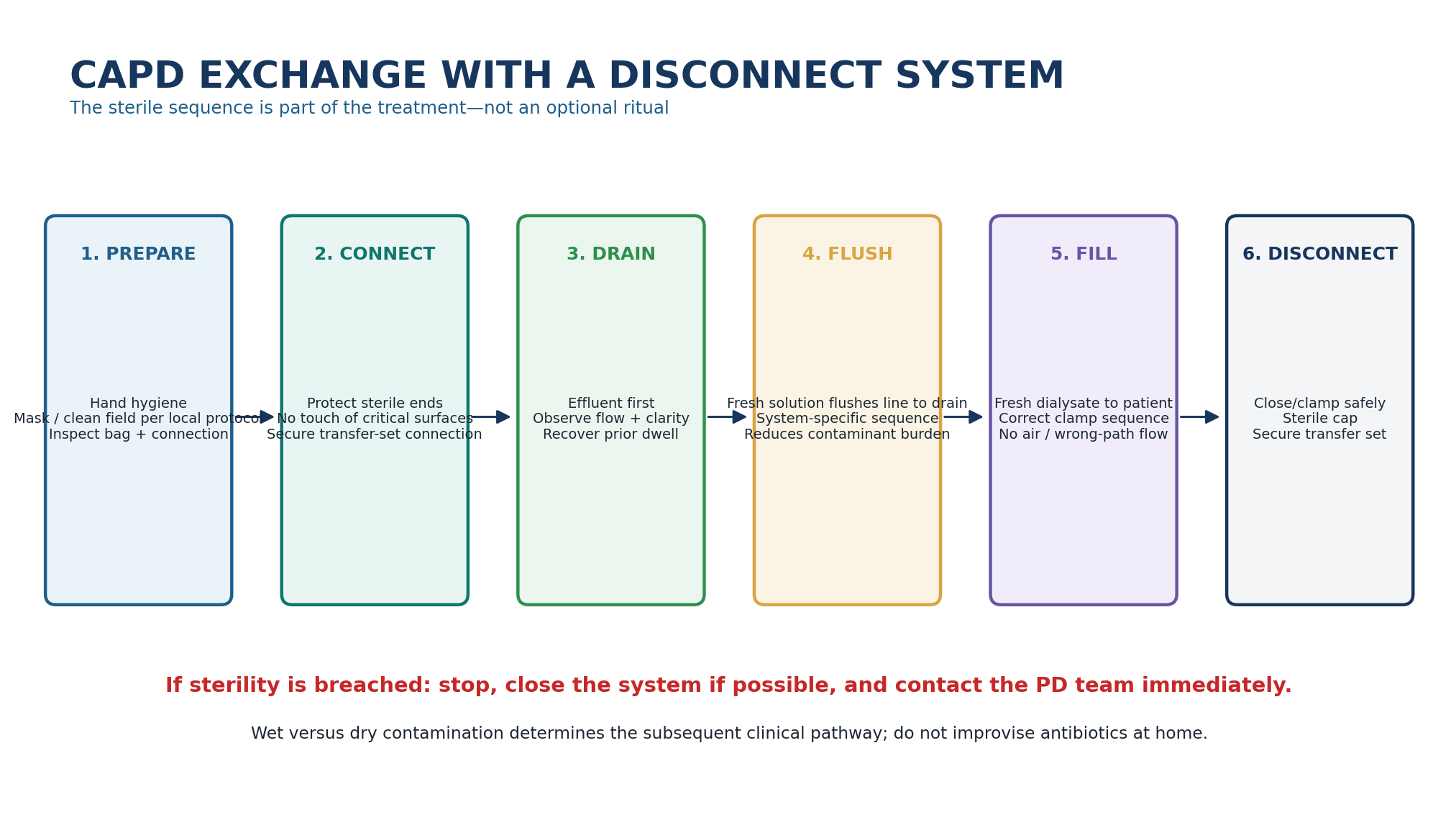
The manual exchange is best understood as fluid routing. The previous dwell is drained first. In disconnect systems, fresh solution is then used to flush the common line toward the drain pathway before the fill phase, helping remove air and potential contaminants from the connection path. The fresh solution is then allowed to fill the abdomen and the patient is disconnected using the system-specific sterile sequence. [8,9]
Table 2.11 - Read a CAPD exchange by phase.
| Phase | What should happen | What an abnormality suggests |
|---|---|---|
| Preparation | Correct bag; clean environment; transfer set inspected | System error before fluid ever moves |
| Drain | Previous dwell exits freely; effluent can be observed | Position, constipation, catheter or drain-path problem |
| Flush | Fresh solution directed to drain per system design | Wrong clamp sequence or connection error |
| Fill | Prescribed fresh volume enters without major pain/resistance | Kink, catheter position, high pressure, wrong clamp |
| Disconnect | Patient-side system closed and recapped aseptically | Contamination risk if sequence breached |
| Dwell | No external tubing connection until next exchange | Leak, discomfort or clinical problem becomes the focus |
SYSTEM RULE The exact hand movements, clamp order and connector sequence must be taught using the patient’s actual system. Generic diagrams teach logic; the manufacturer/local PD program teaches the executable procedure.
11. Contamination: dry versus wet is the first clinical distinction
ISPD 2022 specifically distinguishes contamination outside a closed system (“dry contamination”) from contamination involving an open fluid pathway (“wet contamination”). Examples of wet contamination include a leak or break proximal to the clamp, touch contamination of the connection during an exchange, or infusion after contamination. Patients should contact the PD team immediately whenever sterility is breached. [2]
Table 2.12 - Contamination classification and immediate logic.
| Situation | Likely category | Immediate principle |
|---|---|---|
| Disconnection occurs distal to a securely closed clamp with no open path to patient | Dry contamination | Keep system closed and contact PD team for device-specific advice |
| Critical connection touched while system is open | Wet contamination | Stop exchange; close if possible; contact PD team immediately |
| Tubing leak/break proximal to clamp | Wet contamination | Close pathway; do not continue using compromised set |
| Dialysate infused after contamination | Wet contamination | Urgent PD-team management; peritonitis prophylaxis pathway may be required |
| Unclear whether clamp was open | Treat as wet for safety | Do not assume “probably dry” |
ISPD suggests prophylactic antibiotics after wet contamination, with effluent cell count/culture preferably obtained and close monitoring. Because there is no single universal prophylaxis regimen, this chapter deliberately stops at recognition and escalation rather than supplying an at-home antibiotic recipe. [2]
DO NOT IMPROVISE After wet contamination, the correct action is not “replace the cap and continue.” Close the system if possible, contact the PD team immediately, and follow the program’s transfer-set and prophylaxis pathway. If there is uncertainty about whether the pathway was open, manage it as wet contamination. [2]
12. APD cycler architecture: a programmed fluid-routing machine
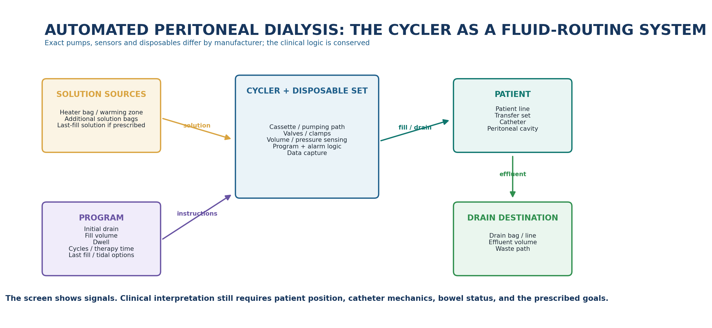
An APD cycler automates the same fundamental exchange phases performed manually, usually overnight. It does not change the underlying membrane physics; it changes the timing, repetition, fluid routing and data capture. Modern devices may use cassettes or integrated disposable sets with valves/pumping pathways and sensors to estimate volume, pressure or flow. Exact mechanisms are device-specific. [1,10,11]
Table 2.13 - Cycler component -> clinical interpretation.
| Cycler element | What it does | What the clinician should ask |
|---|---|---|
| Heater / warming zone | Brings selected solution toward target temperature | Was the correct bag placed and was warming completed safely? |
| Supply lines | Connect one or more prescribed solutions | Are the right solutions attached to the right lines? |
| Disposable cassette/set | Routes fluid through valves/pumps | Is it correctly seated/primed and free of kinks? |
| Patient line | Connects machine to transfer set/catheter | Is it clamped, kinked, tensioned or contaminated? |
| Drain line/bag | Receives effluent | Is the route low, unobstructed and appropriate? |
| Sensors/software | Detects flow/pressure/volume deviations | What physical condition could generate this signal? |
| Data/remote layer | Stores or transmits treatment metrics | Who reviews it, how often, and what triggers action? |
MASTER TRANSLATION A cycler alarm is a signal generated by a fluid-routing system. It is not a diagnosis of “catheter dysfunction,” “constipation” or “membrane failure.” Localise the phase and the physical pathway first.
13. APD program vocabulary: know what each variable changes
Table 2.14 - APD program terms without vendor jargon.
| Variable | Operational meaning | Clinical consequence |
|---|---|---|
| Initial drain | Removal of fluid present before nocturnal cycles | Unexpectedly large/small volume can alter the first cycle and raise safety questions |
| Fill volume | Volume intended to enter each cycle | Affects contact area, pressure, tolerance and clearance |
| Dwell time | Time fluid remains relatively undisturbed for transport | Shortening/lengthening changes solute and UF behavior |
| Drain | Removal phase after a dwell | Slow/incomplete drainage consumes therapy time and alters net UF |
| Number of cycles | How often gradients are renewed | More cycles are not automatically more effective if dwell becomes too short |
| Total therapy time | Clock time allocated to automated treatment | Includes non-dwell phases and may be eroded by alarms/slow flow |
| Last fill | Fluid left in abdomen after cycler treatment | Creates a daytime dwell and must match the intended solution/volume |
| Day exchange | Manual or automated exchange outside the main nocturnal session | Adds clearance/UF but also burden |
| Tidal mode | Leaves a residual intraperitoneal volume between partial drains | May reduce drain pain/flow interruption but changes volume accounting |
PRESCRIPTION RULE Do not translate APD into “number of cycles” alone. A cycle is fill + dwell + drain. If the drain phase repeatedly expands, the actual dwell architecture may be very different from the prescription.
14. Lost dwell time: treatment duration is not the same as effective dwell duration
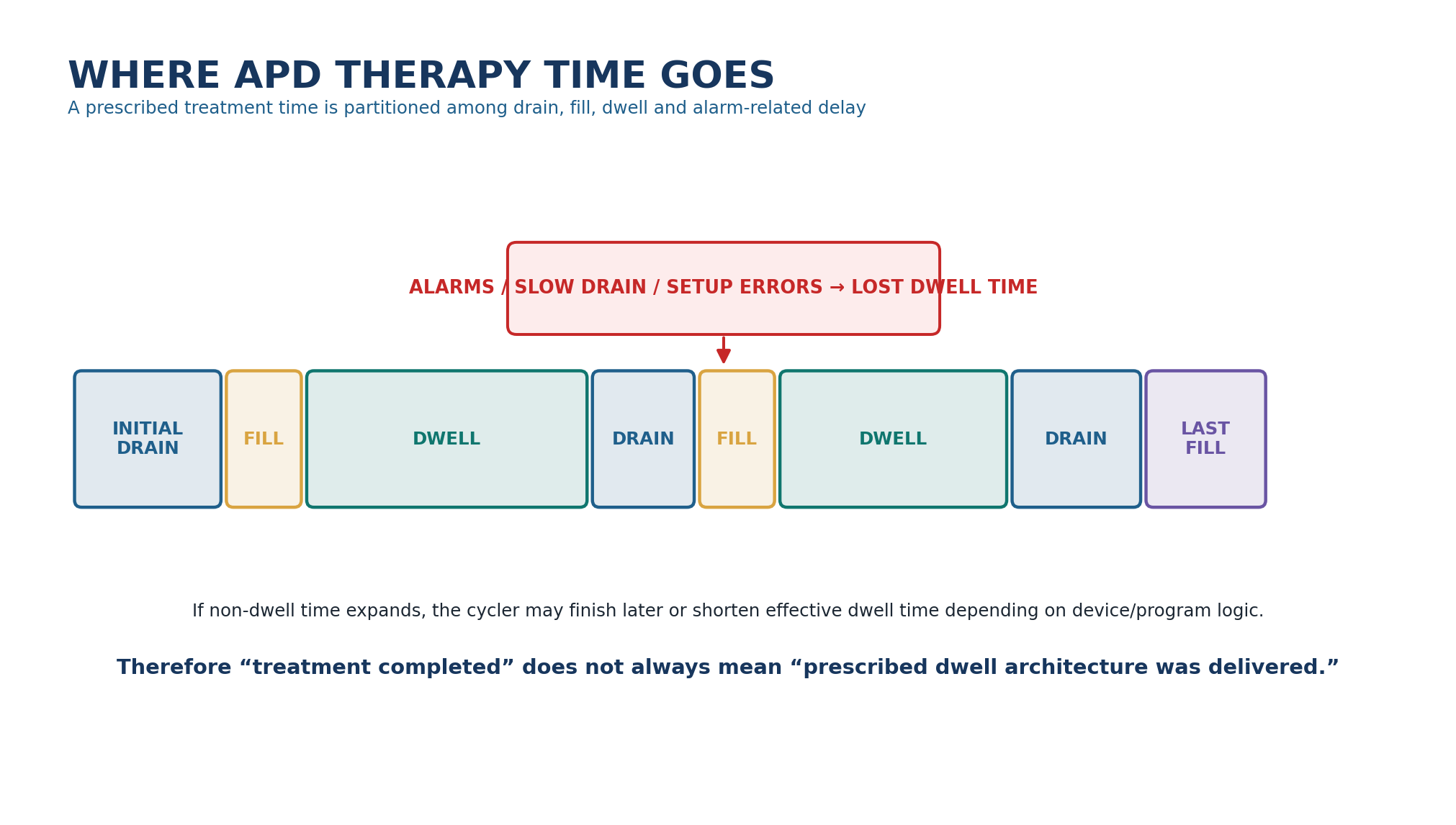
In APD, the membrane only transports according to the time the dialysate is actually in the cavity. Filling, draining, alarm handling and repeated troubleshooting occupy clock time without providing the intended dwell. In a 2024 inpatient study, slow outflow, inadequate drain volumes, patient-line occlusion and priming errors were prominent contributors to lost dwell time. The exact frequency in stable home populations may differ, but the principle is general: non-dwell time can materially change delivered therapy. [10]
Table 2.15 - Lost dwell time: cause -> consequence -> response.
| Cause | Consequence | Response direction |
|---|---|---|
| Slow drain | Longer non-dwell phase; delayed completion | Position/bowel/catheter/drain-path assessment |
| Repeated drain alarms | User interruptions and possible early cycle termination | Localise mechanism; do not normalize resetting |
| Line occlusion/kink | Interrupted fill or drain | Inspect disposable set, patient line and body position |
| Priming/setup error | Treatment starts late or repeatedly alarms | Correct setup; retraining if recurrent |
| Wrong program | Delivered dwell/cycle structure differs from intended | Prescription reconciliation before next treatment |
BEDSIDE TRANSLATION If a patient has worsening biochemical control or a much longer overnight treatment, ask for the cycler log and actual dwell time before prescribing more volume. The missing dose may be time, not dialysate.
15. Drain alarms: mechanics before membrane
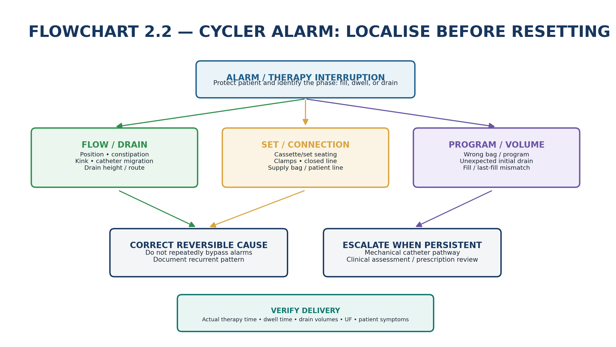
Table 2.16 - Common APD alarm phenotypes.
| Pattern | First hypotheses | Do not jump to |
|---|---|---|
| Slow drain after turning in bed | Position-dependent catheter flow; line kink | Permanent membrane failure |
| Slow drain nightly with constipation | Bowel-related catheter compression/migration | More hypertonic glucose |
| Fill alarm immediately after setup | Clamp/line/cassette/supply problem | Catheter surgery before checking set |
| Large unexpected initial drain | Carry-over from prior dwell; overfill; inaccurate prior record | Assuming cycler overfilled without reconciliation |
| Repeated incomplete drains + pain | Catheter position, suction/drain pain, constipation, pelvic mechanics | Repeated forceful bypass |
| Alarm only after new disposable setup | Priming/seating/tubing issue | New peritoneal transport problem |
ALARM DISCIPLINE Never widen limits, repeatedly bypass a drain phase or normalize line manipulation simply to finish the session. Recurrent alarms are clinical data. Document the pattern and correct the mechanism.
16. Tidal PD: a fluid-management tool, not a separate dialysis modality
Tidal PD leaves a programmed residual volume in the abdomen after each partial drain, then refills on top of that residual volume. It can reduce painful or prolonged attempts to drain the abdomen completely during every cycle and can help selected patients with recurrent drain discomfort or flow problems. Exact tidal percentages, safety limits and end-of-treatment drain logic are device- and protocol-specific and should not be generalized. [1,10]
Table 2.17 - Tidal PD: why it helps and what it can hide.
| Potential advantage | Mechanism | Trade-off / caution |
|---|---|---|
| Less drain pain | Avoids repeated near-empty pelvic suction | Residual volume remains between cycles |
| Fewer prolonged drain attempts | Partial drains may bypass slow terminal drainage | Net UF accounting can be less intuitive |
| More stable cycle timing | Less time spent chasing complete drains | If residual volume progressively accumulates, overfill risk must be prevented by device logic/protocol |
| May improve tolerance | Useful in selected mechanical phenotypes | Does not correct constipation, migration or true obstruction |
THRESHOLD DISCIPLINE There is no universal tidal percentage that belongs in a textbook order. Use the device-specific protocol, know the programmed residual volume and final drain strategy, and reassess pain, drainage, UF and treatment time.
17. CAPD versus APD: system choice follows the person and the physiology
CAPD and APD use the same peritoneal membrane but package the exchange differently. CAPD distributes manual exchanges across the day. APD concentrates repeated automated exchanges into a defined treatment window and may add a daytime/last-fill dwell. ISPD goal-directed prescribing explicitly treats modality, exchange volume/frequency, dwell length, cycler type, connectology and remote monitoring as adjustable elements of an individualized prescription. [1]
Table 2.18 - CAPD and APD as system designs.
| Dimension | CAPD | APD |
|---|---|---|
| Exchange engine | Gravity/manual disconnect system | Programmed cycler + disposable set |
| Dwell pattern | Usually longer daytime and overnight manual dwells | Repeated nocturnal cycles + optional daytime dwell |
| Human workload | Multiple manual sterile connections each day | Nightly setup plus cycler interaction/alarms |
| Sleep impact | Usually no machine overnight | Noise, alarms and drain interruptions may affect sleep |
| Data availability | Manual logs unless digitally supported | Detailed treatment logs often available; remote connectivity may be possible |
| Transport fit | Can suit slower transport and long-dwell strategies | Can exploit shorter repeated dwells in selected fast transporters |
| Choice | Lifestyle, dexterity, support, work/travel, physiology | Same - no universal “better modality” |
DO NOT AUTOMATE A fast transporter does not automatically require APD, and APD is not automatically superior for a working patient. Integrate membrane behavior, residual kidney function, volume needs, sleep, employment, care partner burden and patient preference. Detailed modality selection belongs to Chapter 4 and prescription optimization to Chapter 9.
18. Remote monitoring: data are useful only when someone owns the response
Contemporary cyclers may transmit prescribed and delivered treatment information, including adherence, therapy time, drain/fill behavior, ultrafiltration and alarms. Reviews through 2026 describe potential benefits for earlier troubleshooting and reduced treatment failure/hospital use in some programs, but implementation remains heterogeneous and depends on staffing, alert thresholds, privacy, broadband access and response protocols. [11,12]
Table 2.19 - Remote data: signal -> possible use -> governance question.
| Data element | Clinical use | Governance question |
|---|---|---|
| Missed/shortened therapy | Adherence or technical-support trigger | Who reviews and by when? |
| Repeated drain alarms | Early mechanical troubleshooting | What frequency triggers contact? |
| UF trend | Volume-management context | How is weight/BP/urine integrated? |
| Therapy time / lost dwell | Detect delivery erosion | Does the platform show actual dwell versus clock time? |
| Prescription change | Remote adjustment in supported systems | Who authorizes and verifies implementation? |
| Connectivity gap | May represent no data rather than no treatment | How are offline patients safeguarded? |
CONTINUITY NOTE Chapter 18 will develop remote monitoring, connected cyclers, alert governance, data overload and adaptive PD. Here, the rule is simple: a remote dashboard does not replace clinical ownership.
19. Prescribed therapy versus delivered therapy: verify before redesign
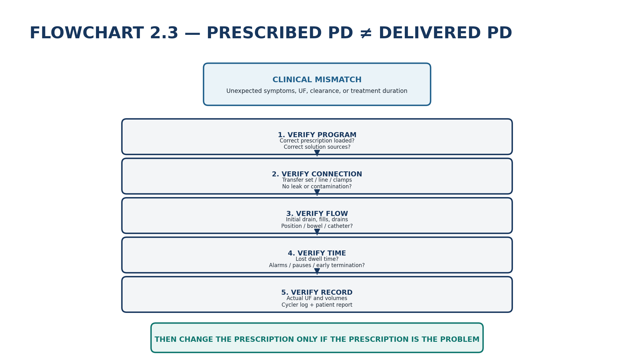
Table 2.20 - A delivered-treatment audit.
| Question | Data to obtain | What failure means |
|---|---|---|
| Was the correct prescription loaded? | Cycler/program record or written CAPD schedule | Programming error, not membrane failure |
| Were the correct bags used? | Solution type, concentration, volume, last-fill product | Wrong osmotic/electrolyte strategy |
| Was every exchange completed? | Missed cycles, early disconnects, bypassed drains | Underdelivery |
| Was dwell time preserved? | Actual dwell/lost dwell time | Clearance/UF can differ from planned |
| Were fills and drains plausible? | Volume log, initial drain, drain alarms | Mechanical or measurement issue |
| Was the patient clinically stable? | Weight, BP, edema, symptoms, urine | Machine success may coexist with clinical failure |
GOAL-DIRECTED RULE Change the prescription only after confirming that the prescription - rather than the bag, connection, program or catheter flow - is the failing component.
20. Clinical pearls
Table 2.21 - Ten system pearls worth carrying to the bedside.
| Pearl | Why it matters |
|---|---|
| The bag label is part of the prescription | Glucose percentage alone does not specify buffer, calcium, pH/GDP profile, volume or product architecture. |
| A two-chamber bag is not ready until it is mixed | The final clinically intended solution does not exist until the internal seal is opened and contents are mixed. |
| Connectology is clinical care | Aseptic connection/disconnection technique directly modifies peritonitis risk and deserves the same attention as drug prescribing. |
| Wet contamination is an exposure, not a cosmetic breach | Once an open pathway has been contaminated or fluid infused after contamination, immediate PD-team management is required. |
| An alarm describes a phase failure, not its cause | Start with fill/drain/dwell/program localization before deciding on catheter pathology or prescription change. |
| Treatment time is not dwell time | Slow drains, repeated alarms and setup delays can consume the clock while reducing effective dwell. |
| Icodextrin has a hospital-safety signature | The glucose-meter interaction must follow the patient across emergency, ward and procedural settings. |
| Tidal PD is a tool, not a diagnosis | It may reduce terminal-drain pain or selected flow problems but should not hide constipation or catheter dysfunction. |
| Remote data require clinical ownership | A dashboard improves care only when someone is responsible for review, contact and escalation. |
| Verify delivery before intensifying therapy | A correctly written prescription cannot compensate for wrong bags, missed cycles, lost dwell or poor drainage. |
21. Mini-cases: system decisions, not trivia
Case 1 - The “same” glucose bag
A patient transfers from another country with a written prescription using 2.5% glucose. The local bags are labelled 2.27% glucose. A staff member assumes the local concentration is weaker and plans to substitute a more hypertonic bag.
REASONING Stop and decode the labeling convention. Some products express anhydrous glucose while others are colloquially described by glucose-monohydrate-equivalent strengths. Confirm the product formulation rather than escalating osmotic strength because the printed number looks smaller. [15,16]
Case 2 - The two-chamber bag that was never mixed
A neutral-pH, low-GDP solution is warmed and connected, but the internal chamber seal has not been fully opened.
REASONING Do not infuse. The two chambers were separated to preserve chemical stability; the final prescribed buffer/electrolyte/glucose composition exists only after complete mixing. Replace or correctly prepare the bag according to product instructions and retrain the setup step.
Case 3 - A touched connector
During CAPD, the patient touches the exposed connection and then continues the fill. There is no abdominal pain and the effluent was clear before the event.
REASONING This is wet contamination because the sterile open pathway was breached and fluid was infused after contamination. Stop improvisation, close the system if possible and contact the PD team immediately. ISPD recommends a wet-contamination prophylaxis pathway; exact antibiotics are program-specific. [2]
Case 4 - The cycler takes two extra hours
An APD patient reports that a formerly 9-hour treatment now lasts almost 11 hours. The machine ultimately says treatment complete. Multiple slow-drain alarms occur most nights and constipation has worsened.
REASONING Completion does not prove delivered dwell architecture. Recurrent slow drains are consuming clock time and may reduce effective dwell. Treat constipation, review position/catheter/drain path and inspect the treatment log before adding cycles or fluid volume. [10]
Case 5 - False hyperglycaemia in hospital
A diabetic patient using icodextrin is admitted to a ward. Point-of-care glucose readings are unexpectedly high and repeated correction insulin is ordered despite minimal symptoms.
REASONING Treat the meter-method question as an emergency safety issue. Icodextrin-derived maltose can falsely elevate glucose readings with susceptible methods and lead to dangerous insulin administration. Use a glucose-specific compatible method and alert the admitting team. [17]
Case 6 - Drain pain on every APD cycle
A patient has sharp pelvic discomfort at the end of each drain. Flow is otherwise adequate, imaging is unrevealing and constipation has been addressed.
REASONING The pain may reflect terminal near-empty drainage rather than true obstruction. A device-specific tidal strategy can be considered to avoid repeated complete drains, while ensuring residual volume and final-drain safety are controlled. Do not prescribe a universal tidal percentage from memory.
22. Common pitfalls - and the correction
Table 2.22 - High-frequency errors in PD system care.
| Pitfall | Why it fails | Correction |
|---|---|---|
| Prescribing only “1.5 / 2.5 / 4.25%” | Label conventions and other solution components vary | Specify the local formulation/product and volume |
| Assuming two glucose bags are interchangeable | Calcium, buffer, pH/GDP and bag design may differ | Read the full label |
| Failing to mix a two-chamber bag | Final solution chemistry is wrong | Open seals and mix completely before connection |
| Microwaving PD fluid | Uneven heating/container injury and product warnings | Use approved dry-heat warming method |
| Treating transfer-set sterility as “nursing detail” | Touch contamination causes peritonitis | Make connectology part of medical review |
| Replacing a cap and ignoring wet contamination | Open-path contamination can seed peritonitis | Contact PD team immediately; follow wet-contamination pathway |
| Resetting recurrent drain alarms | Masks constipation, position or catheter problem and erodes dwell time | Localise and correct the mechanism |
| Calling APD complete because the screen is green | Delivered dwell and volumes may differ from prescribed | Review logs, lost dwell, UF and symptoms |
| Using tidal PD to hide obstruction | Partial drains can reduce alarms without fixing pathology | Treat bowel/catheter causes first |
| Assuming remote monitoring equals active care | Data without response ownership do not improve safety | Define review and escalation responsibilities |
23. Active recall: retrieve before you re-read
MUST MEMORIZE
Table 2.23 - Must memorize.
| Prompt | Answer |
|---|---|
| What are the three functional elements of a PD solution? | Electrolyte frame, buffer system, and osmotic/nutritive agent. |
| Why can 1.36% and 1.5% glucose refer to the same osmotic rung? | Some labels express anhydrous glucose while the familiar alternative reflects glucose-monohydrate equivalent; confirm the product label. |
| Why do many neutral-pH/low-GDP bags have two chambers? | To separate components during sterilization/storage and reduce GDP formation; they must be fully mixed before use. |
| Generic CAPD disconnect sequence? | Prepare/connect -> drain -> flush-to-drain -> fill -> secure sterile disconnection -> dwell. |
| Dry versus wet contamination? | Dry contamination remains outside a closed sterile pathway; wet contamination involves an open pathway or infusion after contamination. |
| What is tidal PD? | APD that intentionally leaves a residual intraperitoneal volume between partial drains. |
| Key icodextrin safety interaction? | Maltose can falsely elevate glucose with susceptible meters, risking inappropriate insulin and severe hypoglycaemia. |
MUST REASON
Table 2.24 - Must reason.
| Clinical prompt | Reasoning target |
|---|---|
| The cycler says complete, but treatment now takes 2 h longer. | Ask how much clock time was lost to fills/drains/alarms and whether effective dwell was preserved. |
| A long-dwell patient is switched from glucose to icodextrin. | Explain the sustained osmotic rationale, then identify product-specific safety monitoring rather than calling it simply “stronger fluid.” |
| Repeated slow-drain alarms occur only supine. | Localize position/constipation/catheter/drain mechanics before changing osmotic prescription. |
| A patient touches the open connector but remains asymptomatic. | Classify the breach, stop further exposure and activate the wet-contamination pathway; absence of symptoms does not neutralize exposure. |
| A two-chamber bag was connected without complete mixing. | Recognize a solution-preparation error: stop use and correct the bag process rather than interpreting symptoms as membrane failure. |
| Remote monitoring shows recurrent alarms but no missed sessions. | Decide whether alarms are eroding dwell/UF and who owns follow-up; adherence alone does not establish adequate delivery. |
USE AS REFERENCE
REFERENCE, NOT MEMORY Keep product-specific glucose-label conventions, exact electrolyte concentrations, warming instructions, transfer-set steps, cycler button sequences, alarm limits, tidal settings and contamination antibiotic regimens in the current local product/device/program reference. Memorize the physiologic and safety logic; verify the operational detail.
24. Flashcards: spaced-repetition deck
1. Q: What does a PD solution label need beyond glucose percentage? A: Product/formulation, osmotic agent, buffer/electrolyte composition, volume and relevant bag architecture.
2. Q: What hard-outcome claim can you make for neutral-pH/low-GDP solutions? A: Trials/meta-analysis support better preservation of residual kidney function/urine; mortality and technique-survival benefit remain uncertain.
3. Q: Best physiologic role for icodextrin? A: A sustained long-dwell osmotic strategy in appropriate patients.
4. Q: Role of amino-acid PD solution? A: A selected nutritional/glucose-sparing tool, not a universal replacement for glucose exchanges.
5. Q: Why inspect a bag before warming/connection? A: Wrong product, leak, damaged seal, particles or cloudiness must be found before infusion.
6. Q: Can a microwave be used to warm PD fluid? A: No; use the product/local approved warming method, commonly dry heat.
7. Q: What is the transfer set? A: The reusable patient-side interface linking the catheter to each disposable exchange system.
8. Q: Which connection technology has randomized evidence for less peritonitis than old spike systems? A: Disconnect Y-set/twin-bag systems.
9. Q: What should happen after any sterility breach? A: Stop/close the pathway if possible and contact the PD team immediately for contamination classification and action.
10. Q: Is a cycler alarm a diagnosis? A: No. It is a signal; localize the treatment phase and physical cause.
11. Q: Why can APD “finish” yet deliver less effective dialysis? A: Slow fills/drains and alarms can consume time intended for dwell.
12. Q: When can tidal PD help? A: Selected terminal-drain pain or flow problems after reversible mechanical causes are addressed.
13. Q: What must be reviewed before escalating APD? A: Program, bags, connection, fill/drain performance, lost dwell, UF and patient clinical status.
14. Q: What is the governance question in remote monitoring? A: Who reviews which data, when, and what trigger produces an action?
15. Q: First principle when prescribed and delivered therapy disagree? A: Correct the system failure before intensifying the prescription.
25. Final revision sheet
TEN TAKE-HOME RULES 1) The PD system is a sterile therapy chain. 2) A glucose percentage is not the whole solution prescription. 3) Check whether glucose is labelled as anhydrous or monohydrate equivalent. 4) Multi-compartment neutral-pH fluids must be completely mixed before use. 5) Icodextrin is a long-dwell strategy with a critical glucose-meter interaction. 6) Disconnect systems reduce peritonitis risk but aseptic technique remains decisive. 7) Wet contamination requires immediate PD-team management. 8) A cycler alarm is a localisation problem, not a diagnosis. 9) Lost dwell time can turn a completed APD session into an underdelivered prescription. 10) Verify delivered therapy before redesigning the prescription.
Table 2.25 - One-minute bedside synthesis.
| If you see... | Think... | Do now... |
|---|---|---|
| Bag percentage looks different from previous country/system | Glucose-label convention mismatch | Check anhydrous vs monohydrate label and full formulation |
| Two-chamber bag not fully mixed | Unsafe final chemistry | Do not connect/infuse; prepare correctly or replace |
| Cloudy unused solution bag | Product integrity problem | Do not use; quarantine/investigate |
| Touched open connector during exchange | Wet contamination | Close if possible; contact PD team immediately |
| Repeated slow-drain alarms | Position/constipation/catheter/drain-path problem | Localise mechanics; review dwell-time loss |
| APD session much longer than usual | Non-dwell time expanding | Review alarm log, drain time and actual dwell |
| Icodextrin patient + implausibly high capillary glucose | Meter interference possible | Use compatible glucose-specific method urgently |
| Drain pain only near end of each APD drain | Terminal drain/suction phenotype | Address mechanics; consider device-specific tidal strategy |
| Cycler says complete but patient clinically worse | Delivered therapy may not match prescribed | Audit bags, program, volumes, dwell and UF |
| Remote dashboard shows repeated alarms | Actionable trend only if ownership exists | Contact patient per protocol; define clinical response |
Table 2.26 - Before and after any PD-system change.
| Check | Pass criterion |
|---|---|
| Prescription identity | Correct solution, concentration convention, volume and intended long-dwell/last-fill agent documented |
| Bag integrity | In date, intact, clear, fully mixed if required and safely warmed |
| Connection | Transfer set/cap intact; trained aseptic pathway used |
| Program | Correct APD prescription loaded; supply lines match intended fluids |
| Flow | Fills/drains occur without persistent unexplained alarms or pain |
| Delivery | Actual dwell, cycles, volumes and UF are plausible and recorded |
| Patient | Symptoms, weight/volume, BP and treatment burden remain acceptable |
| Learning | Any contamination/alarm/setup error produces retraining or system correction when indicated |
FINAL MENTAL MODEL Prescription -> correct solution -> intact/mixed/warmed bag -> sterile connection -> drain/flush/fill or cycler routing -> dwell -> drain -> verify volumes/time/UF -> inspect patient and effluent -> correct the failing system component -> only then redesign the prescription.
Rapid oral viva
Explain why “2.5% glucose” is not a complete PD solution prescription.
Explain why a neutral-pH/low-GDP bag may require two compartments and what happens if it is not mixed.
Compare glucose, icodextrin and amino-acid solutions by mechanism and clinical role.
Walk through a CAPD disconnect exchange without using brand-specific connector names.
Distinguish dry from wet contamination and state the immediate action after wet contamination.
Trace an APD cycler from solution source to patient and drain, then localise a slow-drain alarm.
Explain lost dwell time and why a completed cycler session may still be underdelivered.
Explain tidal PD, one situation in which it may help and one way it can conceal a mechanical problem.
Name the icodextrin/glucose-meter safety issue and how you would prevent it during hospital admission.
List the data you would review before escalating an APD prescription for worsening clearance or volume control.
SCOPE BOUNDARY This chapter teaches solutions, connectology and delivery systems. Formal membrane testing is Chapter 3; patient/modality selection Chapter 4; catheter placement/function Chapter 5; initial prescription Chapter 6; adequacy/RKF Chapter 7; volume/UF failure Chapter 8; CAPD/APD prescription optimization Chapter 9; peritonitis and exit-site/tunnel infection Chapters 10-11; mechanical complications Chapter 12; and connected cyclers/remote monitoring Chapter 18.
26. Selected authoritative references
1. Brown EA, Blake PG, Boudville N, Davies S, de Arteaga J, Dong J, et al. International Society for Peritoneal Dialysis practice recommendations: Prescribing high-quality goal-directed peritoneal dialysis. Perit Dial Int. 2020;40(3):244–253. https://doi.org/10.1177/0896860819895364.
2. Li PK-T, Chow KM, Cho Y, et al. ISPD peritonitis guideline recommendations: 2022 update on prevention and treatment. Perit Dial Int. 2022;42(2):110–153. https://doi.org/10.1177/08968608221080586. Corrigenda published 2023 and 2024.
3. Chow JSF, Brunier G, Figueiredo AE, et al. Teaching peritoneal dialysis: A position paper for the International Society for Peritoneal Dialysis. Perit Dial Int. 2025;45(6):327–343. https://doi.org/10.1177/08968608251375512. PMID: 40966019.
4. Morelle J, Stachowska-Pietka J, Oberg C, et al. ISPD recommendations for the evaluation of peritoneal membrane dysfunction in adults: Classification, measurement, interpretation and rationale for intervention. Perit Dial Int. 2021;41(4):352–372. https://doi.org/10.1177/0896860820982218. PMID: 33563110.
5. Htay H, Johnson DW, Wiggins KJ, Badve SV, Craig JC, Strippoli GFM, Cho Y. Biocompatible dialysis fluids for peritoneal dialysis. Cochrane Database Syst Rev. 2018;10:CD007554. https://doi.org/10.1002/14651858.CD007554.pub3. PMID: 30362116.
6. Johnson DW, Brown FG, Clarke M, et al; balANZ Trial Investigators. Effects of biocompatible versus standard fluid on peritoneal dialysis outcomes. J Am Soc Nephrol. 2012;23(6):1097–1107. https://doi.org/10.1681/ASN.2011121201. PMID: 22440906.
7. Goossen K, Becker M, Marshall MR, et al. Icodextrin versus glucose solutions for the once-daily long dwell in peritoneal dialysis: an enriched systematic review and meta-analysis of randomized controlled trials. Am J Kidney Dis. 2020;75(6):830–846. https://doi.org/10.1053/j.ajkd.2019.10.004. PMID: 32033860.
8. Daly C, Cody JD, Khan I, Rabindranath KS, Vale L, Wallace SA. Double bag or Y-set versus standard transfer systems for continuous ambulatory peritoneal dialysis in end-stage kidney disease. Cochrane Database Syst Rev. 2014;(8):CD003078. https://doi.org/10.1002/14651858.CD003078.pub2. PMID: 25117423.
9. Strippoli GFM, Tong A, Johnson D, Schena FP, Craig JC. Catheter-related interventions to prevent peritonitis in peritoneal dialysis: a systematic review of randomized, controlled trials. J Am Soc Nephrol. 2004;15(10):2735–2746. https://doi.org/10.1097/01.ASN.0000141463.95561.79. PMID: 15466279.
10. Browne MC, Elavia N, Flowers A, et al. Lost dwell time and cycler alarms in inpatient automated peritoneal dialysis at a tertiary care hospital. Ren Fail. 2024;46(2):2408432. https://doi.org/10.1080/0886022X.2024.2408432. PMID: 39352771.
11. Lew SQ, Ronco C. Use of eHealth and remote patient monitoring: a tool to support home dialysis patients, with an emphasis on peritoneal dialysis. Clin Kidney J. 2024;17(Suppl 1):i53-i61. https://doi.org/10.1093/ckj/sfae081. PMID: 38846414.
12. El Shamy O. Remote monitoring in peritoneal dialysis: an underutilized tool. Curr Opin Nephrol Hypertens. 2026;35(1):80–84. https://doi.org/10.1097/MNH.0000000000001127. PMID: 41123424.
13. Flythe JE, Chang TI, Gallagher MP, et al. Blood pressure and volume management in dialysis: conclusions from a Kidney Disease: Improving Global Outcomes (KDIGO) Controversies Conference. Kidney Int. 2020;97(5):861–876. https://doi.org/10.1016/j.kint.2020.01.046. PMID: 32278617.
14. Szeto CC, Chow KM, Lam CWK, et al. Clinical biocompatibility of a neutral peritoneal dialysis solution with minimal glucose-degradation products - a 1-year randomized control trial. Nephrol Dial Transplant. 2007;22(2):552–559. https://doi.org/10.1093/ndt/gfl559. PMID: 17005526.
15. Vantive Limited. Dianeal PD4 Glucose 2.27% w/v / 22.7 mg/mL: Summary of Product Characteristics. electronic Medicines Compendium. Updated 4 June 2025. Accessed 1 September 2026.
16. Vantive Limited. PHYSIONEAL 40 Glucose 1.36% w/v / 13.6 mg/mL: Summary of Product Characteristics. electronic Medicines Compendium. Updated 4 June 2025. Accessed 1 September 2026.
17. Vantive Limited. EXTRANEAL (Icodextrin 7.5%) solution for peritoneal dialysis: Summary of Product Characteristics. electronic Medicines Compendium. Accessed 1 September 2026.
18. Vantive Limited. Nutrineal PD4 with 1.1% amino acids: Summary of Product Characteristics. electronic Medicines Compendium. Updated 15 January 2026. Accessed 1 September 2026.
19. Jones M, Hagen T, Boyle CA, et al. Treatment of malnutrition with 1.1% amino acid peritoneal dialysis solution: results of a multicenter outpatient study. Am J Kidney Dis. 1998;32(5):761–769. https://doi.org/10.1016/S0272-6386(98)70131-3. PMID: 9820445.
20. Cullis B, Al-Hwiesh A, Kilonzo K, et al. ISPD guidelines for peritoneal dialysis in acute kidney injury: 2020 update (adults). Perit Dial Int. 2021;41(1):15–31. PMID: 33267747.
SOURCE NOTE ISPD guidance, KDIGO dialysis-volume conclusions, PubMed-indexed trials/reviews and current manufacturer product information were checked 1 September 2026. Exact bag formulations, glucose-label conventions, warming instructions, transfer-set steps, cycler setup, alarm limits, tidal settings and remote-monitoring functions vary by product and country. The current local product label, device manual and PD-program protocol take precedence over generic teaching diagrams.
CHAPTER 2 COMPLETE
NEXT: CHAPTER 3 - PERITONEAL MEMBRANE TRANSPORT AND THE PERITONEAL EQUILIBRATION TEST