Applied Nephrology Master Series
Chapter 20
Final Review: Algorithms, Cases, High-Yield Tables and Master Revision
Membrane | Prescription | Adequacy | Volume | Access | Infection | Complications | Special Populations | Troubleshooting | Transition
| CHAPTER MISSION Convert the entire Applied Peritoneal Dialysis volume into a fast bedside retrieval system. The goal is not to memorize twenty isolated chapters; it is to recognize the clinical phenotype, identify the dominant PD system that is failing, choose the smallest mechanism-matched intervention, and verify that the patient-important goal has improved. |
|---|
| MASTER PRINCIPLE Start with the patient goal and the dominant failure domain—not with Kt/V, dextrose concentration, cycler settings or a reflex modality switch. A technically “adequate” prescription can still be clinically poor, and a clinically successful prescription may be intentionally individualized. |
|---|
0. One-page chapter map
Table 20.1 — The eight decisions that govern final PD reasoning.
| Decision | Core question | Bedside output |
|---|---|---|
| 1. Safety | Is there a time-critical syndrome that cannot wait for routine PD troubleshooting? | Emergency pathway activated or stable pathway confirmed. |
| 2. Phenotype | What exactly is failing: flow, volume, clearance, pain, effluent, infection, nutrition, support or life participation? | Problem stated in observable terms. |
| 3. Validate | Was treatment actually delivered and were the measurements trustworthy? | Artifact/delivery failure separated from physiology. |
| 4. Localize | Person, membrane, prescription, access, infection or delivery/support system? | Dominant failure domain identified. |
| 5. Mechanism | What causal mechanism best explains the pattern? | Mechanism named before intervention. |
| 6. Change | What is the smallest effective corrective lever? | Targeted treatment or prescription change. |
| 7. Reassess | What outcome should improve, by when, and what would count as failure? | Explicit clinical reassessment endpoint. |
| 8. Transition | If PD goals remain unmet or burden is unacceptable, what is the planned next pathway? | Shared, documented continuation, adaptation or HD transfer/other transition. |
| RETENTION ANCHOR Safety → phenotype → validation → localization → mechanism → targeted change → reassessment → planned transition. |
|---|
Learning outcomes
Integrate PD physiology, PET, prescription, residual kidney function, access, infection, nutrition, patient capacity and treatment burden into one decision architecture.
Recall the small set of current guideline definitions and numerical anchors that genuinely change bedside action, while avoiding obsolete universal targets.
Solve common syndromes—poor drainage, low ultrafiltration, edema, inadequate clearance, abdominal pain, cloudy effluent, catheter-site abnormalities and treatment burden—using mechanism-based reasoning.
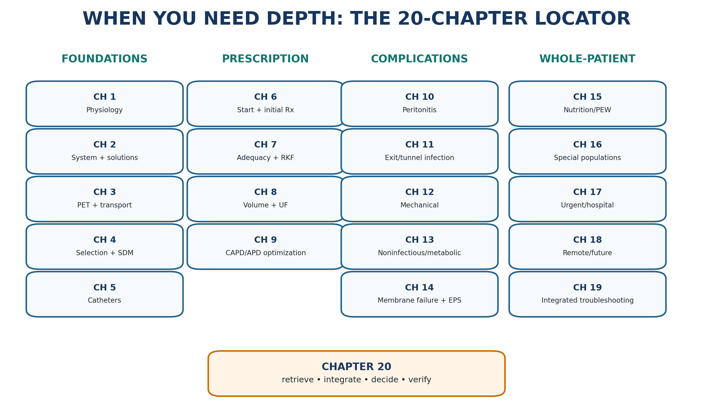
Use the correct chapter or guideline when a problem requires depth rather than overloading the final-review chapter with duplicate detail.
Distinguish a correctable PD problem from a multidomain pattern that warrants planned modality re-decision.
Document loss from PD using the 2026 ISPD HD-transfer framework rather than the historically inconsistent label “technique failure.”
| EVIDENCE POSTURE This master review is anchored in current ISPD goal-directed prescribing (2020), membrane-dysfunction recommendations (2021), peritonitis guidance (2022), catheter-related infection guidance (2023), access guidance (2019), PD teaching guidance (2025), acute-PD guidance, KDOQI nutrition guidance and the 2026 ISPD position statement on loss from PD. Historical adequacy and transport studies are retained only where they remain foundational. [1–18] |
|---|
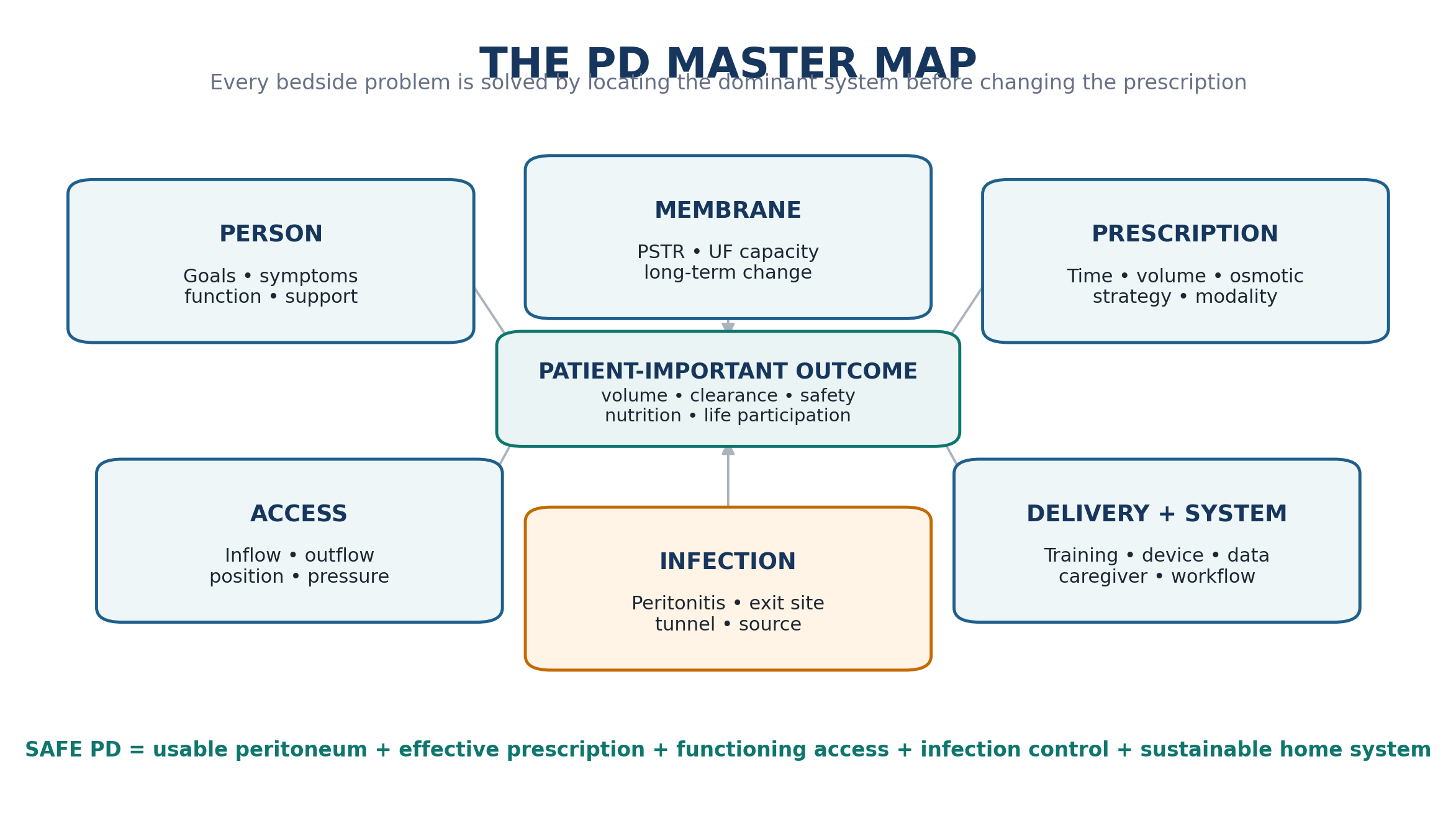
1. The master mental model: six systems, one patient
Peritoneal dialysis succeeds only when six systems cooperate. The peritoneum must transport solute and water appropriately; the prescription must exploit that physiology; the catheter must permit reliable inflow and drainage; infection prevention and source control must protect the cavity and access; the patient must have adequate nutrition, residual kidney function and physiologic reserve; and the home-care system must reliably deliver the treatment. A problem in any one domain can masquerade as failure in another.
Table 20.2 — Six-system PD mental model.
| System | What “working” looks like | Typical failure signal | Best first question |
|---|---|---|---|
| Person | Symptoms controlled; acceptable burden; nutrition/function preserved; goals met. | Fatigue, frailty, poor intake, treatment refusal, caregiver collapse. | What matters most to the patient now, and what has changed? |
| Membrane | Appropriate PSTR; sufficient UF capacity; stable long-term function. | Fast transport with long-dwell UF loss, UF insufficiency, escalating glucose need. | Is the prescription mismatched to transport, or has membrane function changed? |
| Prescription | Delivered treatment matches clearance, volume and life goals. | Inadequate clearance, sodium/volume problems, excessive glucose or burden. | Which lever is mismatched: time, volume, osmotic agent, frequency or modality? |
| Access/mechanics | Comfortable fill, complete drainage, intact abdominal boundary. | Alarms, poor drain, leaks, hernias, genital edema, hydrothorax, pain. | Is the problem lumen, position, extrinsic compression, pressure or a structural defect? |
| Infection | No peritonitis; healthy exit site/tunnel; rapid response when infection occurs. | Cloudy effluent, pain, pus, tunnel tenderness, recurrent organism. | Is there infection, how deep/source-linked is it, and is source control needed? |
| Delivery/support | Technique reliable; supplies, training, cognition and caregiver capacity adequate. | Missed/incomplete treatments, contamination, unexplained data gaps, burnout. | Could the problem be delivery, environment, training or support rather than physiology? |
| BEDSIDE TRANSLATION A low drain volume is not “inadequate PD.” It may be constipation, catheter migration, leak, cycler setup, posture or a true prescription issue. A high Kt/V is not “successful PD” if the patient is overloaded, malnourished or overwhelmed by treatment. |
|---|
2. The numbers worth memorizing—and the numbers not to worship
Table 20.3 — High-value current numerical anchors.
| Anchor | Current standard / definition | How to use it | Do not misapply it |
|---|---|---|---|
| Peritonitis diagnosis | At least 2 of 3: compatible symptoms/cloudy effluent; effluent WBC >100/µL after dwell ≥2 h with >50% PMN; positive culture. | Trigger diagnosis/treatment using ISPD 2022 context; APD rapid cycles may have lower absolute WBC with PMN predominance. | Do not wait for culture positivity or severe pain before acting on a cloudy bag. |
| Peritonitis program target | Overall rate ≤0.40 episodes per patient-year at risk; >80% of patients peritonitis-free per year. | Program quality improvement and benchmarking. | Not an individual patient “acceptable infection quota.” |
| Exit-site infection program target | Overall ESI rate ≤0.40 episodes per year at risk. | Program quality metric under ISPD 2023. | Do not diagnose ESI from erythema alone; purulent discharge is the defining clinical sign of definitive ESI. |
| PET timing | Formal PSTR assessment early in treatment, typically 6–12 weeks, and subsequently when clinically indicated; standard 4-h PET methods. | Establish transport phenotype and guide dwell/modal strategy. | Do not repeat PET mechanically when the clinical question can be answered otherwise. |
| PD nutrition: protein | KDOQI 2020: about 1.0–1.2 g/kg ideal body weight/day for metabolically stable adults on PD. | Starting nutrition framework; individualize for illness, intake, body composition and losses. | Do not interpret serum albumin as a direct protein-intake meter. |
| PD nutrition: energy | KDOQI 2020: about 25–35 kcal/kg ideal body weight/day, accounting for absorbed dialysate glucose. | Estimate energy needs in context of age, activity, body composition and metabolic goals. | Do not add dialysate glucose calories on top of an already excessive energy plan. |
| Elective catheter break-in | ISPD access guidance favors a break-in of at least 2 weeks for elective starts; earlier starts use urgent-start, low-pressure strategies. | Distinguish elective from urgent-start risk management. | Do not treat “2 weeks” as a reason to deny needed kidney replacement therapy. |
| NUMERICAL HUMILITY Current ISPD goal-directed PD deliberately moves away from defining dialysis quality by a single small-solute clearance threshold. Clearance remains important, but symptoms, volume, residual kidney function, nutrition, biochemical control, burden and life participation belong in the same judgment. |
|---|
3. Membrane physiology and PET: the four predictions that solve most prescription questions
Table 20.4 — Transport prediction table.
| If you see... | Mechanism to recall | Prediction | Prescription implication |
|---|---|---|---|
| Fast PSTR | Rapid solute equilibration and faster glucose dissipation. | Short dwells clear small solutes efficiently but long glucose dwells may lose osmotic force. | Consider shorter effective dwells/APD strategy and icodextrin for appropriate long dwell; assess UF capacity rather than labeling the membrane “good” or “bad.” |
| Slow PSTR | Slower small-solute equilibration. | Longer dwells may be needed for solute transfer. | Avoid excessively short cycles that sacrifice clearance; CAPD or longer APD dwells may fit. |
| Early sodium sieving | AQP1-mediated free-water transport precedes sodium equilibration. | Short hypertonic glucose dwells can remove proportionally more water than sodium. | Interpret water removal and sodium removal separately; persistent volume problems may be sodium problems. |
| Late long-dwell reabsorption | Osmotic gradient dissipates; lymphatic/intraperitoneal absorption continues. | Net UF can fall or reverse during prolonged glucose dwells. | Match dwell length and osmotic agent to physiology; icodextrin is designed for a long dwell. |
| Loss of UF capacity | Structural/functional membrane change, rapid transport, free-water transport defect or other mechanism. | Escalating glucose may yield diminishing benefit and more burden. | Reassess membrane phenotype, sodium, leak/mechanics and modality sustainability; cross-reference Chapters 8 and 14. |
| MEMORY ANCHOR Fast transport is not “fast dialysis” in every sense. It can improve small-solute equilibration while shortening the useful glucose osmotic window. |
|---|
4. Prescription architecture: change one lever for one stated reason
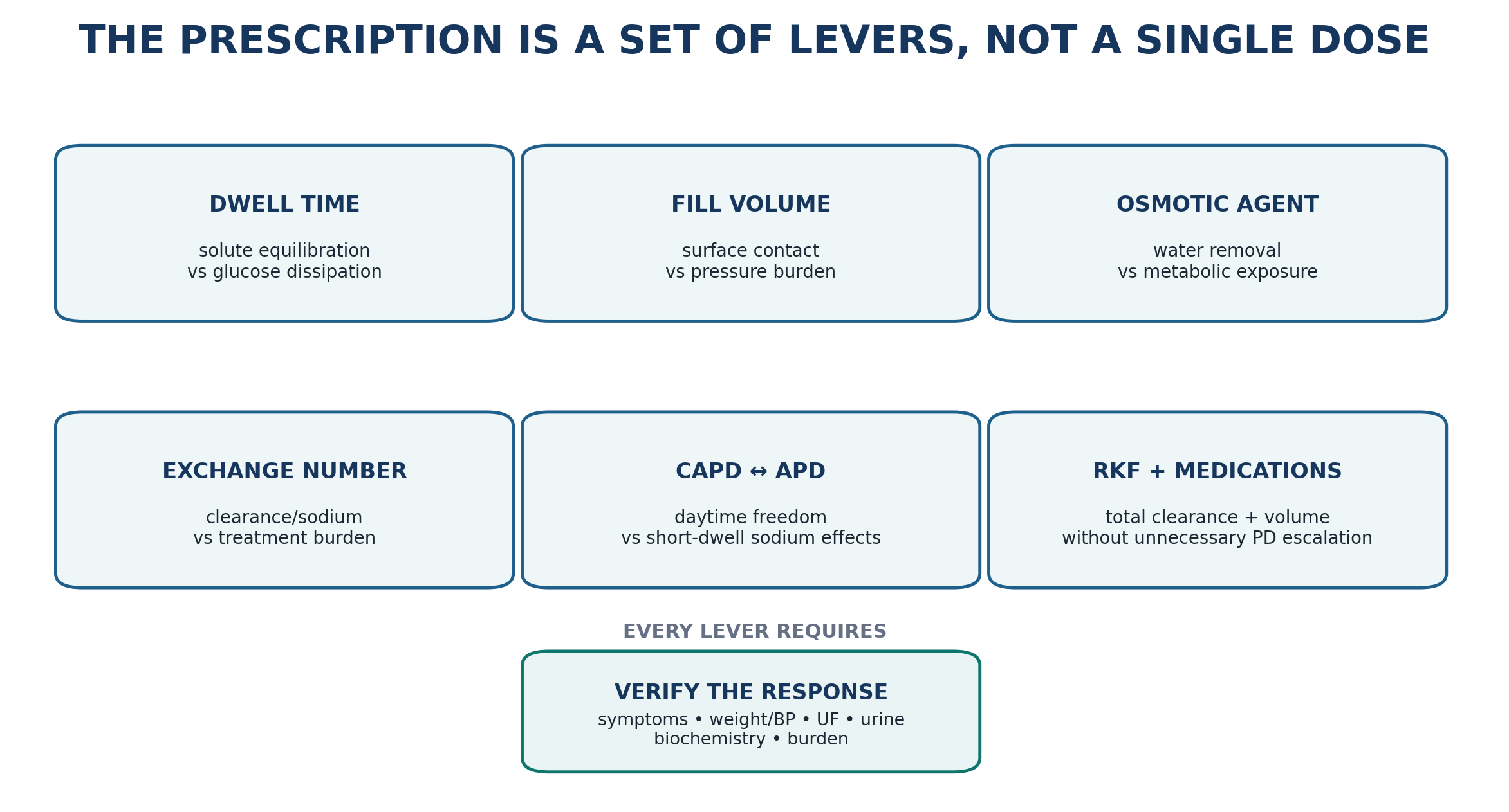
Table 20.5 — Prescription lever → expected benefit → trade-off.
| Lever | Best used when | Expected effect | Trade-off / safety check |
|---|---|---|---|
| Dwell time | Transport-dwell mismatch. | Changes solute equilibration, glucose dissipation, sodium removal and absorption. | Shortening can improve long-dwell UF in fast transport but reduce clearance in slow transport. |
| Fill volume | Need more effective peritoneal surface exposure or need lower pressure. | Higher volume may increase effective exchange; lower volume reduces intraperitoneal pressure. | Large upright fills can worsen pain, leaks and hernias; small fills can underdeliver therapy. |
| Glucose strength | Need stronger crystalloid osmotic force after reversible causes assessed. | Increases early water removal. | Metabolic exposure, glucose absorption, membrane burden; not a substitute for sodium/RKF/mechanical assessment. |
| Icodextrin long dwell | Long-dwell UF problem or need glucose-sparing strategy in appropriate patient. | Sustained colloid osmotic UF during long dwell. | Medication/device glucose-testing safety; assess volume response rather than assuming benefit. |
| Exchange number / total time | Need additional solute clearance or sodium/water opportunity. | Increases treatment opportunity. | Treatment burden, sleep disruption, drain exposure, caregiver workload. |
| CAPD ↔︎ APD redesign | Need different dwell distribution or daytime freedom. | Changes dwell pattern, sodium dynamics and lifestyle fit. | Cycler short dwells may worsen sodium removal in some phenotypes; CAPD increases daytime manual burden. |
| Diuretic/RKF strategy | Meaningful urine output remains. | Supports sodium/water balance and total clearance without extra PD burden. | Monitor volume, blood pressure, kidney function and electrolytes; avoid assuming RKF is static. |
5. Adequacy and residual kidney function: ask whether the patient is adequately treated
Adequacy is a clinical state, not a laboratory checkbox. A patient with acceptable urea kinetics may still have uncontrolled volume, potassium/acid-base abnormalities, anorexia, sleep disruption, severe treatment burden or declining life participation. Conversely, a patient with substantial residual kidney function may require less peritoneal dose than a clearance-only model would imply. The practical task is to identify the failed goal and determine whether the cause is underdelivery, RKF loss, membrane/prescription mismatch, access dysfunction, systemic disease or non-dialysis symptom burden.
Table 20.6 — Adequacy problem solver.
| Problem | Check first | Then ask | Action frame |
|---|---|---|---|
| Uremic symptoms | Was PD delivered? Is the symptom plausibly uremic? | RKF decline? clearance collection quality? nutrition/inflammation? sleep/depression/medications? | Correct underdelivery; quantify RKF/clearance when needed; increase dose only if the mechanism supports it. |
| Hyperkalemia/acidosis | Diet/medications, missed exchanges, constipation, catabolism. | Is clearance genuinely insufficient or is there acute illness? | Correct reversible cause, ensure adequate dialysis delivery, escalate therapy if biochemical safety requires. |
| Low measured clearance | Collection validity, actual prescription, body-water estimation, urine collection. | Does the patient have symptoms or another failed goal? | Do not chase a number blindly; identify why the metric is low and whether changing PD improves a patient-important outcome. |
| Loss of urine | Trend, medications, hemodynamics, intercurrent illness. | Has volume/clearance need changed? | Recalculate the whole prescription rather than simply adding a fixed amount of PD. |
| High treatment burden | Number/timing of exchanges, alarms, caregiver tasks, sleep. | Can the same goals be met with a simpler regimen? | Simplify, assist or redesign; treatment burden is part of quality. |
| RKF RULE Residual kidney function is not a bonus line on the adequacy report. It is a dynamic component of total kidney replacement therapy and should influence both clearance and volume decisions. |
|---|
6. Volume, sodium and ultrafiltration: localize before stronger dextrose
The common failure mode is to treat edema as “not enough glucose.” Volume status integrates sodium intake, fluid intake, residual urine, peritoneal sodium/water removal, membrane transport, dwell design, medication, cardiac/hepatic disease, leaks and drainage. Net UF is only one part of that system.
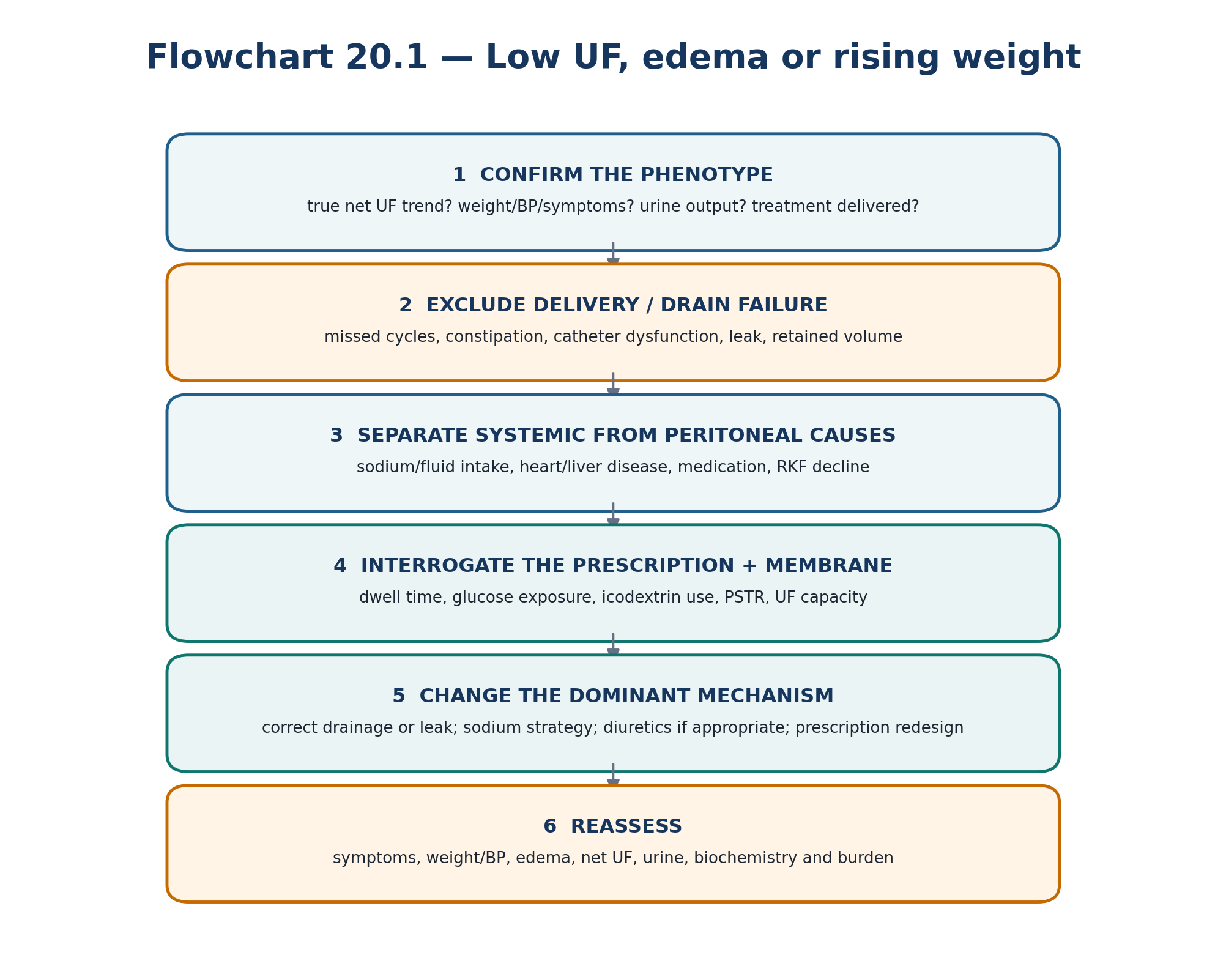
Table 20.7 — Volume pattern recognition.
| Pattern | Think first | Do next |
|---|---|---|
| Edema + rising weight + preserved urine | Sodium intake, cardiac disease, medication, insufficient total sodium removal. | Review sodium/fluid strategy, urine/diuretic plan, UF pattern and prescription. |
| Low measured UF + drain alarms | Mechanical/drain failure before membrane failure. | Troubleshoot flow, constipation, position, leak and retained volume. |
| Fast PSTR + poor long-dwell glucose UF | Rapid glucose absorption. | Shorten effective glucose dwell and consider long-dwell icodextrin when appropriate. |
| Strong dextrose use escalating over months | Progressive membrane/prescription mismatch or sodium/RKF change. | Structured UF/membrane assessment; do not continue dose escalation without mechanism. |
| Dyspnea + unilateral pleural effusion | Hydrothorax/pleuroperitoneal communication as well as heart failure. | Drain/reduce PD while evaluating; chest imaging and fluid/anatomic assessment as indicated. |
| Hypotension/cramps + weight loss | Possible over-removal, low intake, illness or cardiac/autonomic problem. | Reassess volume target and osmotic prescription; do not celebrate high UF in a depleted patient. |
7. Catheter and mechanical complications: solve the anatomy
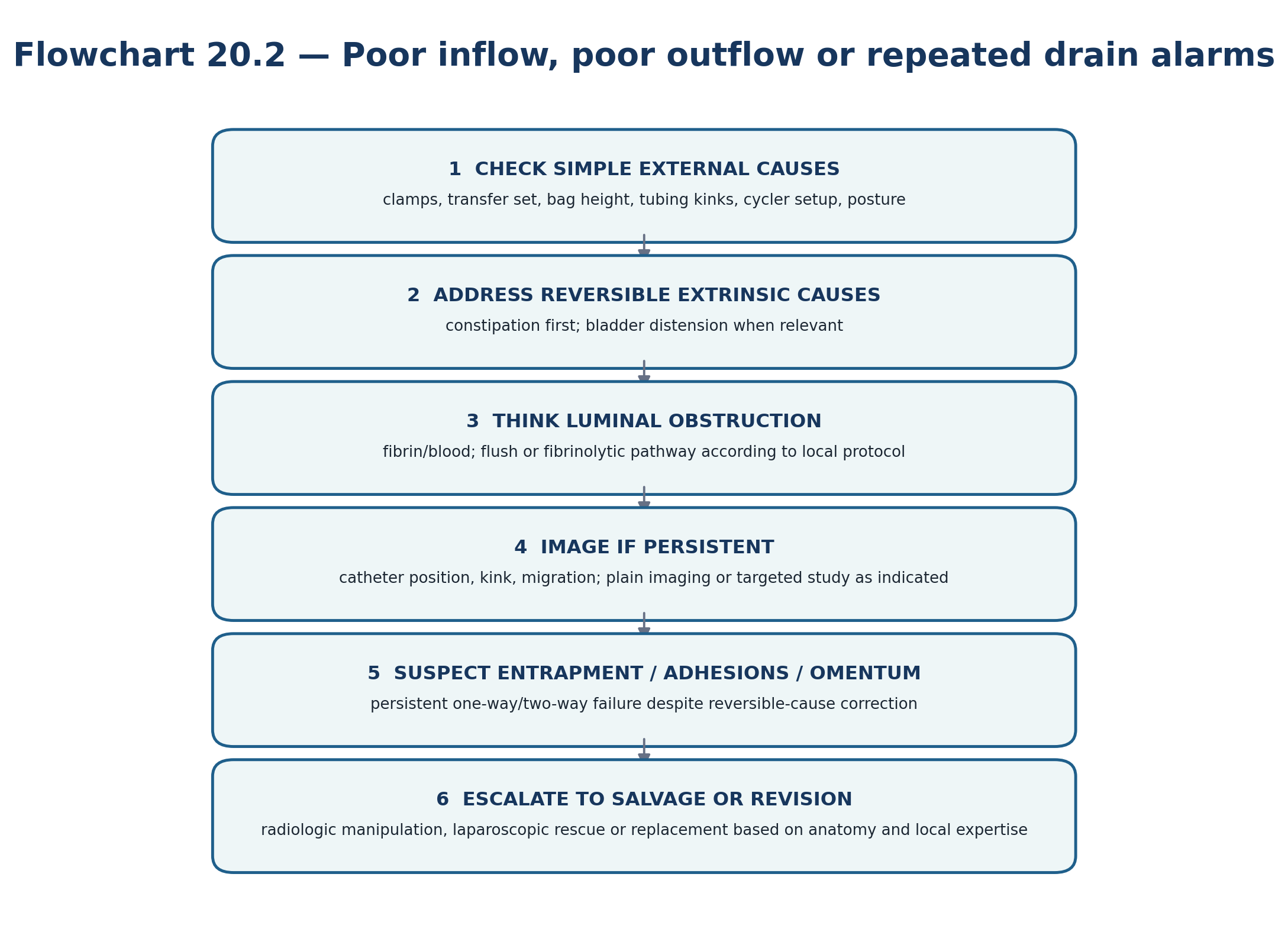
Table 20.8 — Mechanical complication one-look table.
| Presentation | Most useful localization | High-yield clue | Definitive direction |
|---|---|---|---|
| Two-way poor flow | Lumen/extrinsic compression/kink. | Constipation, fibrin/blood, tubing or catheter kink. | Bowel/bladder correction → flush/fibrinolytic pathway → imaging/procedural salvage if persistent. |
| One-way poor drain | Position, tissue entrapment, pressure-dependent drainage. | Works only in certain positions; recurrent low drain alarms. | Imaging and salvage/revision when persistent. |
| Pericatheter/wall swelling | Dialysate leak. | Temporal relation to fills; reduced drain recovery; edema tracking. | Lower intraperitoneal pressure/peritoneal rest as appropriate; localize defect if persistent. |
| New reducible bulge | Hernia. | Worse upright/with cough or fill. | Surgical evaluation/repair with pressure-aware PD plan. |
| Tender irreducible hernia | Incarceration/strangulation risk. | Pain, nonreducibility, obstructive symptoms. | Urgent surgical assessment—do not continue routine troubleshooting. |
| Right-sided effusion after PD | Pleuroperitoneal communication. | Effusion temporally related to dwell; dialysate-like pleural chemistry may support diagnosis. | Suspend/reduce PD, confirm cause, then pleural/surgical strategy or modality transition if unresolved. |
8. Peritonitis: diagnose quickly, then treat the organism and the source
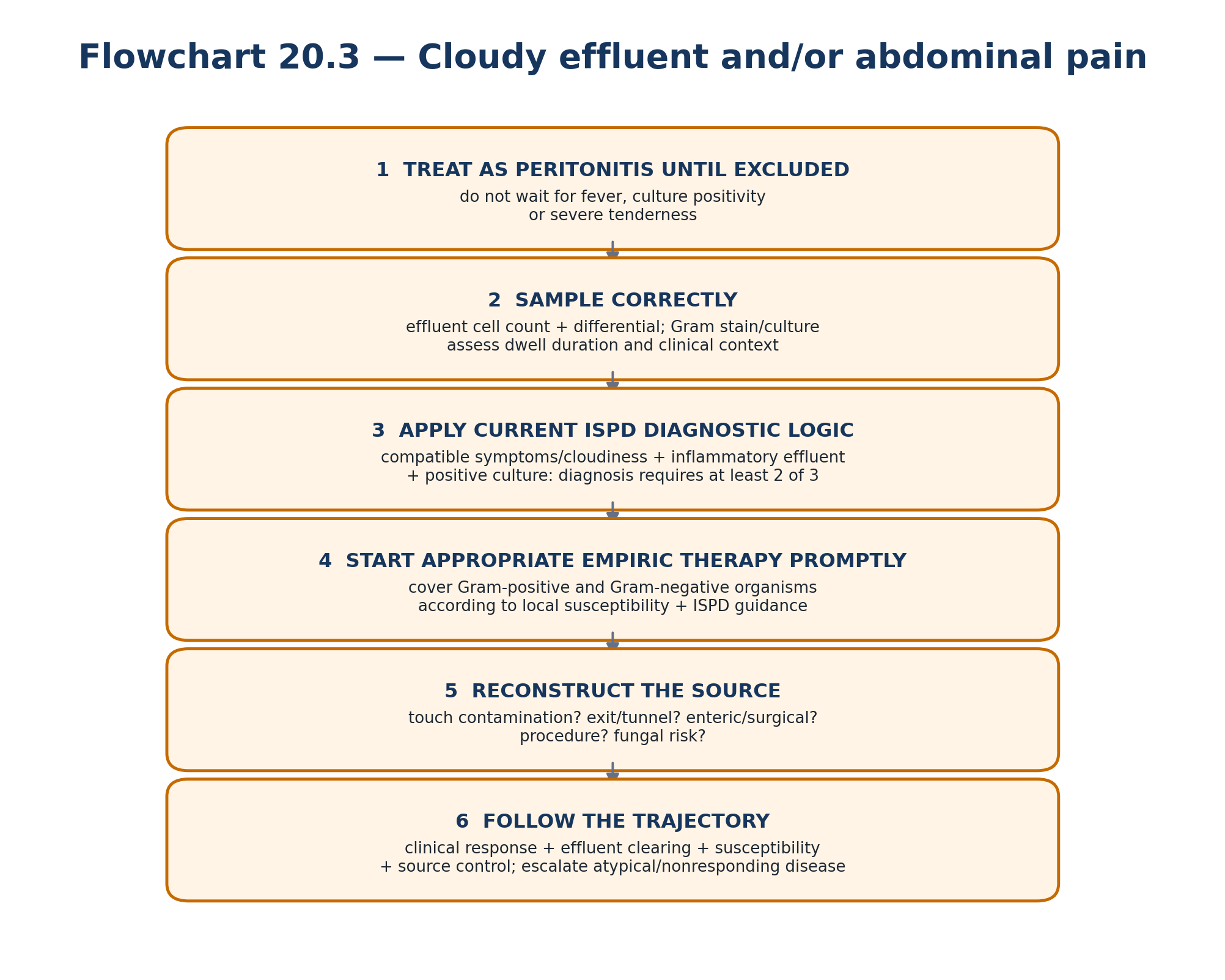
Table 20.9 — Peritonitis organism-to-source reminders.
| Pattern | What it should make you think | Why it matters |
|---|---|---|
| Coagulase-negative staphylococci | Touch contamination/connection technique. | Treatment plus technique review/retraining may prevent recurrence. |
| S. aureus | Catheter-related source or skin colonization; inspect exit/tunnel carefully. | Source control threshold is lower when catheter infection coexists. |
| Pseudomonas | More aggressive infection; catheter-related disease must be sought. | Requires organism-specific ISPD treatment strategy and close source assessment. |
| Mixed enteric organisms | Intra-abdominal/surgical source. | Imaging and surgical assessment may be as important as intraperitoneal antibiotics. |
| Fungus | High-risk infection often after antibiotic exposure. | Prompt catheter-removal/source-control pathway under ISPD guidance. |
| Culture negative with atypical course | Sampling/antibiotic effect, enteric inflammation, chemical/eosinophilic mimic, unusual organism. | Do not equate “no growth” with uncomplicated bacterial disease. |
| CLOUDY-BAG RULE A cloudy PD bag is an urgent diagnostic problem. Noninfectious mimics exist, but they are diagnoses considered after appropriate peritonitis evaluation—not reasons to delay it. |
|---|
9. Exit-site and tunnel infection: depth and source control determine the plan
Table 20.10 — Catheter-related infection final-review matrix.
| Finding | Interpretation | Action principle |
|---|---|---|
| Purulent discharge at exit site | Definitive exit-site infection under ISPD 2023, with or without erythema. | Culture when possible; organism-directed therapy and early response review. |
| Erythema/crust/granuloma without purulence | Not automatically definitive ESI. | Examine for trauma/dermatitis/colonization; do not prescribe antibiotics by redness alone. |
| Tenderness/induration along tunnel | Tunnel infection phenotype. | Assess extent; ultrasound can map a clinical question but a negative study does not overrule convincing clinical tunnel disease. |
| Pseudomonas or poor response | Higher-risk catheter phenotype. | Follow longer organism-specific treatment and source-control recommendations. |
| Same-organism catheter infection + peritonitis | Linked source. | Catheter removal/source control is generally required. |
| Persistent disease despite effective therapy | Foreign-body/biofilm or deep source. | Cross the source-control threshold deliberately; repeated antibiotics alone may not cure an infected catheter. |
10. Noninfectious and metabolic complications: interpret the bag and the glucose burden
Table 20.11 — Abnormal effluent and metabolic quick reference.
| Pattern | Likely noninfectious possibilities | Do not miss |
|---|---|---|
| Pink/red effluent | Ovulation/menstruation, catheter trauma, recent procedure, anticoagulation. | Persistent/heavy bleeding, hemodynamic compromise, intra-abdominal pathology. |
| Milky effluent | Chylous dialysate/lymphatic process. | Peritonitis can also look cloudy; send appropriate studies when symptomatic/uncertain. |
| Eosinophil-rich cloudy effluent | Early catheter/material/air/drug-related eosinophilic reaction. | Bacterial infection must be excluded before reassurance. |
| Fibrin strands | Inflammation, blood, postoperative state, peritonitis recovery. | Luminal obstruction and ongoing infection. |
| Rising glucose/weight | Absorbed dialysate glucose + insulin resistance + dietary factors. | Volume gain versus adipose gain; unnecessary hypertonic exposure. |
| Hypokalemia | K-free dialysate exposure plus low intake, diarrhea, insulin, medications. | Arrhythmia/weakness; peritonitis risk association; search cause rather than reflex potassium restriction. |
11. Long-term membrane failure and EPS: recognize the change in phenotype
Long PD vintage alone is not an EPS diagnosis and is not, by itself, a reason to stop PD. The warning pattern is a change: escalating osmotic requirement, loss of UF capacity, altered transport, recurrent inflammation, persistent gastrointestinal symptoms, weight loss, malnutrition, abdominal pain, nausea/vomiting or obstructive episodes. EPS is a clinicoradiologic syndrome; neither CT abnormalities nor transport change alone is sufficient.
Table 20.12 — Long-term membrane warning signals.
| Signal | What it may mean | Response |
|---|---|---|
| Escalating dextrose requirement | Reduced UF efficiency, sodium/RKF change, leak or prescription mismatch. | Structured volume + membrane assessment before further escalation. |
| New UF insufficiency | Membrane dysfunction or non-membrane cause. | Confirm delivery/mechanics; characterize PSTR/UF capacity when indicated. |
| Recurrent inflammatory injury | Risk of structural membrane change. | Treat source, reduce preventable infection, review long-term technique sustainability. |
| Progressive anorexia/weight loss + abdominal symptoms | Possible EPS or other gastrointestinal/systemic disease. | Urgent diagnostic evaluation; do not attribute to “uremia” without evidence. |
| Obstruction pattern in long-vintage/former PD patient | EPS must enter differential. | Cross-sectional imaging + specialist multidisciplinary management. |
12. Nutrition and protein-energy wasting: treat the cause, not the albumin
Table 20.13 — Nutrition/PEW decision framework.
| Finding | Interpretation | Next action |
|---|---|---|
| Low albumin alone | Inflammation, losses, volume and illness may dominate; not a stand-alone intake marker. | Assess intake, weight/body composition, inflammation/infection, protein losses and volume. |
| Weight loss + poor intake | High risk for PEW, especially with inflammation or hospitalization. | Dietitian assessment; search reversible causes; oral supplementation when counseling cannot meet needs. |
| Obesity + low muscle | Sarcopenic obesity possible. | Do not use BMI alone; assess function, intake, muscle/strength and metabolic burden. |
| Increasing dextrose exposure | More absorbed glucose calories. | Include dialysate calories in nutrition/metabolic plan and revisit volume strategy. |
| Peritonitis / hospitalization | Catabolic event with appetite and protein-loss consequences. | Reassess nutrition after the event; temporary requirements may differ from stable targets. |
| NUTRITION MEMORY PD nutrition is not “eat more protein.” It is intake + dialysate losses + absorbed glucose + inflammation + body composition + residual kidney function + treatment burden. |
|---|
13. Special populations: redesign the system around the constraint
Table 20.14 — Special-population memory anchors.
| Population / constraint | Dominant risk | Design response |
|---|---|---|
| Older/frail/cognitive impairment | Unsafe self-care, falls, delirium, caregiver burden. | Assisted PD/caregiver plan, simplified regimen, home safety, goals-of-care review. |
| Diabetes | Dialysate glucose burden, vision/dexterity, autonomic/vascular disease. | Glucose-aware prescription, safe glucose monitoring with icodextrin, assistance when needed, volume-first interpretation. |
| Obesity | Exit-site visibility, pressure/mechanical risk, clearance interpretation. | Seated exit-site planning/extended catheter when appropriate; pressure-aware prescription; avoid BMI as blanket contraindication. |
| Heart failure | Congestion with hemodynamic fragility. | Gentle sodium/water removal strategy; preserve RKF; judge success by symptoms/hospitalization/volume rather than clearance alone. |
| Cirrhosis/ascites | Infection, protein loss, leak, hypotension, complex volume physiology. | Selected patients can receive PD; specialist, nutrition and infection strategy required. |
| Limited caregiver/support capacity | Technique failure risk is social/systemic rather than membrane-related. | Assistance, respite, retraining, simplified prescription, planned backup modality. |
14. Urgent-start and hospitalized PD: lower pressure, increase surveillance, preserve options
Table 20.15 — Acute-scenario memory table.
| Scenario | Core principle | Safety endpoint |
|---|---|---|
| Urgent-start PD before standard healing period | Use low-volume supine/recumbent exchanges with pressure-conscious escalation and close leak/mechanical monitoring. | Escalate or temporarily use another KRT when urgent biochemical/volume goals cannot be safely met. |
| Hospitalized established PD patient | Reconstruct the home prescription, delivered therapy, residual urine, volume, infection/access status and acute illness before “continuing usual PD.” | Daily clinical reassessment; medication/procedure/position/nutrition changes can alter PD needs. |
| ICU / AKI setting | PD can be a suitable AKI modality in appropriate settings; prescription intensity and delivery method must match catabolic/volume needs. | Do not persist with inadequate PD when life-threatening solute/volume control requires another modality. |
| Procedure or surgery | Risk depends on anatomy, wound, contamination and intra-abdominal pressure. | Coordinate PD rest/low-volume strategy or temporary alternate KRT with surgical context. |
| Acute respiratory compromise | Large fills can worsen diaphragmatic mechanics; hydrothorax must be considered. | Stabilize respiration first; modify/hold PD while cause is evaluated. |
15. Remote monitoring and connected cyclers: data must lead to governed action
Table 20.16 — Digital PD: what the technology can and cannot prove.
| Signal | Useful for | Cannot prove by itself | Safe response |
|---|---|---|---|
| Treatment completion / alarms | Delivery visibility, recurrent drain problems. | Adequate clearance, adherence intent, catheter diagnosis. | Verify context; contact nonjudgmentally; troubleshoot mechanics/support. |
| Delivered UF trend | Trajectory recognition. | Euvolemia or the cause of low UF. | Confirm weight/BP/symptoms/urine/intake and prescription. |
| Weight/BP/home symptoms | Context for volume and acute illness. | Specific diagnosis from one measurement. | Check technique and trend; escalate clinically significant change. |
| Effluent optical/turbidity signal | Potential earlier peritonitis signal in investigational/validated systems. | Exclusion of infection when negative. | Symptoms/cloudy effluent still trigger standard ISPD diagnostic pathway. |
| Prediction model score | Potential prioritization. | Causal diagnosis or safe autonomous prescription change. | Require external/prospective validation, human review, auditability and override. |
| Missing upload | Connectivity/platform problem possible. | Nonadherence. | Check network/device/caregiver context before labeling behavior. |
| DIGITAL SAFETY Connected cycler ≠ remote monitoring ≠ remote patient management ≠ adaptive PD. Fully autonomous prescription change remains investigational; every clinically meaningful alert requires an accountable response pathway. |
|---|
16. Integrated troubleshooting: one algorithm for almost every complaint
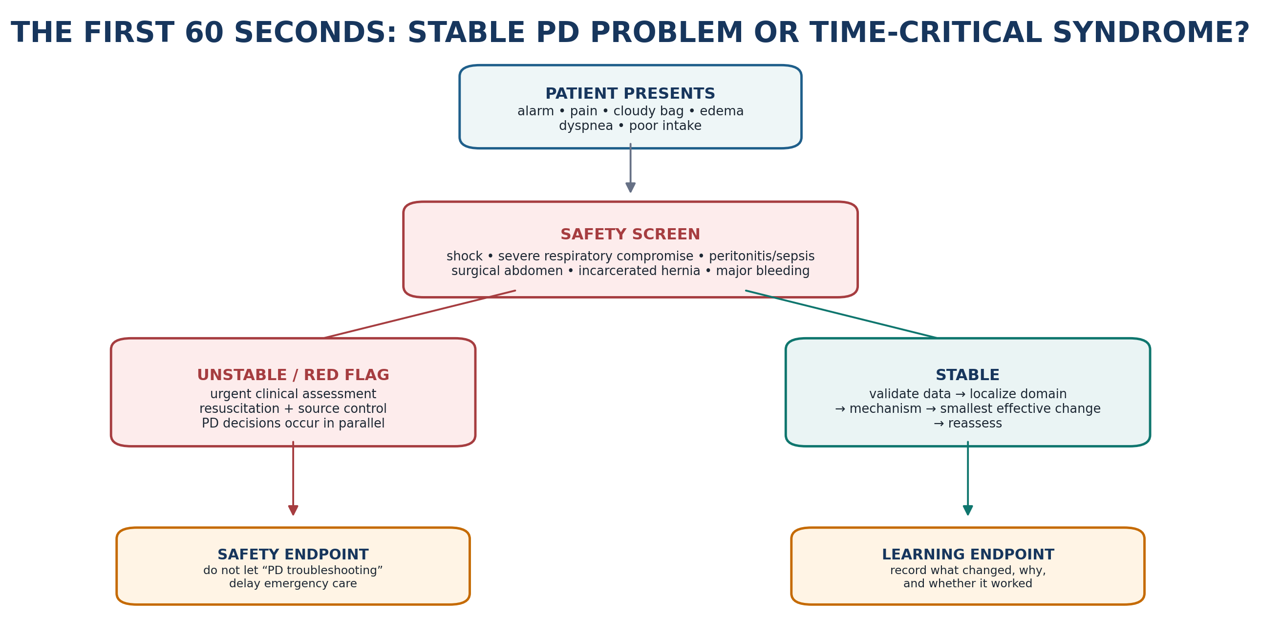
Table 20.17 — Complaint → dominant domain → first discriminator.
| Complaint | Dominant domains to consider | First discriminator |
|---|---|---|
| Poor drain | Delivery/access/mechanical. | External setup + constipation + posture before imaging. |
| Low UF / edema | Delivery, access, membrane, prescription, systemic/RKF. | Was treatment delivered/drained and is the patient truly volume expanded? |
| Inadequate clearance | Delivery, RKF, membrane/prescription, systemic illness. | What clinical goal is failing and is the measurement valid? |
| Abdominal pain | Peritonitis, drain/fill mechanics, leak/hernia, surgical abdomen. | Cloudy effluent/systemic signs/localized peritoneal signs first. |
| Cloudy/bloody/milky bag | Infection vs noninfectious effluent syndrome. | Cell count/differential/culture when peritonitis possible; phenotype the color/timing. |
| Exit-site abnormality | Infection, trauma/dermatitis, granuloma, cuff issue. | Purulence? tunnel involvement? same-organism peritonitis? |
| Weight loss/fatigue | Nutrition/PEW, inflammation, underdialysis, depression/systemic disease, EPS. | Intake + inflammation/infection + symptoms + delivered therapy + GI red flags. |
| Repeated missed treatments | Delivery/support, burden, cognition, technical problems. | Ask why without blame; distinguish inability, access, caregiver, device and preference issues. |
17. Transition, HD transfer and the safety boundary
A transition away from PD is not automatically a failure of the patient, clinician or modality. The 2026 ISPD position statement recommends standardized tracking of loss from PD therapy and uses causes such as HD transfer, death, transplantation, recovery and other outcomes rather than relying on the inconsistent historical phrase “technique failure.” For bedside practice, the critical step is to document the immediate cause, contributing causes, reversibility, urgency and whether the transition was planned.
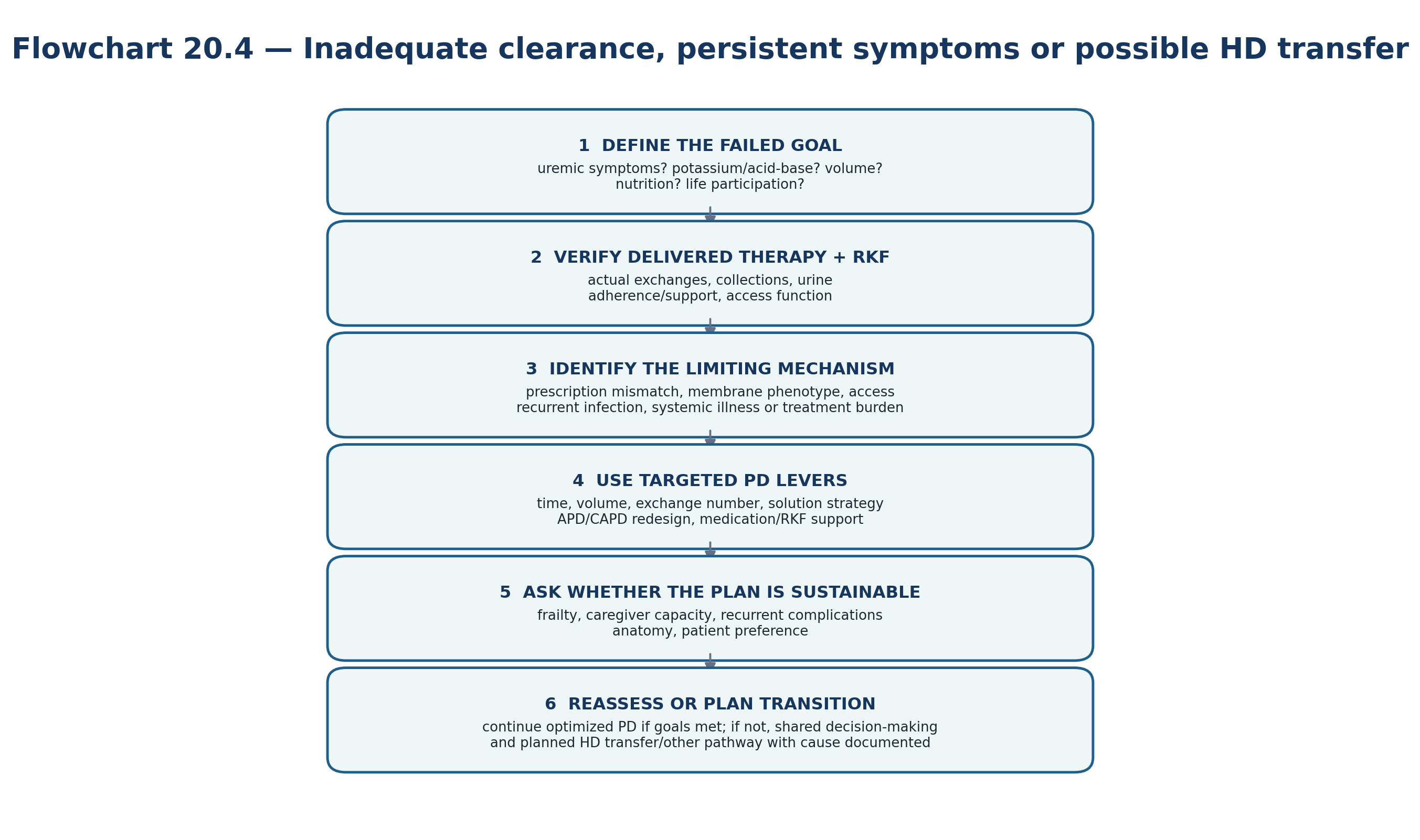
Table 20.18 — When to consider a planned modality re-decision.
| Pattern | Question | Reasonable direction |
|---|---|---|
| Recurrent severe infection/source-control events | Can the infection risk be made acceptably low with retraining, catheter intervention or support? | Continue only if source and prevention plan are credible; otherwise plan alternate KRT. |
| Irreparable/recurrent mechanical problem | Can anatomy be repaired or PD pressure redesigned safely? | Surgical salvage if feasible; otherwise planned transition. |
| Progressive membrane failure | Can goals be met without excessive glucose/burden or unsafe volume? | Prescription redesign; if ineffective, shared decision-making for transition. |
| Multidomain decline | Are nutrition, function, support and symptom goals all worsening despite optimization? | Reassess the entire care plan, not one PD variable. |
| Caregiver/patient burden unacceptable | Can assistance/simplification restore sustainability? | Assist/simplify if desired; otherwise offer alternate modality/supportive pathway. |
| Patient preference changes | Does the patient understand feasible options and trade-offs? | Respect informed preference; plan access and timing before crisis. |
18. Major clinical algorithms
19. Retention tables: pattern recognition
Table 20.19 — One-look “problem → mechanism → response” matrix.
| Problem | Likely dominant mechanism | Immediate response frame |
|---|---|---|
| Poor drainage | Constipation/extrinsic compression, fibrin, migration, entrapment. | Fix reversible causes → image → procedural salvage/revision. |
| Low UF | Delivery/drain failure, leak, sodium/RKF issue, fast transport, membrane dysfunction. | Verify delivery → localize → mechanism-matched volume/prescription strategy. |
| Edema | Sodium/water imbalance, cardiac/hepatic disease, RKF decline, inadequate UF. | Treat whole volume system, not UF number alone. |
| Cloudy effluent | Peritonitis until excluded. | Sample + prompt empiric therapy + source reconstruction. |
| Purulent exit discharge | Definitive ESI. | Culture/organism-directed therapy + tunnel/source assessment. |
| Milky effluent | Chylous process vs infection. | Phenotype + cell count/culture if infection possible + triglyceride/etiologic work-up. |
| Progressive glucose need | UF/membrane/prescription or sodium/RKF problem. | Structured reassessment before stronger glucose. |
| Low albumin | Inflammation/losses/volume/intake. | Nutrition + inflammation + volume assessment; do not prescribe to albumin alone. |
| Repeated treatment gaps | Support/device/cognition/burden or preference. | Nonjudgmental root-cause assessment + assistance/simplification/transition as appropriate. |
Table 20.20 — What to memorize vs what to look up.
| Memorize | Why | Look up / verify |
|---|---|---|
| Cloudy-bag urgency and 2-of-3 peritonitis framework. | Delay is harmful and the decision is time-sensitive. | Organism-specific antibiotic doses/durations and local resistance. |
| Purulent drainage defines definitive ESI; tunnel disease changes risk. | Prevents treating redness alone and missing deep disease. | Organism-specific treatment duration and catheter-intervention details. |
| Fast transport can coexist with poor long-dwell glucose UF. | Core prescription physiology. | Exact PET protocol/units and center laboratory calculations. |
| Constipation is a common reversible cause of poor drain. | Prevents premature invasive intervention. | Local fibrinolytic, fluoroscopic and surgical protocols. |
| Goal-directed adequacy is multidimensional. | Prevents Kt/V-only medicine. | Collection method, laboratory equations and reimbursement thresholds. |
| HD transfer is the current neutral reporting term. | Improves communication and quality reporting. | Registry-specific coding and analytic definitions. |
20. Clinical pearls
| CLINICAL PEARL A machine alarm is a symptom of the PD system—not a diagnosis of the catheter. |
|---|
| CLINICAL PEARL Low UF is a measurement; volume overload is a clinical state; neither tells you the mechanism. |
|---|
| CLINICAL PEARL Fast solute transfer and good long-dwell glucose UF are not synonymous. |
|---|
| CLINICAL PEARL Residual kidney function belongs inside the prescription, not at the bottom of the adequacy report. |
|---|
| CLINICAL PEARL When edema worsens, ask “where is the sodium?” before simply asking “which stronger bag?” |
|---|
| CLINICAL PEARL Purulent exit-site drainage matters more diagnostically than erythema intensity. |
|---|
| CLINICAL PEARL A right pleural effusion in a PD patient deserves a pleuroperitoneal communication in the differential. |
|---|
| CLINICAL PEARL A negative sensor, culture or ultrasound does not overrule a convincing high-risk clinical syndrome. |
|---|
| CLINICAL PEARL Albumin is a risk marker and illness marker; it is not a direct dietary protein prescription target. |
|---|
| CLINICAL PEARL “Nonadherence” should be the end of an assessment, not the beginning: check device, cognition, caregiver, cost, technique, symptoms and burden. |
|---|
| CLINICAL PEARL The safest prescription change is the smallest change that targets the named mechanism and has a defined reassessment endpoint. |
|---|
| CLINICAL PEARL A planned modality transition is better PD care than an emergency transfer after weeks of futile escalation. |
|---|
21. Common pitfalls — and the correction
| PITFALL Pitfall: treating every edema episode with stronger dextrose. Correction: confirm delivery/drainage, sodium intake, residual urine, systemic disease and membrane/prescription phenotype first. |
|---|
| PITFALL Pitfall: calling a red exit site “infection.” Correction: use the ISPD definition; purulence defines definitive ESI, while erythema alone has a broader differential. |
|---|
| PITFALL Pitfall: using Kt/V as the definition of adequate PD. Correction: interpret clearance with symptoms, volume, RKF, nutrition, biochemistry, burden and patient goals. |
|---|
| PITFALL Pitfall: escalating fill volume in a patient with leak, hernia or pressure symptoms. Correction: fill volume is a pressure lever as well as a clearance lever. |
|---|
| PITFALL Pitfall: assuming culture-negative cloudy effluent is benign. Correction: culture can be negative in true peritonitis; atypical noninfectious/enteric causes also require active evaluation. |
|---|
| PITFALL Pitfall: interpreting a remote-data gap as nonadherence. Correction: verify connectivity, device, caregiver and workflow before behavioral attribution. |
|---|
| PITFALL Pitfall: changing several prescription variables at once. Correction: make the smallest effective change and define what response should occur. |
|---|
| PITFALL Pitfall: persisting with antibiotics when a foreign-body source is not controlled. Correction: recognize the catheter source-control threshold. |
|---|
| PITFALL Pitfall: viewing HD transfer as personal or technical failure. Correction: document immediate and contributing causes and make transition a planned shared decision when possible. |
|---|
| PITFALL Pitfall: forgetting that a hospitalized PD patient is not physiologically “the same prescription.” Correction: acute illness, nutrition, position, procedures, medications and residual urine can all change PD requirements. |
|---|
22. Mini-cases: decisions, not trivia
Case 1 — Nightly drain alarms A stable APD patient reports 4 nights of slow-drain alarms. UF is lower. No pain or cloudy fluid. He has not opened his bowels for 4 days. Dominant domain? Access/mechanics, with constipation as the most likely reversible extrinsic cause. Treat constipation and validate response before imaging or prescription escalation. |
|---|
Case 2 — Edema despite “strong bags” A patient has progressively stronger glucose exchanges, falling urine output, edema and increasing weight. Drain volumes are technically complete. What was missed? The whole sodium/water system and RKF trajectory. Reassess sodium intake, urine/diuretics, PSTR/UF capacity and systemic causes; do not continue osmotic escalation by reflex. |
|---|
Case 3 — Cloudy effluent but little pain An APD patient notices a cloudy morning drain and feels only mildly unwell. Next move? Peritonitis evaluation now: effluent cell count/differential/culture and prompt empiric treatment per local/ISPD pathway. Mild pain does not make the bag safe. |
|---|
Case 4 — Red exit site There is erythema and crusting but no purulent drainage, no tunnel tenderness and no systemic symptoms. Is this definitive ESI? Not by the ISPD 2023 definition. Examine for trauma, dermatitis, granuloma and colonization; monitor closely and culture/treat if infection criteria emerge. |
|---|
Case 5 — Low measured Kt/V The patient feels well, is euvolemic, has substantial urine output and good appetite; the adequacy collection is unexpectedly low. First response? Validate the collection and delivered prescription. Do not intensify treatment from one number without integrating RKF and patient-important adequacy. |
|---|
Case 6 — Dyspnea with right effusion A PD patient develops new dyspnea and a large right-sided pleural effusion soon after a prescription increase. Key mechanical diagnosis? Pleuroperitoneal communication/hydrothorax. Modify/hold PD while evaluating; distinguish from cardiac volume overload. |
|---|
Case 7 — Weight loss in long-vintage PD A patient with many years on PD has poor appetite, intermittent vomiting, abdominal discomfort and progressive weight loss. What must not be missed? EPS or another obstructive/intra-abdominal process. This is not a routine “increase protein” problem; urgent diagnostic evaluation is required. |
|---|
Case 8 — Missed treatments in frailty An 84-year-old has repeated incomplete treatments after a fall. The daughter reports exhaustion managing the cycler. Dominant domain? Delivery/support and frailty. Reassess function/cognition/home safety/caregiver capacity; assisted PD, simplification or planned modality re-decision may be more appropriate than admonishment. |
|---|
Case 9 — Connected cycler shows falling UF The platform flags a 5-day UF decline, but weight and symptoms are unchanged. Should the prescription auto-change? No. Confirm measurement quality and clinical context first; connected data support triage, not autonomous treatment. |
|---|
Case 10 — Recurrent S. aureus peritonitis + exit-site infection The same organism appears at the exit site and in effluent. What changes the problem? A linked catheter source: source control becomes central; repeated antibiotics without addressing the catheter are unlikely to be durable. |
|---|
Case 11 — Hyperglycemia and weight gain A diabetic patient is using increasing hypertonic glucose for edema and now has worse glycemia and weight gain. What is the systems error? The prescription is being used to treat an incompletely localized volume problem while creating metabolic harm. Reconstruct sodium/RKF/membrane/mechanical causes and reduce unnecessary glucose exposure. |
|---|
Case 12 — PD no longer fits After recurrent mechanical procedures, nutrition decline and caregiver burnout, the patient says the treatment has become intolerable. What is the correct endpoint? A structured modality re-decision. If optimization cannot restore safe, acceptable goals, planned HD transfer or another pathway is good care—not “failure.” |
|---|
23. Active recall
Table 20.21 — MUST MEMORIZE.
| Question | Answer anchor |
|---|---|
| What is the universal PD troubleshooting sequence? | Safety → phenotype → validate → localize → mechanism → smallest effective change → reassess → transition if needed. |
| What does “goal-directed PD” reject? | The idea that one clearance number alone defines high-quality dialysis. |
| What does fast PSTR predict? | Rapid solute equilibration and shorter effective glucose osmotic dwell; long-dwell glucose UF may be poor. |
| First reversible cause of poor drainage to actively seek? | Constipation, alongside external setup/posture problems. |
| How is peritonitis diagnosed? | At least 2 of 3 current ISPD criteria: compatible clinical/cloudy effluent; inflammatory effluent; positive culture. |
| Defining sign of definitive ESI? | Purulent discharge at the catheter-epidermal interface, with or without erythema. |
| What must be integrated with peritoneal clearance? | Residual kidney function, symptoms, volume, nutrition/biochemistry, burden and goals. |
| What is the 2026 neutral term replacing “technique failure”? | HD transfer, with standardized cause tracking/reporting. |
| What is the remote-monitoring safety principle? | Validate signal → clinical context → accountable human review → action → reassessment; no direct sensor-to-prescription shortcut. |
| When is transition appropriate? | When patient-important goals cannot be met safely/acceptably despite reversible causes and reasonable PD optimization, or when informed preference changes. |
USE AS REFERENCE
Organism-specific peritonitis regimens, doses and durations: Chapter 10 + current ISPD/local antibiogram.
Exit/tunnel organism-specific therapy and catheter interventions: Chapter 11 + ISPD 2023.
Fibrinolytic dwell, radiologic manipulation and surgical catheter protocols: Chapters 5 and 12 + local access pathway.
PET protocol, UF-capacity testing and membrane classification: Chapters 3, 8 and 14.
Detailed nutrition prescription/supplementation: Chapter 15 + current dietitian/KDOQI pathway.
Acute/urgent-start prescription detail: Chapter 17 + current local acute-PD/ISPD protocols.
Commercial cycler, solution and remote-platform settings: manufacturer instructions + local governance; never infer a universal device setting from this book.
24. Flashcards: spaced repetition
Table 20.22 — Flashcards.
| Front | Back |
|---|---|
| What is the first question before changing a PD prescription? | What patient-important goal is failing, and is there an urgent safety issue? |
| Why can low UF be misleading? | It can result from incomplete drainage, leak, prescription timing, sodium/RKF/systemic factors or membrane dysfunction. |
| Why can constipation reduce PD drainage? | A distended colon can compress/displace the catheter and impair outflow. |
| Why can APD short cycles worsen sodium removal? | Early free-water transport can predominate before sodium equilibrates, especially with very short dwells. |
| Why is icodextrin useful? | It provides sustained colloid osmotic UF during a long dwell and can reduce glucose exposure in appropriate patients. |
| What does a negative culture mean in cloudy effluent? | It does not exclude peritonitis and should not stop consideration of unusual or noninfectious causes if the course is atypical. |
| What links S. aureus/Pseudomonas peritonitis to catheter care? | They should increase concern for a catheter-related source and lower the threshold for source-control thinking. |
| Why is albumin an imperfect nutrition target? | It is strongly influenced by inflammation, volume and protein losses as well as intake. |
| Why should BMI not exclude PD? | Obesity is primarily a technical/access/pressure/metabolic planning issue, not a universal contraindication. |
| What is the safest use of connected-cycler alerts? | Prioritize review, confirm context, then act through a defined clinical pathway. |
| What is a good reassessment endpoint after a prescription change? | The specific outcome that motivated the change: symptoms, weight/BP, UF, urine, biochemistry, clearance or burden. |
| What is the final test of a PD prescription? | Whether it safely meets the person’s goals with acceptable burden and sustainable delivery. |
25. Rapid differential / troubleshooting
Table 20.23 — One-minute bedside differential.
| Presentation | Common causes | Dangerous / important alternatives | First move |
|---|---|---|---|
| Poor drain | Constipation, posture, setup, fibrin. | Migration, kink, omental entrapment, leak. | Check external setup + bowel/bladder + symptoms; image if persistent. |
| Edema/weight gain | Sodium/fluid excess, RKF decline, insufficient UF. | Heart/liver disease, leak, severe membrane dysfunction. | Confirm true volume state and delivery before stronger glucose. |
| Abdominal pain | Peritonitis, drain/fill pain, constipation. | Surgical abdomen, incarcerated hernia, pancreatitis/ischemia. | Cloudy bag/systemic/localized signs → urgent sampling/imaging as indicated. |
| Cloudy bag | Bacterial peritonitis. | Eosinophilic/chemical/chylous/hemoperitoneum/enteric process. | Peritonitis pathway first. |
| Dyspnea | Volume overload. | Hydrothorax, pneumonia, PE, diaphragmatic splinting. | Stabilize; chest/volume assessment; consider relation to dwell. |
| Exit-site redness | Trauma/dermatitis/granuloma. | Definitive ESI/tunnel infection. | Look for purulence and tunnel features; culture/treat when infection phenotype present. |
| Fatigue/poor appetite | Uremia, PEW, anemia, depression, sleep, inflammation. | EPS/obstruction, systemic disease, recurrent infection. | Validate delivered therapy + nutrition/inflammation + red flags; do not assume underdialysis. |
| Frequent alarms/missed therapy | Device/setup, drain problem, cognition/support. | True inability to deliver safe home therapy. | Root-cause analysis and support redesign before labeling nonadherence. |
| TROUBLESHOOTING RULE If the first intervention does not improve the predefined endpoint, do not simply repeat it harder. Re-open the localization step and ask whether the original mechanism was wrong or whether more than one domain is failing. |
|---|
26. Final revision sheet
| CORE CONCEPT Peritoneal dialysis is a patient-centered control system. A living membrane, a chosen prescription, a functioning catheter, infection control, residual kidney function, nutrition, training and support interact continuously. Mastery means identifying which part of that system is limiting the patient today. |
|---|
One-minute revision
Safety before troubleshooting.
Validate delivery before interpreting physiology.
Localize the failure domain before changing the prescription.
Fast transport can mean poor long-dwell glucose UF.
Low UF is not the same as volume overload.
Clearance adequacy is not a single number.
Constipation is a first-line drain differential.
Cloudy effluent is peritonitis until appropriately excluded.
Purulent exit-site discharge defines definitive ESI.
Recurrent infection may be a source-control problem.
Fill volume changes pressure as well as clearance.
Nutrition assessment must integrate inflammation and dialysate losses/glucose.
Remote data need human-governed validation and action.
HD transfer is a planned clinical outcome when PD no longer safely meets goals.
TEN TAKE-HOME RULES
1. Name the failed patient goal before naming the dialysis change.
2. Never let routine PD troubleshooting delay emergency assessment.
3. Confirm what was actually delivered, drained, measured and recorded.
4. Think in systems: person, membrane, prescription, access, infection and delivery/support.
5. Use physiology to select the smallest effective lever, then reassess.
6. Protect residual kidney function and interpret it as part of total therapy.
7. Treat infection by organism, source, trajectory and source control—not antibiotic name alone.
8. Treat mechanical problems anatomically and pressure problems by reducing pressure plus correcting the defect.
9. Treat nutrition, frailty and caregiver burden as dialysis outcomes, not side notes.
10. Plan transition early when PD can no longer meet safe, acceptable goals.
| ONE-MINUTE BEDSIDE SYNTHESIS What is the patient telling me? Is it dangerous? Was PD truly delivered? Which system is failing? What mechanism explains it? What one change targets that mechanism? What will I measure afterward? If the system remains unsafe or unacceptable, what is the planned next pathway? |
|---|
| FINAL MENTAL MODEL The expert PD clinician does not memorize a bag concentration for every problem. The expert recognizes the phenotype, reconstructs the mechanism, changes the correct lever and verifies the consequence. |
|---|
Rapid oral viva
Table 20.24 — Rapid oral viva.
| Question | Model answer |
|---|---|
| Why is Kt/V insufficient as the definition of PD adequacy? | Because high-quality PD is goal-directed and also requires acceptable symptoms, volume, RKF, nutrition/biochemistry, burden and life participation. |
| What are the first three actions for low UF? | Confirm the phenotype/delivery, exclude drainage/leak/mechanical problems, then assess the whole sodium-water system before membrane/prescription escalation. |
| What is the first action for cloudy effluent? | Urgent peritonitis sampling and treatment pathway; do not wait for culture. |
| What changes the meaning of an exit-site infection? | Depth/tunnel involvement, organism, response trajectory and linked peritonitis/source control. |
| When is HD transfer good care? | When PD cannot safely or acceptably meet patient goals despite reasonable correction/optimization, or when informed preference changes; it should be planned and cause-documented when possible. |
| SAFETY BOUNDARY This chapter is a retrieval and reasoning tool, not a substitute for current local protocols, device instructions, antimicrobial susceptibility data, surgical judgment or patient-specific clinical assessment. When a numerical dose, drug regimen, device setting or procedural threshold is not explicitly standardized in current guidance, verify it rather than infer it. |
|---|
27. Selected authoritative references
1. Brown EA, Blake PG, Boudville N, et al. International Society for Peritoneal Dialysis practice recommendations: Prescribing high-quality goal-directed peritoneal dialysis. Perit Dial Int. 2020;40(3):244–253. https://doi.org/10.1177/0896860819895364. PMID: 32063219.
2. Morelle J, Stachowska-Pietka J, Öberg C, et al. ISPD recommendations for the evaluation of peritoneal membrane dysfunction in adults: classification, measurement, interpretation and rationale for intervention. Perit Dial Int. 2021;41(4):352–372. https://doi.org/10.1177/0896860820982218. PMID: 33563110.
3. Li PKT, Chow KM, Cho Y, et al. ISPD peritonitis guideline recommendations: 2022 update on prevention and treatment. Perit Dial Int. 2022;42(2):110–153. https://doi.org/10.1177/08968608221080586. PMID: 35264029.
4. Chow KM, Li PKT, Cho Y, et al. ISPD Catheter-related Infection Recommendations: 2023 Update. Perit Dial Int. 2023;43(3):201–219. https://doi.org/10.1177/08968608231172740. PMID: 37232412.
5. Crabtree JH, Shrestha BM, Chow KM, et al. Creating and Maintaining Optimal Peritoneal Dialysis Access in the Adult Patient: 2019 Update. Perit Dial Int. 2019;39(5):414–436. https://doi.org/10.3747/pdi.2018.00232. PMID: 31028108.
6. Chow JSF, Brunier G, Figueiredo AE, et al. Teaching peritoneal dialysis: A position paper for the International Society for Peritoneal Dialysis. Perit Dial Int. 2025;45(6):327–343. https://doi.org/10.1177/08968608251375512. PMID: 40966019.
7. Hurst H, Figueiredo AE, Perez Moran D, et al. Peritoneal dialysis training and interventions: A narrative review. Perit Dial Int. 2026;46(1):6–15. https://doi.org/10.1177/08968608251328517. PMID: 40221963.
8. Quinn RR, Johnson DW, Mehrotra R, et al. 2026 ISPD Position Statement on tracking and reporting loss from PD therapy. Perit Dial Int. 2026 Jun 17:8968608261461639. https://doi.org/10.1177/08968608261461639. PMID: 42307417.
9. Ikizler TA, Burrowes JD, Byham-Gray LD, et al. KDOQI Clinical Practice Guideline for Nutrition in CKD: 2020 Update. Am J Kidney Dis. 2020;76(3 Suppl 1):S1–S107. https://doi.org/10.1053/j.ajkd.2020.05.006. PMID: 32829751.
10. Cullis B, Al-Hwiesh A, Kilonzo K, et al. ISPD guidelines for peritoneal dialysis in acute kidney injury: 2020 update (adults). Perit Dial Int. 2021;41(1):15–31. PMID: 33267747.
11. KDIGO Diabetes Work Group. KDIGO 2022 Clinical Practice Guideline for Diabetes Management in Chronic Kidney Disease. Kidney Int. 2022;102(5S):S1–S127. https://doi.org/10.1016/j.kint.2022.06.008. PMID: 36272764.
12. Twardowski ZJ, Nolph KD, Khanna R, et al. Peritoneal equilibration test. Perit Dial Int. 1987;7(3):138–148. https://doi.org/10.1177/089686088700700306.
13. Paniagua R, Amato D, Vonesh E, et al. Effects of increased peritoneal clearances on mortality rates in peritoneal dialysis: ADEMEX, a prospective, randomized, controlled trial. J Am Soc Nephrol. 2002;13(5):1307–1320. https://doi.org/10.1681/ASN.V1351307.
14. Bargman JM, Thorpe KE, Churchill DN; CANUSA Peritoneal Dialysis Study Group. Relative contribution of residual renal function and peritoneal clearance to adequacy of dialysis: a reanalysis of the CANUSA study. J Am Soc Nephrol. 2001;12(10):2158–2162. https://doi.org/10.1681/ASN.V12102158.
15. Perl J, Davies SJ, Lambie M, et al. The Peritoneal Dialysis Outcomes and Practice Patterns Study (PDOPPS): unifying efforts to inform practice and improve global outcomes in peritoneal dialysis. Perit Dial Int. 2016;36(3):297–307. https://doi.org/10.3747/pdi.2014.00288.
16. Paniagua R, Ramos A, Ávila M, et al. Remote monitoring of automated peritoneal dialysis reduces mortality, adverse events and hospitalizations: a cluster-randomized controlled trial. Nephrol Dial Transplant. 2025;40(3):588–597. https://doi.org/10.1093/ndt/gfae188.
17. Anutrakulchai S, Tatiyanupanwong S, Kananuraks S, et al. Effect of the Chronic Kidney Disease-Peritoneal Dialysis (CKD-PD) app on improvement of overhydration treatment in patients on peritoneal dialysis: randomized controlled trial. J Med Internet Res. 2025;27:e70641. https://doi.org/10.2196/70641.
18. Chung MC, Yu TM, Cheng BC, et al. Remote patient monitoring automated peritoneal dialysis and clinical outcomes: a nationwide real-world cohort study from Taiwan. Clin J Am Soc Nephrol. 2026. Online ahead of print. https://doi.org/10.2215/CJN.0000001112.
| SOURCE NOTE Guideline status and key contemporary evidence were checked 4 September 2026. This final-review chapter intentionally avoids inventing universal drug doses, cycler settings, PET cutoffs, fill volumes, ultrafiltration targets or clearance thresholds where current guidance requires individualized clinical judgment or local protocol. |
|---|