Applied Nephrology Master Series
Chapter 19
Integrated Troubleshooting:
From Clinical Problem to Prescription Change
Symptoms | Localization | Mechanism | Minimal Effective Change | Reassessment | Safety | Transition
| CHAPTER MISSION Convert the entire PD volume into one bedside reasoning system. For any new symptom, alarm, abnormal effluent, volume problem, biochemical drift or treatment-burden signal, identify the safety priority, verify what was actually delivered, localize the dominant failure domain, name the mechanism, make the smallest corrective change, and define how success will be reassessed. |
|---|
| MASTER PRINCIPLE Do not change the PD prescription until you can state what problem you are treating and which mechanism the change is expected to correct. If the mechanism is uncertain, gather the next discriminating piece of information rather than escalating therapy by reflex. |
|---|
0. One-page chapter map
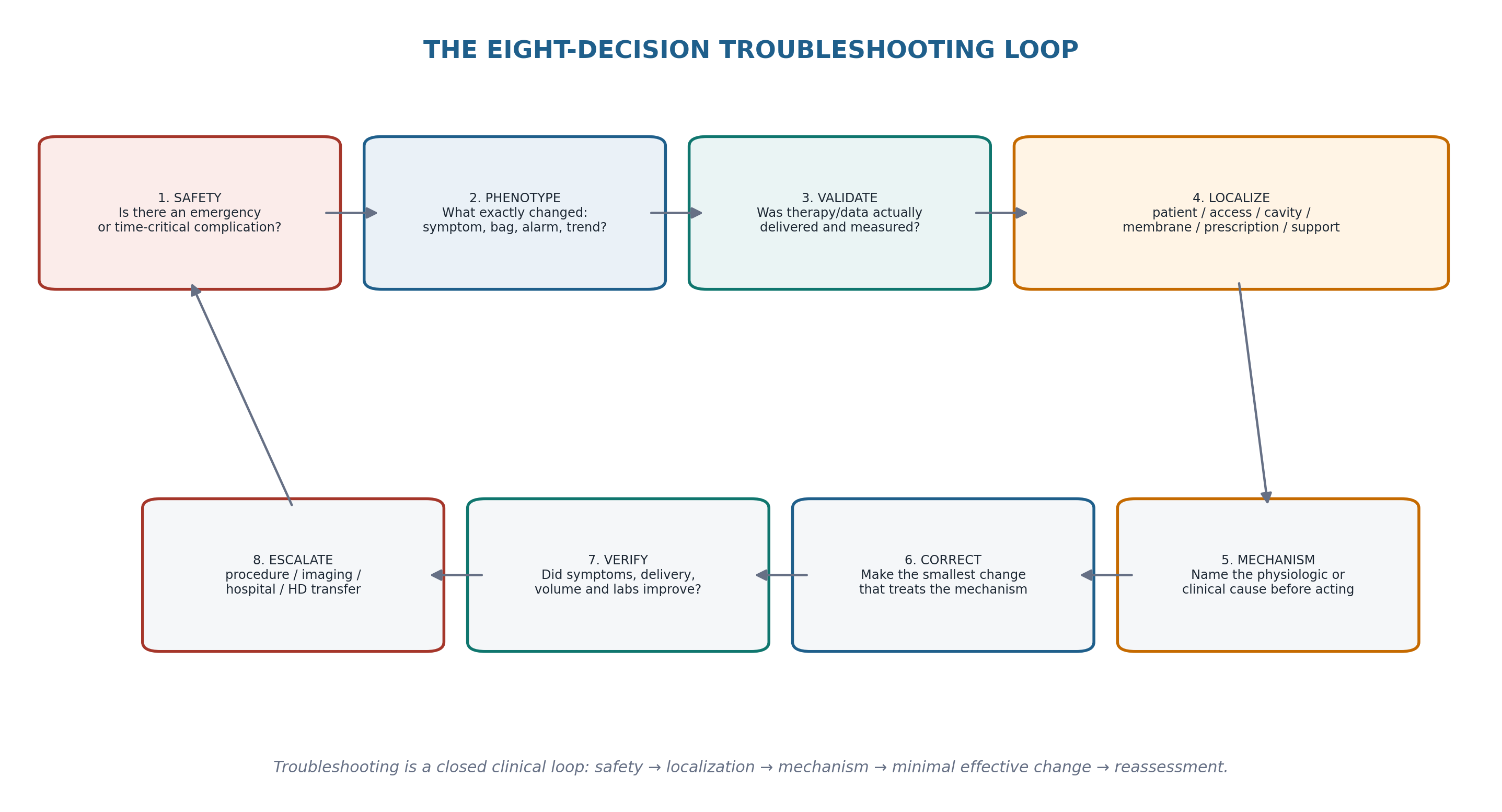
Table 19.1 — The eight decisions that govern integrated PD troubleshooting.
| Decision | Core question | Bedside output |
|---|---|---|
| 1. Safety | Is there a time-critical threat independent of the PD mechanism? | Urgent stabilization/escalation |
| 2. Phenotype | What exactly changed: symptom, bag appearance, alarm, weight, BP, lab or delivered treatment? | Problem stated precisely |
| 3. Validate | Was therapy delivered, measured and documented correctly? | Real problem separated from data/delivery artifact |
| 4. Localize | Which domain is failing: delivery, access, infection, membrane/prescription, patient physiology or support? | Failure domain named |
| 5. Mechanism | What causal pathway best explains the phenotype? | Mechanistic diagnosis |
| 6. Correct | What is the smallest effective change that treats that mechanism? | Targeted intervention |
| 7. Verify | What symptom, treatment metric, examination or laboratory change should improve? | Reassessment endpoint |
| 8. Escalate | If unresolved or unsafe, what specialist, procedure, imaging, hospitalization or HD-transfer plan is needed? | Safe next level of care |
Learning outcomes
Use a single troubleshooting sequence across common PD complaints without collapsing every problem into “more dialysis.”
Distinguish data/delivery failure from catheter mechanics, infection, membrane dysfunction, prescription mismatch, systemic illness and treatment-burden problems.
Recognize time-critical patterns that require urgent assessment before routine PD troubleshooting.
Build a differential from the phenotype of poor drainage, low ultrafiltration, volume overload, inadequate clearance, pain or abnormal effluent.
Choose prescription levers by mechanism—time, volume, osmotic strategy, drain strategy, RKF/medication plan or support—not by habit.
Define a reassessment endpoint before making a PD change and avoid simultaneous multi-variable changes that obscure cause and effect.
Integrate recurrent infection, mechanical dysfunction, membrane failure, nutrition decline, frailty and caregiver burden into multidomain decision-making.
Use the 2026 ISPD terminology of HD transfer and document immediate and contributing causes when PD can no longer be delivered safely or sustainably.
| EVIDENCE POSTURE This chapter is a synthesis rather than a new disease-specific guideline. It is anchored in the current ISPD goal-directed prescribing framework, 2021 membrane-dysfunction recommendations, 2022 peritonitis guideline, 2023 catheter-related infection update, 2019 PD-access guideline and the 2026 ISPD statement on loss from PD therapy. Contemporary adequacy/prescription guidance, PDOPPS outcome data and training/CQI literature are used to support integration. Disease-specific thresholds remain in their dedicated chapters and are not reinvented here. [1–12] |
|---|
1. Troubleshooting is a localization problem, not a prescription problem
A PD complaint often arrives as an output: “the machine alarmed,” “the drain is low,” “I am swollen,” “the fluid is cloudy,” “the creatinine rose,” or “I cannot do this anymore.” These statements are clinically useful only after they are translated into a failure domain. Increasing glucose, adding cycles or extending treatment time may improve one output while worsening the actual problem—for example, more intraperitoneal pressure in a leak, more glucose burden in a patient with metabolic toxicity, or longer nocturnal treatment in a patient whose main problem is recurrent drain pain and sleep disruption.
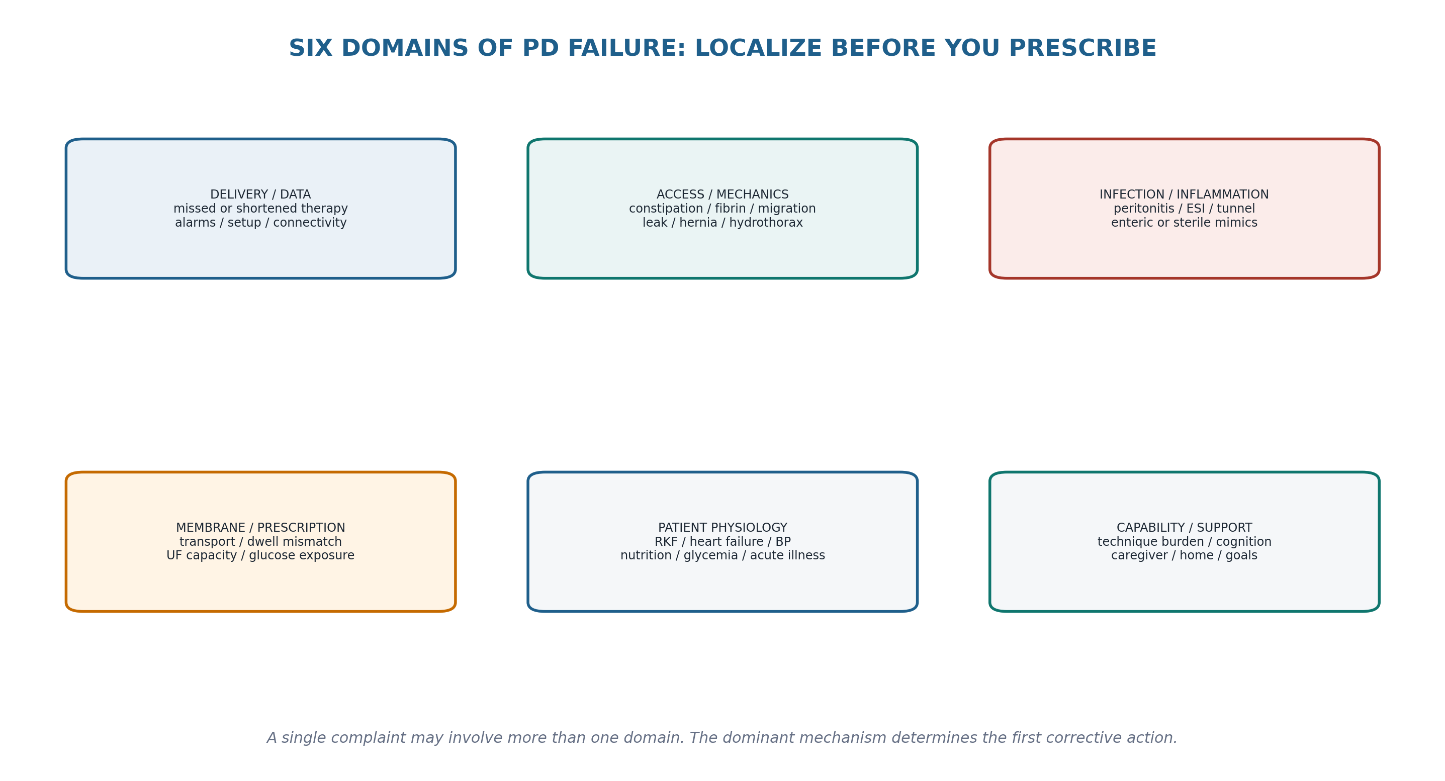
Table 19.2 — Complaint to failure-domain translation.
| What the patient/team reports | First domains to test | Do not assume |
|---|---|---|
| “My UF is low.” | Delivery/drain mechanics; intake/RKF; prescription; membrane | Membrane failure |
| “I am gaining weight.” | Sodium/water balance; glucose calories; edema; RKF; adherence/support | All weight gain is fluid |
| “The cycler keeps alarming.” | Drain mechanics; setup; position; constipation; device/data | Catheter needs replacement |
| “My bag is cloudy.” | Peritonitis first; then noninfectious mimics | Culture-negative means noninfectious |
| “I feel weak and nauseated.” | Uremia; volume; electrolytes; infection; nutrition; medications | Kt/V alone explains symptoms |
| “PD is too much.” | Treatment burden; cognition; caregiver; sleep; symptoms; depression | Nonadherence |
| “Blood pressure is high.” | Volume; sodium; medications; pain/stress; measurement quality | More dextrose is the first step |
| “Clearance is low.” | Collection/delivery error; RKF loss; prescription; membrane transport | Immediate maximal dose escalation |
| LOCALIZATION RULE The first useful sentence in a PD troubleshooting note is not “increase dialysis.” It is “the dominant problem appears to be ___ because ___.” |
|---|
2. First-pass safety screen: identify what cannot wait
Integrated troubleshooting begins by separating a stable home-therapy problem from a medical emergency. PD should not delay resuscitation, urgent laboratory correction, imaging, surgery or transfer to an appropriate acute-care setting. Some dangerous presentations are caused by PD; others simply occur in a person receiving PD. The clinician must recognize both.
Table 19.3 — Safety screen before routine troubleshooting.
| Red-flag phenotype | Potential threat | Immediate direction |
|---|---|---|
| Severe generalized or focal abdominal pain, guarding, shock | Peritonitis, perforation, ischemia, obstruction, bleeding | Urgent clinical/surgical assessment; sample effluent when appropriate |
| Cloudy effluent with systemic illness | PD peritonitis / sepsis | Chapter 10 pathway without delay |
| Acute dyspnea, hypoxemia, large new effusion | Pulmonary edema, hydrothorax, pneumonia, PE, cardiac event | Direct acute evaluation; do not manage from UF data alone |
| Severe hyperkalemia or acid-base instability | Life-threatening biochemical emergency | Stabilize and provide adequate KRT; modality choice follows urgency |
| Tender irreducible hernia / vomiting / obstruction | Incarceration/strangulation or EPS/other obstruction | Urgent surgical pathway |
| Heavy or persistent hemoperitoneum with hemodynamic change | Intra-abdominal or gynecologic bleeding | Urgent diagnostic assessment |
| Fever + tunnel/exit-site findings + peritonitis features | Deep catheter infection with intraperitoneal spread | Chapter 10/11 source-control pathway |
| Rapid functional/cognitive decline affecting sterile technique | Immediate home-treatment safety risk | Assisted pathway, temporary supervision or modality reassessment |
| SAFETY BOUNDARY A “PD problem” becomes an acute medical problem whenever physiology, infection severity, bleeding, respiratory compromise or surgical features make remote or routine troubleshooting unsafe. |
|---|
3. Validate the treatment before interpreting the physiology
The most common avoidable reasoning error is to interpret a number before confirming how it was produced. Delivered dialysis can differ from prescribed dialysis because of alarms, early termination, skipped exchanges, unrecorded manual exchanges, wrong solution, short dwells, retained intraperitoneal volume, poor drain or data-transfer failure. Clearance collections can be incomplete. Weight and blood pressure can be measured inconsistently. A reported urine volume may be estimated rather than measured. Validation is not clerical work; it determines whether the physiological inference is valid. [1,7]
Table 19.4 — Data validation checklist.
| Data stream | Validation questions | Common false inference |
|---|---|---|
| APD treatment | Start/stop time? cycles completed? alarms/bypasses? actual fill/drain? | “Prescription failed” when it was not delivered |
| CAPD log | All exchanges performed? dwell timing? correct bags? | Assuming planned schedule equals delivered schedule |
| Net UF | Was initial drain/retained volume handled correctly? any leak? | Calling retained or leaked dialysate membrane UF failure |
| Weight | Same scale, timing, clothing, fill state? | Treating measurement noise as fluid gain |
| Blood pressure | Correct cuff/position/rest? trend vs single reading? | Escalating volume therapy from a single poor measurement |
| Urine/RKF | Measured collection? interval correct? major diuretic/illness change? | Assuming silent RKF stability |
| Adequacy collection | Complete urine/dialysate collection and correct timing? | Dose escalation for collection error |
| Remote upload | Connected and complete? manual exchanges visible? | Calling missing connectivity nonadherence |
| DELIVERED-THERAPY RULE Prescribed therapy is an intention. Troubleshooting uses delivered therapy. |
|---|
4. Poor inflow, poor outflow and repeated drain alarms
Flow dysfunction is solved by separating external/setup problems, reversible extrinsic compression, intraluminal obstruction, catheter displacement and tissue entrapment. Constipation remains one of the most common reversible causes and should be actively sought. A distended bladder can also impair function. Fibrin or blood can obstruct the lumen. Persistent positional dysfunction, migration, omental wrapping, adhesions or kinking require imaging and procedural expertise rather than repeated prescription escalation. The detailed access pathway is in Chapters 5 and 12. [5]
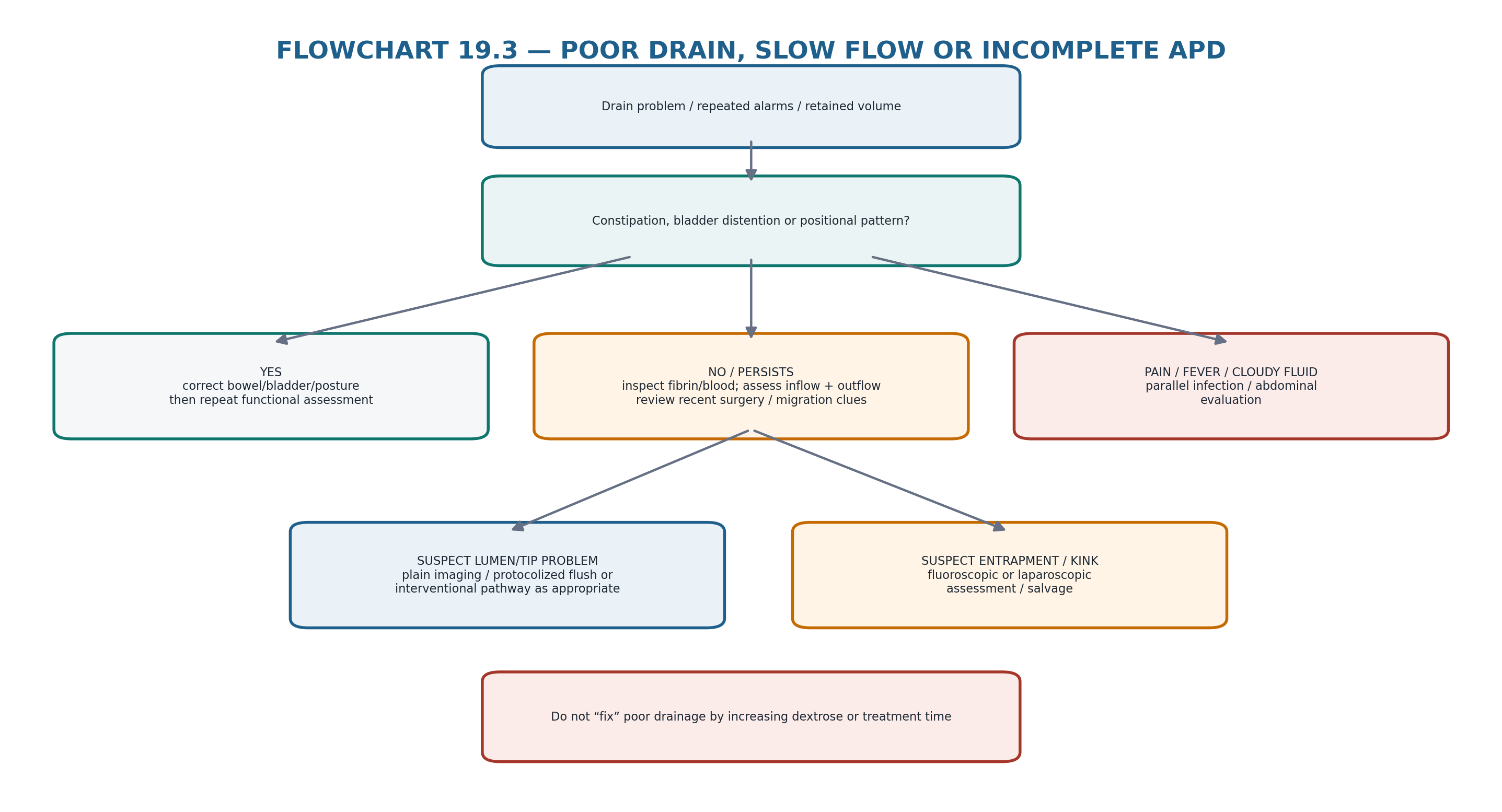
Table 19.5 — Flow phenotype → likely mechanism → next discriminator.
| Pattern | Think first | Next discriminator |
|---|---|---|
| Good inflow, poor outflow | Constipation, position, tip migration, omentum | Bowel history, posture response, imaging if persistent |
| Poor inflow + poor outflow | Kink, clamp/transfer set, fibrin/clot, severe malposition | External inspection + lumen assessment |
| Sudden dysfunction after blood/fibrin | Intraluminal obstruction | Visible fibrin/blood; protocolized catheter assessment |
| Works only in one position | Tip position / entrapment | Reproducible posture effect; imaging |
| Repeated APD drain alarms but manual drains okay | Cycler pressure/drain dynamics, posture, tidal strategy | Event log + manual functional comparison |
| Low drain with edema/swelling near catheter or genitalia | Leak rather than “retained UF” | Physical exam + leak imaging when indicated |
| Poor drain + fever/cloudy fluid | Peritonitis/inflammation plus mechanics | Parallel infection work-up |
| Persistent dysfunction despite conservative correction | Migration, omentum, adhesions, kink | Fluoroscopic/laparoscopic salvage pathway |
| MECHANICAL PITFALL Do not compensate for incomplete drainage by increasing glucose concentration, exchange number or treatment time. That can increase retained volume and intraperitoneal pressure while leaving the mechanical cause untreated. |
|---|
5. Low ultrafiltration, weight gain and edema
“Low UF” is not a diagnosis. First ask whether dialysis was delivered and drained, whether fluid is leaking, whether sodium and fluid intake changed, whether residual kidney function declined, and whether the prescription matches the patient’s transport physiology. Only after these are addressed should persistent low ultrafiltration be classified with formal membrane assessment as described in Chapters 3, 8 and 14. ISPD goal-directed prescribing explicitly requires volume, symptoms, RKF, delivered therapy and patient priorities to be interpreted together rather than reducing adequacy to a single number. [1,2]
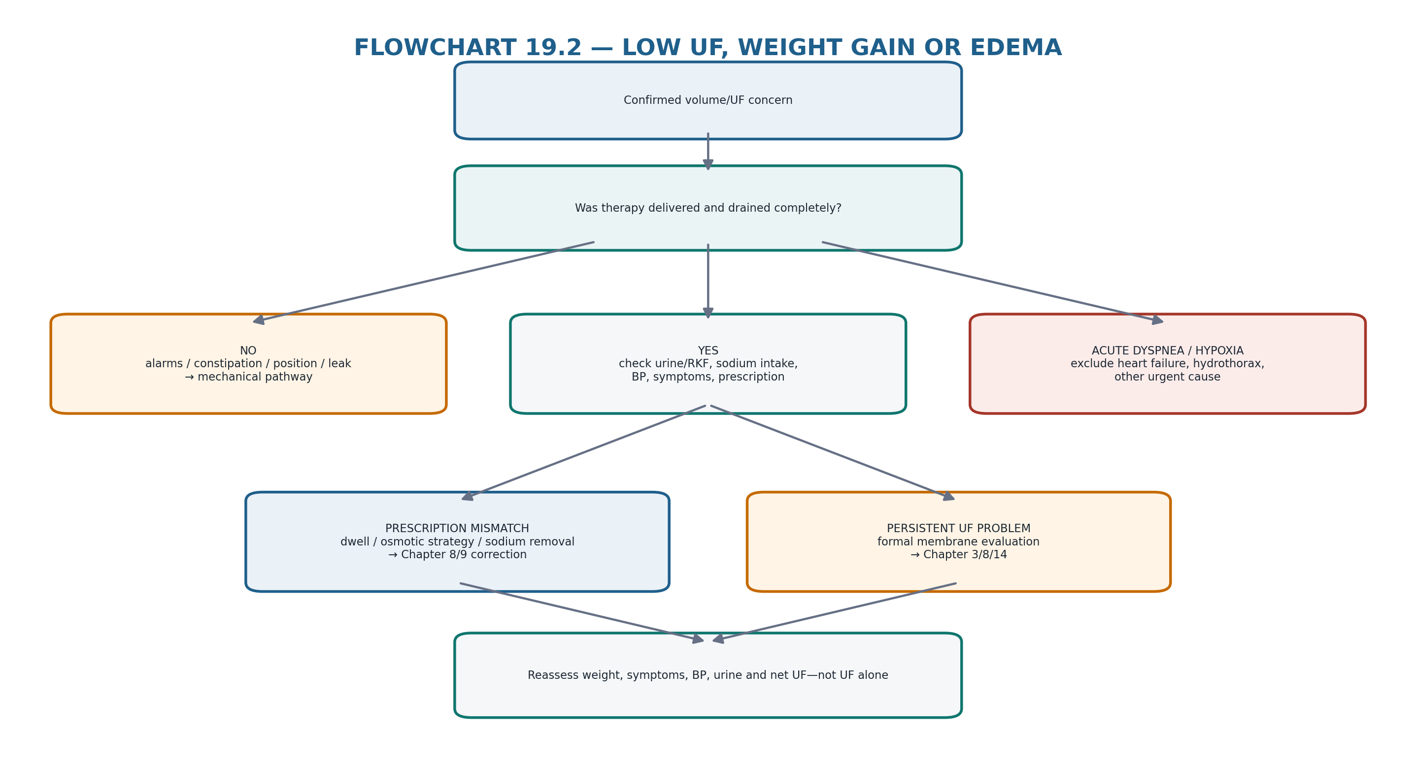
Table 19.6 — Volume troubleshooting matrix.
| Finding | Mechanism candidates | Useful discriminators | Direction |
|---|---|---|---|
| Low net UF + repeated drain alarms | Incomplete drainage / constipation / migration | Event log, retained volume, bowel/posture response | Mechanical pathway |
| Low UF + genital/wall edema | Dialysate leak | Exam, fill-volume relation, imaging when needed | Pressure reduction + source correction |
| Weight ↑ + edema + urine ↓ | RKF loss + sodium/water accumulation | Measured urine, medications, intake | Rebuild total volume plan |
| Weight ↑ without edema | Glucose calories / nutrition / adiposity | Glycemia, intake, longitudinal body composition | Metabolic rather than UF escalation |
| Hypertension + weight ↑ + UF trend ↓ | Likely congestion but confirm | Symptoms, exam, urine, intake, prescription | Volume strategy |
| Dyspnea + unilateral effusion | Hydrothorax / noncardiac effusion | Chest imaging ± pleural evaluation | Chapter 12 pathway |
| Persistent UF problem despite good drainage | Prescription mismatch or membrane dysfunction | PET/UF-capacity assessment per guideline | Chapters 3/8/14 |
| Frequent hypertonic glucose use | Refractory volume problem or prescribing habit | Why high glucose is needed; metabolic cost | Mechanism-based redesign |
| VOLUME RULE Treat the volume phenotype, not the UF number. A patient can have low UF without clinical overload, or overload despite apparently acceptable UF when sodium intake, RKF loss or cardiac disease dominates. |
|---|
6. Hypertension, hypotension, dizziness and cramps
Blood-pressure abnormalities in PD should be interpreted as hemodynamic phenotypes, not automatic instructions to change dialysate strength. Hypertension can reflect extracellular volume excess, sodium intake, medications, sympathetic activation, pain or measurement error. Hypotension and dizziness may follow excessive fluid removal, poor intake, autonomic dysfunction, infection, bleeding or cardiac disease. The prescription change must follow the mechanism.
Table 19.7 — BP symptom troubleshooting.
| Phenotype | High-probability questions | PD-specific response |
|---|---|---|
| Hypertension + edema/weight gain | Sodium, urine decline, missed/short treatments, UF pattern | Chapter 8 volume redesign after validation |
| Hypertension without congestion | Medication, pain, sleep, measurement technique | Do not force osmotic UF without volume evidence |
| Dizziness + weight loss | Excess fluid removal, low intake, diuretics, acute illness | Reassess dry-state assumptions and osmotic intensity |
| Hypotension during/after drains | Volume depletion, cardiac/autonomic disease, pain/vagal response | Clinical assessment; reduce harmful fluid removal if confirmed |
| Cramps with strong dextrose | Rapid/excessive fluid removal or electrolyte issue | Review osmotic exposure and electrolytes |
| BP variability in frailty/diabetes | Autonomic dysfunction, medication timing, inconsistent volume | Use trends and symptoms; individualize goals |
| PRESSURE DISCIPLINE A BP value is a clue, not a prescription. Confirm volume phenotype and competing causes before using PD to chase the number. |
|---|
7. “Inadequate dialysis”: symptoms, clearance and biochemical drift
Troubleshooting inadequate dialysis begins with the clinical question: what patient-important problem is not controlled? Uremic symptoms, hyperkalemia, acidosis, phosphate burden, appetite decline, volume overload and declining life participation are not interchangeable. A low measured clearance should trigger validation of the collection, assessment of delivered therapy and RKF, and review of transport/prescription before dose escalation. Conversely, acceptable numerical clearance does not dismiss persistent symptoms that require another explanation. [1,7]
Table 19.8 — Inadequacy phenotype → mechanism.
| Problem | Check before increasing PD dose | Potential lever |
|---|---|---|
| Low measured clearance | Collection completeness, delivered exchanges, RKF, body-water estimate | Time/cycles/dwell strategy if truly needed |
| New nausea/anorexia | Volume, infection, medications, constipation, nutrition, uremia | Treat dominant cause; dose only if uremic mechanism likely |
| Hyperkalemia | Diet, medications, constipation, missed/incomplete therapy, acidosis | Correct cause + ensure adequate dialysis |
| Persistent acidosis | Delivery, diet/protein, residual function, intercurrent illness | Prescription/medical correction |
| Phosphate burden | Dietary bioavailability, binders/adherence, residual function, dialysis exposure | Integrated nutrition/medication/dialysis plan |
| Fatigue | Anemia, sleep, depression, infection, frailty, volume, uremia | Avoid using clearance as universal explanation |
| Loss of RKF | Measured urine/clearance trend | Recalculate total therapy rather than react to serum creatinine alone |
| GOAL-DIRECTED RULE Adequacy is not a rescue number. The question is whether the whole treatment plan controls symptoms, volume, metabolic needs and life goals with acceptable burden. [1] |
|---|
8. Pain: fill pain, drain pain, abdominal pain and pressure pain
Pain must be classified by timing and relation to the exchange. Infusion pain suggests solution temperature/pH/osmolality, rapid inflow, catheter-tip irritation or intraperitoneal pathology. Drain pain is commonly related to catheter-tip suction against pelvic structures or low residual volume, particularly during APD, but constipation and malposition can contribute. Diffuse abdominal pain—especially with cloudy effluent or systemic features—must be treated as possible peritonitis until evaluated. Focal severe pain or peritoneal signs demand a broader abdominal differential.
Table 19.9 — Pain localization.
| Timing/pattern | Think | Next action |
|---|---|---|
| Pain during inflow | Solution/temperature/flow, catheter irritation, inflammation | Check setup/solution; examine; infection pathway if suspicious |
| Pain late in drain | Suction/drain pain, low residual volume, catheter position | Review drain settings/tidal options and mechanics |
| Pain only upright/full abdomen | Pressure-related mechanical problem | Fill-volume/hernia/leak assessment |
| New diffuse pain + cloudy effluent | Peritonitis | Chapter 10 now |
| Focal pain + guarding/vomiting | Surgical/enteric pathology | Urgent imaging/surgical review |
| Pain + bloody effluent | Hemoperitoneum / abdominal or gynecologic cause | Chapter 13 assessment |
| Chronic pain + treatment burden | Mechanical symptom + psychosocial burden | Treat mechanism and simplify regimen where possible |
| PAIN RULE Timing is a diagnostic test. Ask whether the pain starts with fill, during dwell, at end-drain, with posture, or independently of PD. |
|---|
9. Cloudy, bloody, milky or otherwise abnormal effluent
Cloudy effluent remains infectious until peritonitis is appropriately assessed; the 2022 ISPD diagnostic and treatment framework governs this pathway. Noninfectious abnormalities—hemoperitoneum, eosinophilic reactions, fibrin, chylous effluent, chemical inflammation or biliary/enteric contamination—are considered only after the clinical safety context is clear. Chapter 10 contains the peritonitis criteria and organism-specific management; Chapter 13 contains the noninfectious effluent differential. [3]
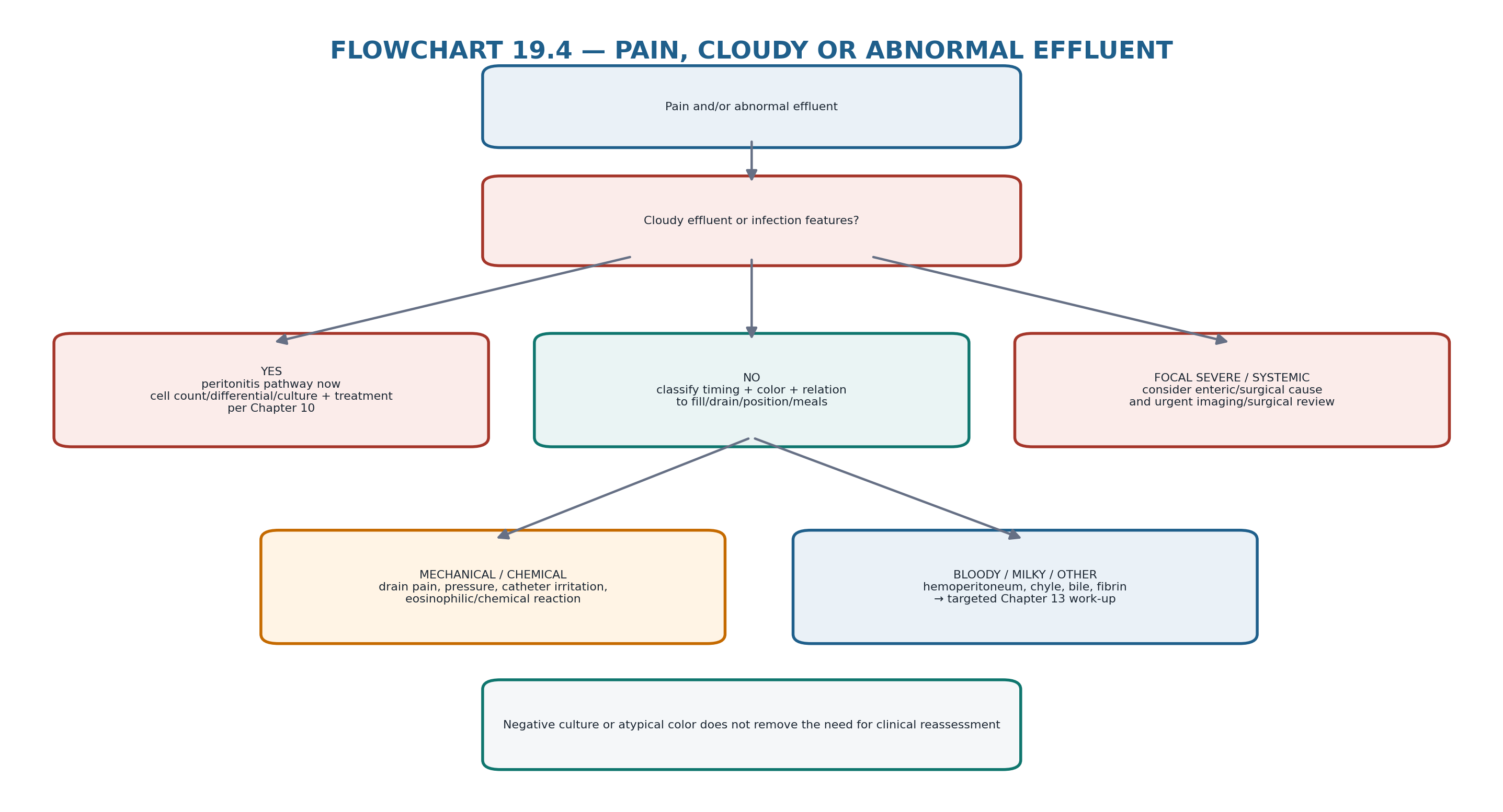
Table 19.10 — Abnormal bag: rapid routing.
| Appearance/setting | Priority | Route |
|---|---|---|
| Cloudy + pain | Peritonitis | Chapter 10 |
| Cloudy without pain | Still assess peritonitis; consider mimics by differential | Chapter 10 → Chapter 13 if excluded |
| Pink/red after procedure or gynecologic timing | Hemoperitoneum often benign but context-dependent | Chapter 13; escalate if persistent/heavy/systemic |
| Milky/white | Chyle vs infectious cloudiness | Triglyceride/cell/culture evaluation as indicated |
| Fibrin strands | Inflammation, blood, postoperative state | Assess infection/bleeding and catheter patency |
| Green/brown/bile-like | Enteric/biliary pathology | Urgent abdominal assessment |
| New unusual odor/color + systemic illness | Contamination, infection, enteric source | Do not reassure from appearance alone |
| CLOUDY-BAG RULE When cloudiness is present, do not let an attractive noninfectious explanation delay appropriate peritonitis assessment. [3] |
|---|
10. Exit-site and tunnel abnormalities
The catheter exit site is a source-control question, not simply a skin-care problem. Purulent drainage is central to the ISPD definition of definitive exit-site infection; erythema alone is not equivalent to infection. Tunnel pain, induration, swelling or ultrasound abnormalities increase concern for deeper disease. Recurrent or refractory infection changes the catheter-salvage threshold and may coexist with peritonitis. Chapter 11 contains the full definitions, antibiotic-duration logic and intervention options. [4]
Table 19.11 — Catheter-site routing.
| Finding | Interpretation | Action direction |
|---|---|---|
| Purulent exit-site drainage | Definitive ESI phenotype | Culture + guideline-directed treatment |
| Erythema only | Trauma/dermatitis/early infection possible | Examine trend; do not automatically label ESI |
| Tunnel tenderness/induration | Tunnel infection possible | Clinical + ultrasound assessment |
| Same organism in catheter infection + peritonitis | Linked source more likely | Lower source-control threshold |
| External cuff extrusion + persistent infection | Foreign-body/source issue | Catheter intervention pathway |
| Repeated infection despite appropriate therapy | Biofilm/deep disease/organism issue | Reassess anatomy, microbiology and source control |
| SOURCE-CONTROL RULE Repeated antibiotics without re-localizing the source is not troubleshooting. |
|---|
11. Metabolic and nutrition signals: hyperglycemia, weight gain, hypokalemia and PEW
The PD prescription is also a metabolic exposure. Hyperglycemia and weight gain may reflect glucose absorption; hypokalemia may reflect low intake, gastrointestinal loss, insulin effect or medications; appetite decline and low albumin require evaluation for inflammation, infection, undernutrition, fluid status and protein losses. Chapter 13 handles noninfectious metabolic complications and Chapter 15 provides the nutrition/PEW framework. The troubleshooting task is to avoid solving one problem by worsening another—for example, escalating hypertonic glucose in a patient with recurrent metabolic burden when the actual low-UF cause is constipation or leak.
Table 19.12 — Metabolic troubleshooting.
| Signal | Do not assume | Check |
|---|---|---|
| Glucose rises after PD intensification | Diabetes “progression” alone | Dialysate glucose exposure, illness, medication plan |
| Weight gain | Fluid overload alone | Edema/volume vs caloric gain |
| Hypokalemia | Dietary restriction is appropriate | Intake, diarrhea, insulin, diuretics, magnesium, infection risk |
| Albumin falls | Protein intake alone | Inflammation, peritonitis, volume, protein losses, liver disease |
| Poor appetite | Uremia alone | Constipation, infection, depression, glucose satiety, GI disease |
| PEW/frailty pattern | More dialysis always helps | Treatment burden, inflammation, intake, RKF, goals |
| METABOLIC TRADE-OFF Every osmotic strategy has a metabolic price. Escalate glucose only when it treats the mechanism you have actually identified. |
|---|
12. Treatment burden, capability and the “nonadherence” trap
A technically correct prescription can fail in practice because it exceeds the patient’s or caregiver’s capacity. Recurrent missed exchanges, early APD termination, contamination, alarm fatigue or reluctance to use prescribed solutions may reflect pain, cognitive change, poor vision/dexterity, work or sleep disruption, caregiver burnout, cost, supply problems or misunderstanding. The 2025–2026 training literature emphasizes repeated competency assessment and support rather than assuming one-time training is permanent. [8]
Table 19.13 — When the prescription is not being delivered.
| Observed pattern | Possible hidden cause | Response |
|---|---|---|
| Missed exchanges | Work/life burden, depression, misunderstanding | Nonjudgmental cause-finding; simplify/assist |
| Cycler stopped early | Drain pain, alarms, sleep disruption | Event-log + symptom review; mechanical/prescription correction |
| Wrong bag/concentration used | Complex regimen / label confusion / vision | Simplify; retrain; caregiver support |
| Repeated contamination | Technique drift, cognition, home environment | Competency reassessment + retraining/support |
| No data upload | Connectivity/device failure | Verify before labeling nonadherence |
| Caregiver no longer available | Support failure | Assisted-PD/social plan or modality reassessment |
| Patient says “I cannot continue” | Treatment burden/goals have changed | Shared decision-making, redesign or planned transition |
| LANGUAGE RULE Use “delivered treatment differs from the plan” before using “nonadherent.” The cause may be clinical, mechanical, cognitive, social or system-level—and therefore correctable. |
|---|
13. Hospitalization and acute illness: the baseline PD prescription may no longer fit
Acute illness changes the assumptions behind a home prescription. Oral intake, glycemia, blood pressure, urine output, ventilation, medications, nutrition and bowel function can change rapidly. Procedures may interrupt exchanges; supine immobilization can alter drain behavior; constipation and opioids are common; infection may change membrane transport. Chapter 17 contains the detailed inpatient and acute-care pathways. The troubleshooting principle is to re-state the current clinical goal rather than automatically reproducing the home prescription.
Table 19.14 — Inpatient reset questions.
| Question | Why it matters |
|---|---|
| What is the current KRT goal? | Volume, potassium, acid-base, solute control or maintenance may dominate |
| What changed since home? | Acute illness can invalidate the prior prescription |
| Can PD be delivered safely now? | Surgery, ventilation, leaks, abdominal pathology or instability may limit PD |
| Is bowel function adequate? | Constipation can create mechanical failure |
| What medications/fluids are being given? | Hospital inputs may overwhelm home volume assumptions |
| What is urine output today? | RKF may fall rapidly during acute illness |
| Who is managing sterile technique? | Staff unfamiliarity can introduce risk |
| What is the escalation threshold? | Temporary HD/CRRT may be appropriate for acuity, not a permanent modality judgment |
| INPATIENT RULE A hospitalized PD patient needs a fresh prescription purpose, not an automatic copy of the home cycler program. |
|---|
14. Multidomain failure: when one problem is not enough
Real patients frequently have coupled failures. Peritonitis can worsen ultrafiltration, appetite and glycemic control. Constipation can impair drainage, reduce delivered dialysis and increase discomfort, which then causes skipped therapy. Loss of RKF can expose a previously adequate prescription, leading to volume overload and higher glucose use, which increases metabolic burden. Frailty or cognitive decline can make otherwise minor technical problems recurrent. Multidomain failure is recognized when a single-domain intervention produces only partial or transient improvement.
Table 19.15 — Common coupled-failure patterns.
| Pattern | Coupled domains | Integrated response |
|---|---|---|
| Peritonitis + low UF + poor appetite | Infection + membrane + nutrition | Treat infection; monitor delivery/volume; nutrition recovery plan |
| Constipation + alarms + low clearance | Mechanical + delivery + adequacy | Correct bowel issue first; then reassess delivered clearance |
| RKF decline + edema + high glucose exposure | Physiology + volume + metabolic | Recalculate total plan; redesign UF/sodium strategy |
| Frailty + contamination + caregiver burnout | Capability + infection risk + support | Assisted/retraining pathway; revisit goals |
| Obesity + hidden exit site + recurrent infection | Anatomy + access + infection | Exit-site/access strategy + infection source control |
| Repeated pain + shortened APD + fatigue | Mechanical/prescription + delivery + burden | Treat pain mechanism; simplify/retune therapy |
| MULTIDOMAIN RULE If one technically correct intervention repeatedly fails, ask what second domain is sustaining the problem. |
|---|
15. From mechanism to prescription change: use the smallest effective lever
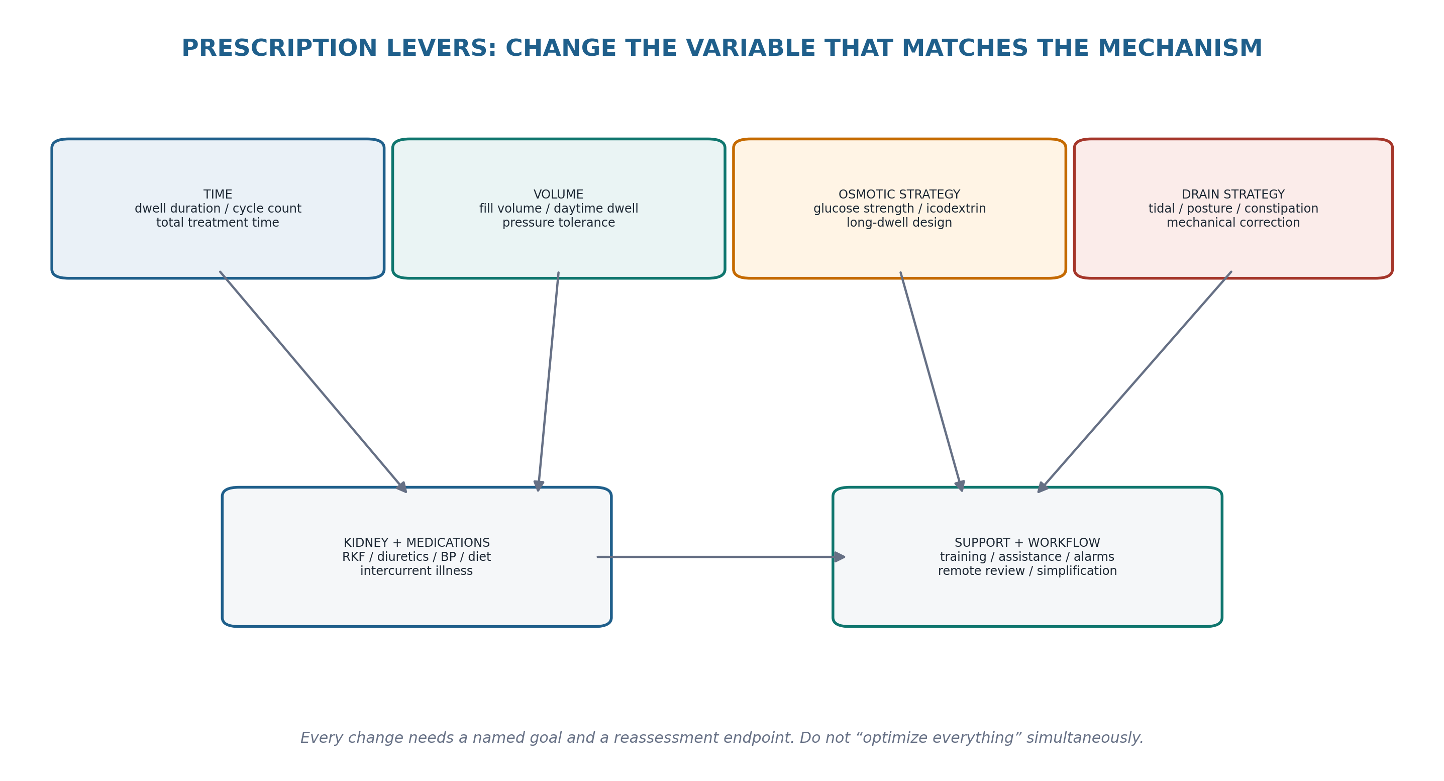
A good PD change is testable. It names the target mechanism, changes as few variables as possible, and predicts what should improve. Simultaneously increasing treatment time, glucose strength, cycle number and fill volume may produce a new number but destroys causal clarity and can increase burden or complications. Goal-directed care favors iterative, patient-centered adjustment rather than maximal prescription intensity. [1,7]
Table 19.16 — Problem → mechanism → smallest useful lever.
| Problem | Mechanism | First targeted lever | Reassessment |
|---|---|---|---|
| Short-dwell fast transport with poor long-dwell UF | Dwell/transport mismatch | Redesign dwell/osmotic strategy | Volume symptoms + delivered UF |
| Drain pain terminating APD | Suction/tip/prescription mechanics | Drain/tidal/posture/mechanical correction | Pain + treatment completion |
| Congestion after RKF loss | Reduced total water/sodium removal | Rebuild total volume plan | Weight/BP/symptoms/urine |
| Low clearance from incomplete APD | Delivery failure due alarms | Fix drain cause | Completion + clearance |
| Metabolic burden from strong glucose | Excess osmotic exposure | Reduce unnecessary exposure after cause correction | Glycemia/weight + volume |
| Caregiver burnout | Support mismatch | Assisted/simplified workflow | Sustainability + treatment completion |
| Persistent catheter dysfunction | Structural access problem | Procedural salvage/revision | Functional inflow/outflow |
| ONE-CHANGE RULE When clinically safe, change one dominant lever at a time. If several changes are necessary because the patient is acutely unwell, document which endpoint each change is intended to affect. |
|---|
16. Reassessment: close the loop or you have not troubleshot the problem
The timing of reassessment depends on severity and mechanism. A severe infection or respiratory problem requires immediate or same-day reassessment; a bowel-related drain problem may be reassessed after the corrective intervention; a prescription change may require several exchanges or days to judge; nutrition or support interventions may require longer. There is no single universal interval for every PD adjustment. The key requirement is that an endpoint is specified before the change is made.
Table 19.17 — Reassessment endpoints by domain.
| Domain | What should improve | If it does not |
|---|---|---|
| Mechanical flow | Drain time, alarms, retained volume, pain | Image/procedural escalation |
| Volume | Symptoms, edema, weight trend, BP, net fluid balance | Re-localize intake/RKF/membrane/cardiac cause |
| Peritonitis | Clinical status + effluent inflammatory trajectory per guideline | Organism/source/refractory pathway |
| Catheter infection | Purulence/inflammation/tunnel findings | Source-control reassessment |
| Clearance | Delivered treatment + biochemical/symptom target | Validate collection, RKF, prescription again |
| Pain | Timing/intensity + treatment completion | Re-examine mechanical/infectious/abdominal causes |
| Metabolic | Glycemia, potassium, appetite, weight phenotype | Reassess exposure/intake/illness/medications |
| Support | Treatment completion, contamination, caregiver burden | Escalate assistance or modality planning |
| CLOSED-LOOP RULE An intervention without a planned reassessment is not troubleshooting; it is an experiment without an outcome measure. |
|---|
17. When the problem does not resolve: escalation, HD transfer and quality improvement
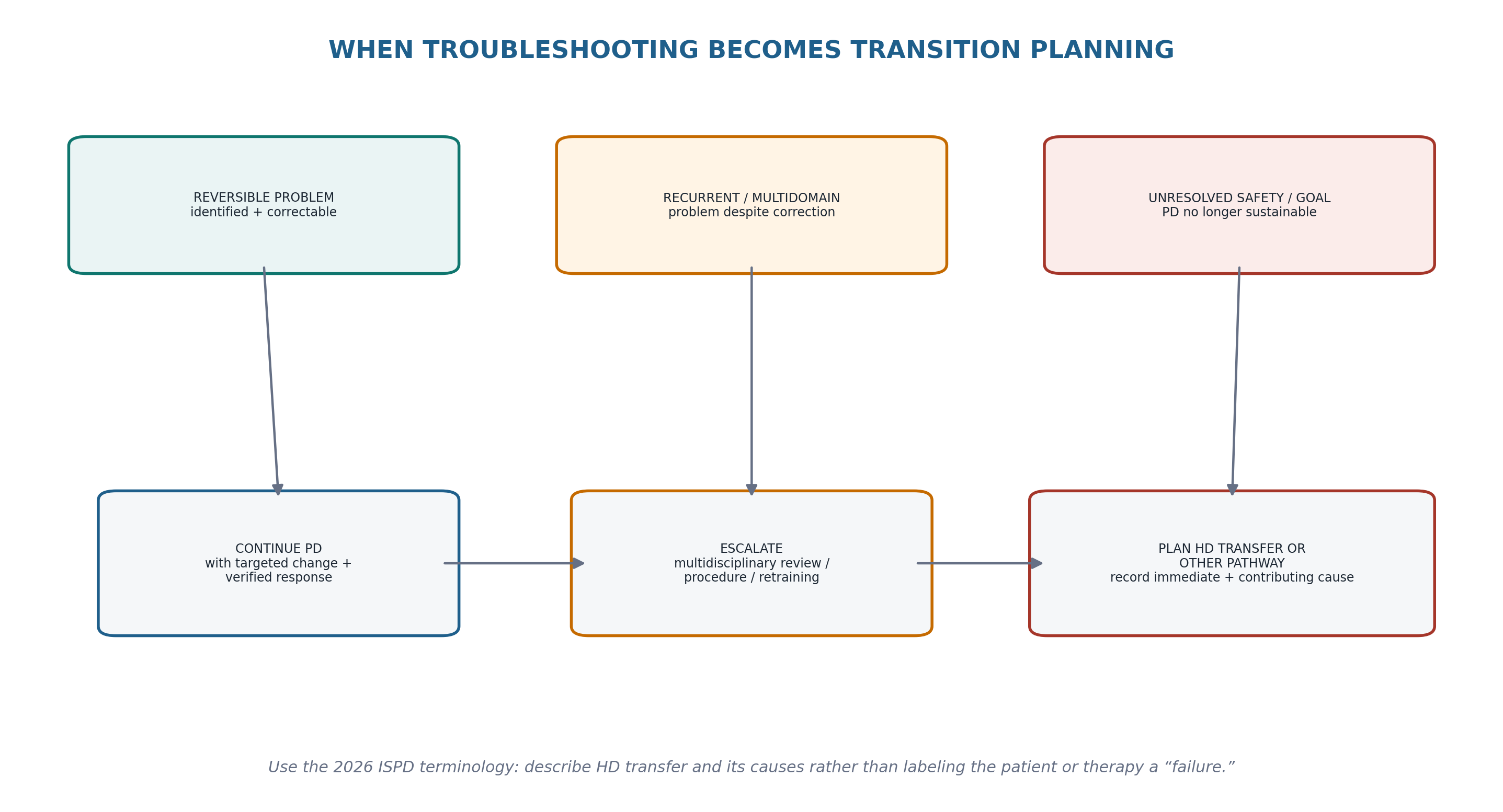
The 2026 ISPD position statement recommends replacing the imprecise terms “technique failure” and “technique survival” with direct reporting of HD transfer and other causes of loss from PD. For bedside troubleshooting, this changes the question from “Has PD failed?” to “What immediate event caused the transfer, what contributing factors were present, which were modifiable, and is return to PD feasible or desired?” The statement encourages programs to distinguish immediate from contributing causes because recurrent problems often reflect treatment burden, capability, access or program-level factors in addition to the final clinical event. [6]
Table 19.18 — Escalation and transition documentation.
| Field | What to document |
|---|---|
| Immediate cause | The event that directly led to HD transfer or PD interruption |
| Contributing causes | Infection history, access problems, membrane dysfunction, frailty, treatment burden, caregiver/system factors |
| Reversibility | Temporary, potentially correctable, or unlikely to resolve |
| PD return potential | Whether PD restart is clinically feasible and aligned with patient goals |
| Intervention already attempted | Medical, prescription, retraining, access or surgical steps |
| Patient priorities | Preference, burden, life participation, risk tolerance |
| Transition plan | Temporary HD, permanent HD, transplant, conservative/supportive pathway or other |
| Program learning | What CQI action could prevent a similar avoidable transfer? |
Quality improvement should use recurrent patterns rather than isolated anecdotes. Infection rates, access dysfunction, early HD transfer, repeated alarms, missed training competencies, hospitalization and treatment burden can reveal program-level failure modes. PDOPPS data show substantial international variation in time on PD and identify infection as a leading cause of HD transfer, supporting structured program review rather than attributing every transfer to patient-level factors. [9,10,12]
| TRANSITION RULE A planned HD transfer can be the safest and most patient-centered outcome of excellent troubleshooting. The failure is not changing modality; the failure is allowing an unresolved problem to become an avoidable emergency. |
|---|
18. Major clinical algorithms
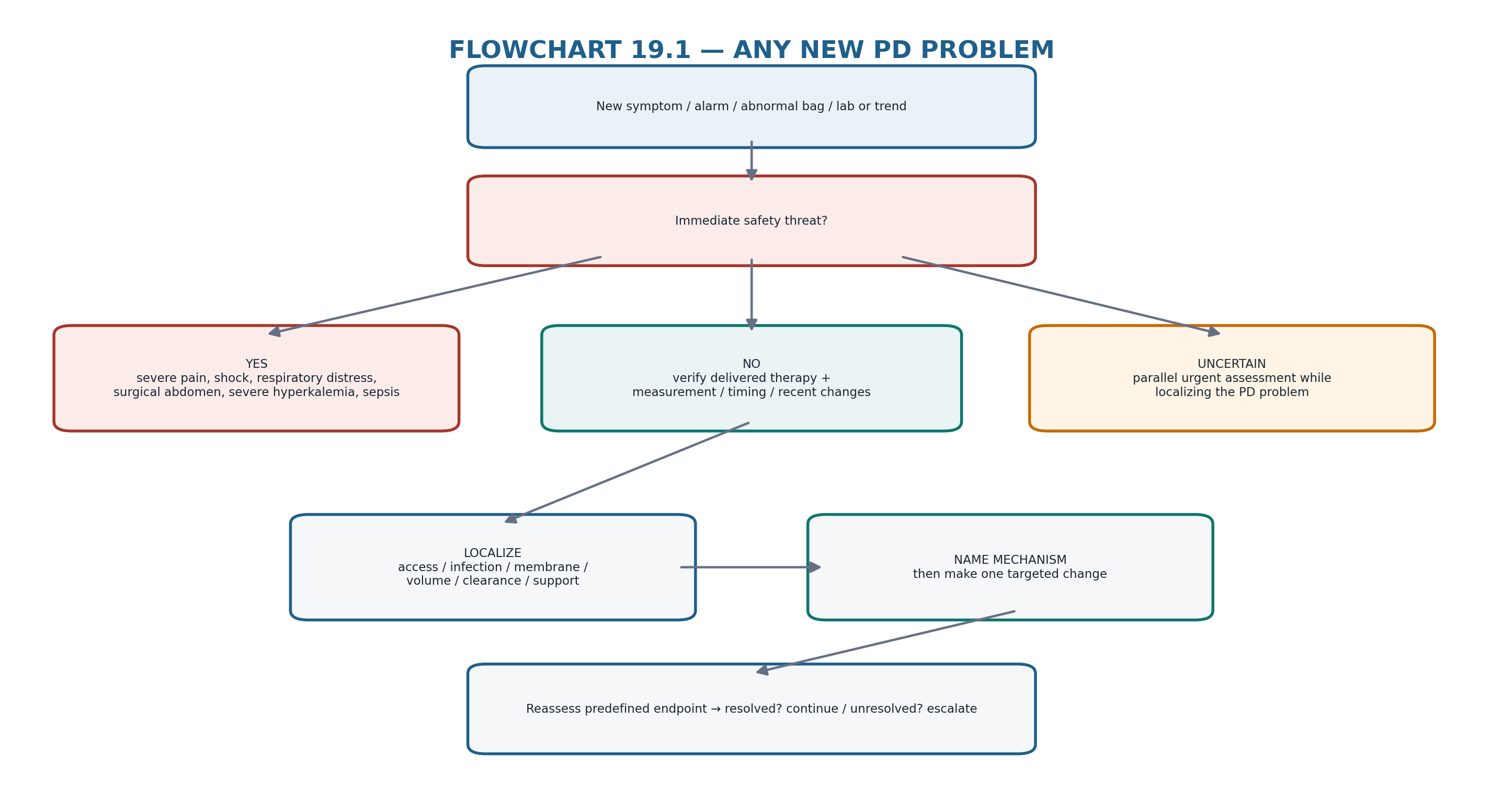
19. Retention tables: pattern recognition
Table 19.19 — If you see this, think this first.
| Pattern | First thought | Do now |
|---|---|---|
| Low UF + drain alarms | Delivery/mechanical problem may masquerade as membrane failure | Check constipation, position, retained volume, event log |
| Low UF + good drainage + urine decline | Total water removal fell because RKF declined | Rebuild volume plan |
| Cloudy effluent | Peritonitis until assessed | Chapter 10 pathway |
| Red exit site without pus | Not automatically definitive ESI | Examine trend/trauma/dermatitis; Chapter 11 definitions |
| Purulent exit drainage | Definitive ESI phenotype | Culture/treat per Chapter 11 |
| Dyspnea + right pleural effusion | Hydrothorax vs cardiac/other effusion | Chapter 12 evaluation |
| Repeated low clearance with short APD nights | Delivered-dose problem | Fix alarms/burden before dose escalation |
| High glucose use + weight/glycemia rise | Metabolic cost may be treating an unlocalized volume problem | Re-localize why UF is inadequate |
| Missed therapy + caregiver exhaustion | Support failure | Assisted/simplified plan; Chapter 16 |
| Recurrent multidomain problems | PD sustainability question | Multidisciplinary review; transition planning if needed |
Table 19.20 — High-yield “do not confuse” pairs.
| Do not confuse | With | Correction |
|---|---|---|
| Prescribed PD | Delivered PD | Verify actual treatment |
| Low UF | Volume overload | Phenotype clinically |
| Low UF | Membrane failure | Exclude drainage/leak/RKF/prescription first |
| Alarm count | Catheter diagnosis | Use pattern + clinical mechanics |
| Cloudy effluent | Culture-positive infection only | Culture-negative peritonitis exists; mimics also exist |
| Erythema | Definitive exit-site infection | Purulence is central to definitive ESI definition |
| Low Kt/V | Global inadequate care | Use goal-directed assessment |
| High BP | Automatic need for stronger glucose | Confirm volume |
| Missing upload | Nonadherence | Check connectivity/data |
| HD transfer | “Technique failure” | Use neutral 2026 ISPD terminology + causal documentation |
20. Clinical pearls
1. The fastest route to the right intervention is often to ask what changed first: the patient, the catheter, the prescription, the membrane, the kidney, or the support system.
2. If a problem is sudden, think delivery, access, infection or acute illness before chronic membrane failure.
3. If a problem is gradual, trend RKF, transport, nutrition, support and treatment burden rather than reacting to one datapoint.
4. Constipation is a dialysis complication when it causes catheter dysfunction, pain or inadequate delivery.
5. Low UF plus incomplete drainage is a mechanical problem until proved otherwise.
6. Cloudy effluent is a safety signal; diagnose first, theorize later.
7. “Adequate numbers” do not exclude inadequate life participation, nutrition, symptom control or volume management.
8. A repeated prescription change without a reassessment endpoint creates therapeutic noise.
9. When several domains are abnormal, correct the one with the highest safety consequence or causal leverage first.
10. Transition planning is part of PD excellence, not an admission of defeat.
21. Common pitfalls — and the correction
Table 19.21 — Pitfalls and corrections.
| Pitfall | Why it fails | Correction |
|---|---|---|
| Increase dextrose whenever UF is low | Misses retained fluid, leak, constipation, RKF loss, sodium intake and membrane phenotype | Localize first |
| Increase dose whenever clearance is low | Collection/delivery error and RKF loss may be the real issue | Validate before prescribing |
| Treat every cycler alarm as machine trouble | Repeated drain alarms often encode catheter physiology | Read event pattern + assess mechanics |
| Treat every red exit site as infection | Overdiagnosis/antibiotic exposure | Use Chapter 11 definition |
| Treat every culture-negative cloudy bag as noninfectious | Culture-negative infectious peritonitis exists | Use Chapter 10 criteria and trajectory |
| Change several prescription variables together | Cannot tell what worked; increases burden | Use smallest effective lever |
| Ignore treatment burden because labs are acceptable | Delivered care may become unsustainable/unsafe | Assess capability, caregiver and goals |
| Keep retrying PD despite recurrent unresolved safety events | Turns planned transition into emergency HD | Escalate and plan |
| Call HD transfer “technique failure” | Imprecise and stigmatizing; obscures causes | Use 2026 ISPD HD-transfer terminology |
| Document only the final event | Misses modifiable contributing causes | Record immediate + contributing factors |
22. Mini-cases: decisions, not trivia
Table 19.22 — Mini-cases.
| Case | Clinical signal | Reasoning pause | Best next move |
|---|---|---|---|
| Case 1 — Low UF after a “bad week” | APD patient reports low UF, 2-kg weight gain and repeated drain alarms. Abdomen comfortable; urine unchanged. | The low UF may be retained volume from poor drainage, not membrane failure. | Review event log, constipation/posture and retained volume before increasing osmotic strength. |
| Case 2 — “Adequacy fell” | Stable patient has lower weekly clearance after an incomplete collection; symptoms and volume are unchanged. | Measurement validity comes before dose escalation. | Repeat/validate collection and delivered therapy; assess RKF trend. |
| Case 3 — Cloudy bag, minimal pain | Patient notices cloudy effluent but feels only mildly unwell. | Peritonitis can present without dramatic pain. | Immediate Chapter 10 diagnostic/treatment pathway. |
| Case 4 — High BP and no edema | Home BP is repeatedly high, weight stable, no orthopnea, strong UF. | Volume overload is not established. | Validate BP and assess medications/other causes before stronger dextrose. |
| Case 5 — Drain pain ends APD early | Patient terminates treatment 60–90 min early most nights because of late-drain pain. | “Nonadherence” is secondary to a prescription/mechanical symptom. | Treat drain pain/mechanics and redesign the APD drain strategy. |
| Case 6 — Frailty + contamination | Older patient has two contamination events after recent cognitive decline; daughter is exhausted. | Capability/support domain has changed. | Competency reassessment; assisted PD or transition plan rather than repeated blame/retraining alone. |
| Case 7 — Edema after urine loss | Previously stable CAPD patient loses residual urine over months and now needs stronger glucose. | The old prescription has been exposed by RKF loss. | Rebuild total sodium/water removal and formal membrane/prescription review. |
| Case 8 — Recurrent peritonitis + burden | After multiple infections the latest episode resolves, but patient no longer wishes to restart PD. | The immediate infection and treatment-burden context both matter. | Shared decision-making; document immediate/contributing causes and planned HD transfer per 2026 terminology. |
23. Active recall
MUST MEMORIZE
Table 19.23 — Must-memorize troubleshooting anchors.
| Prompt | Answer |
|---|---|
| First action in any PD problem? | Safety screen, then define the phenotype precisely. |
| Before interpreting a treatment metric? | Verify delivered therapy and measurement validity. |
| Six failure domains? | Delivery/data; access/mechanics; infection/inflammation; membrane/prescription; patient physiology; capability/support. |
| Low UF first principle? | It is a finding, not a diagnosis. |
| Poor drain first reversible cause to actively seek? | Constipation (also bladder/position/setup). |
| Cloudy effluent rule? | Peritonitis assessment takes priority. |
| Prescription-change principle? | Match one dominant lever to one identified mechanism when clinically safe. |
| What closes troubleshooting? | A predefined reassessment endpoint. |
| If one intervention repeatedly fails? | Look for a coupled second domain. |
| Preferred 2026 terminology for leaving PD to HD? | HD transfer, with immediate and contributing causes documented. |
USE AS REFERENCE
Chapter 3 for PET and membrane transport classification.
Chapter 5 for catheter placement/function anatomy.
Chapter 8 for volume, sodium balance and UF failure.
Chapter 9 for CAPD/APD prescription optimization.
Chapter 10 for peritonitis diagnosis and treatment.
Chapter 11 for exit-site/tunnel infection definitions and source control.
Chapter 12 for flow dysfunction, leaks, hernias, hydrothorax and mechanical pain.
Chapter 13 for noninfectious abnormal effluent and metabolic complications.
Chapter 14 for long-term membrane failure and EPS.
Chapter 15 for nutrition/PEW.
Chapter 16 for frailty, assistance and difficult prescriptions.
Chapter 17 for hospitalized/urgent acute scenarios.
Chapter 18 for connected-cycler data and remote alert governance.
24. Flashcards: spaced repetition
Table 19.24 — Flashcards.
| Question | Answer |
|---|---|
| Why is “low UF” not a diagnosis? | Because low net UF can arise from incomplete drainage, leak, intake/RKF change, prescription mismatch or membrane dysfunction. |
| What should precede interpretation of a cycler trend? | Verification that the treatment was actually delivered and the data are complete/credible. |
| Why can constipation mimic inadequate dialysis? | It can impair catheter drainage, shorten APD delivery and reduce effective exchanges. |
| What is the safest response to cloudy effluent? | Enter the peritonitis pathway promptly; consider noninfectious mimics only within a safe diagnostic process. |
| Why not change several prescription variables at once? | You lose causal clarity and may increase treatment burden or complications. |
| What does a low clearance result require before dose escalation? | Validation of collection, delivered therapy and residual kidney function. |
| What is the main distinction between hypertension and volume overload? | Hypertension is a sign with multiple causes; volume overload requires a volume phenotype. |
| When does treatment burden become a clinical mechanism? | When it causes incomplete treatment, contamination, early termination, caregiver collapse or goals conflict. |
| What should repeated correct but unsuccessful interventions trigger? | Search for a second coupled failure domain. |
| What does 2026 ISPD recommend instead of “technique failure”? | Describe HD transfer and record its immediate/contributing causes. |
| What is a good reassessment endpoint? | A patient- and mechanism-relevant change such as symptom relief, treatment completion, drain function, volume improvement or effluent trajectory. |
| What is the role of transition planning? | To prevent unresolved PD problems from becoming emergency modality changes. |
25. Rapid differential / troubleshooting
Table 19.25 — One-minute problem sorter.
| Presentation | First three questions | Primary route |
|---|---|---|
| Poor drain | Constipation? position? fibrin/blood? | Ch 5/12 |
| Low UF | Delivered/drained? leak? RKF/intake changed? | Ch 8/12 |
| Edema/weight gain | True fluid? urine decline? sodium/prescription? | Ch 8 |
| High BP | Measurement valid? volume phenotype? meds/pain? | Ch 8/16 |
| Low clearance | Collection valid? delivered therapy? RKF? | Ch 7/9 |
| Abdominal pain | Fill/drain timing? cloudy fluid? focal/systemic? | Ch 10/12/13 |
| Cloudy bag | Pain/systemic? cell/culture? mimic clues? | Ch 10/13 |
| Red exit site | Pus? tunnel findings? trauma/dermatitis? | Ch 11 |
| Hyperglycemia/weight gain | Glucose exposure? fluid vs calories? illness? | Ch 13/15 |
| Missed/short therapy | Pain? alarms? cognition/support? | Ch 16/18 |
| Hospital deterioration | What acute goal? is PD safe? what changed? | Ch 17 |
| Repeated multidomain events | What is immediate cause? contributing causes? reversibility? | Integrated review / transition plan |
| TROUBLESHOOTING SHORTCUT If you are about to change the prescription but cannot say which domain and mechanism are failing, stop and collect the next discriminating piece of information. |
|---|
26. Final revision sheet
| CORE CONCEPT PD troubleshooting is a closed-loop diagnostic discipline: define the phenotype, exclude danger, verify delivery, localize the failing domain, name the mechanism, make the smallest useful change, and reassess. |
|---|
One-minute revision
Safety before troubleshooting.
Delivered therapy before physiology.
Localization before prescription.
Mechanism before lever.
One dominant change when clinically safe.
Reassessment before declaring success.
Multidomain thinking when the problem recurs.
Treatment burden and support are clinical domains, not soft extras.
Planned escalation/procedure can preserve PD; planned HD transfer can also be the correct outcome.
Use neutral causal language: HD transfer, immediate cause, contributing causes, reversibility and learning.
TEN TAKE-HOME RULES
1. Never treat a number before validating how it was generated.
2. Low UF is a finding; membrane failure is one possible diagnosis.
3. Poor drainage can convert a technically adequate prescription into inadequate delivered therapy.
4. Cloudy effluent is a time-sensitive infection signal until properly assessed.
5. Redness alone is not the same as definitive exit-site infection.
6. Volume, clearance, pain and treatment burden must be interpreted as separate but interacting domains.
7. Prescription changes should be mechanism-linked and testable.
8. Recurrent problems often indicate a second uncorrected domain.
9. Treatment that cannot be safely delivered at home is not an adequate prescription, even if it looks ideal on paper.
10. A planned transition is safer than repeated unsuccessful rescue.
One-minute bedside synthesis
State the complaint in one line. Ask whether anyone is in danger. Confirm what treatment was actually delivered. Choose the dominant failure domain. Name the mechanism. Make the smallest safe correction. Decide what should improve and when you will check it. If it does not improve, do not simply intensify the same intervention—re-localize the problem or escalate the level of care.
| FINAL MENTAL MODEL PD succeeds when six systems remain aligned: treatment is delivered, the catheter works, infection is controlled, the membrane/prescription match, the patient’s physiology is supported, and the home plan is sustainable. Troubleshooting is the process of finding which alignment has been lost. |
|---|
Rapid oral viva
“The UF is low.” — What four non-membrane explanations must you exclude first?
“The cycler alarms every night.” — How do you separate machine/data failure from catheter mechanics?
“Kt/V fell.” — What makes the result valid enough to act on?
“The bag is cloudy.” — What is the safety-first pathway?
“BP is high.” — What evidence would make you treat volume rather than the number?
“The patient keeps missing exchanges.” — What capability/support questions come before blame?
“Three correct interventions have not solved the problem.” — What does multidomain failure mean?
“The patient is moving to HD.” — How should this be described and documented in 2026 terminology?
| SAFETY BOUNDARY This chapter is a reasoning and routing framework. Disease-specific diagnostic criteria, drug doses, antimicrobial regimens, PET/UF thresholds, access procedures and urgent KRT decisions remain governed by their dedicated chapters, current guidelines, local protocols and direct clinical assessment. |
|---|
27. Selected authoritative references
1. Brown EA, Blake PG, Boudville N, et al. International Society for Peritoneal Dialysis practice recommendations: Prescribing high-quality goal-directed peritoneal dialysis. Perit Dial Int. 2020;40(3):244–253. https://doi.org/10.1177/0896860819895364. PMID: 32063219.
2. Morelle J, Stachowska-Pietka J, Öberg C, et al. ISPD recommendations for the evaluation of peritoneal membrane dysfunction in adults: classification, measurement, interpretation and rationale for intervention. Perit Dial Int. 2021;41(4):352–372. https://doi.org/10.1177/0896860820982218. PMID: 33563110.
3. Li PKT, Chow KM, Cho Y, et al. ISPD peritonitis guideline recommendations: 2022 update on prevention and treatment. Perit Dial Int. 2022;42(2):110–153. https://doi.org/10.1177/08968608221080586. PMID: 35264029.
4. Chow KM, Li PKT, Cho Y, et al. ISPD Catheter-related Infection Recommendations: 2023 Update. Perit Dial Int. 2023;43(3):201–219. https://doi.org/10.1177/08968608231172740. PMID: 37232412.
5. Crabtree JH, Shrestha BM, Chow KM, et al. Creating and Maintaining Optimal Peritoneal Dialysis Access in the Adult Patient: 2019 Update. Perit Dial Int. 2019;39(5):414–436. https://doi.org/10.3747/pdi.2018.00232. PMID: 31028108.
6. Quinn RW, Johnson DW, Mehrotra R, et al. 2026 ISPD Position Statement on tracking and reporting loss from PD therapy. Perit Dial Int. Published online June 17, 2026. https://doi.org/10.1177/08968608261461639.
7. Borràs Sans M, Ponz Clemente E, Rodríguez Carmona A, et al. Clinical guideline on adequacy and prescription of peritoneal dialysis. Nefrologia (Engl Ed). 2024;44 Suppl 1:1–27. https://doi.org/10.1016/j.nefroe.2024.09.001. PMID: 39341764.
8. Hurst H, Figueiredo AE, Perez Moran D, et al. Peritoneal dialysis training and interventions: A narrative review. Perit Dial Int. 2026;46(1):6–15. https://doi.org/10.1177/08968608251328517. PMID: 40221963.
9. Lambie M, Zhao J, McCullough K, et al. Variation in Peritoneal Dialysis Time on Therapy by Country: Results from the Peritoneal Dialysis Outcomes and Practice Patterns Study. Clin J Am Soc Nephrol. 2022;17(6):861–871. https://doi.org/10.2215/CJN.16341221. PMID: 35641246.
10. Perl J, Fuller DS, Bieber BA, et al. Peritoneal Dialysis-Related Infection Rates and Outcomes: Results From the Peritoneal Dialysis Outcomes and Practice Patterns Study (PDOPPS). Am J Kidney Dis. 2020;76(1):42–53. https://doi.org/10.1053/j.ajkd.2019.09.016. PMID: 31932094.
11. Nataatmadja M, Cho Y, Johnson DW. Continuous Quality Improvement Initiatives to Sustainably Reduce Peritoneal Dialysis-Related Infections in Australia and New Zealand. Perit Dial Int. 2016;36(5):472–477. https://doi.org/10.3747/pdi.2016.00114. PMID: 27659926.
12. Kraft L, Oppold J, Kitterer D, et al. Changing microbiology and outcomes of PD-associated peritonitis over four decades. Clin Kidney J. 2026;19(1):sfaf387. https://doi.org/10.1093/ckj/sfaf387. PMID: 41536568.
13. Blake PG, Bargman JM. Peritoneal dialysis. In: Brenner & Rector's The Kidney. 12th ed. Elsevier; 2024. Foundational physiology, prescription, adequacy and complication framework.
| SOURCE NOTE Current ISPD guideline status and contemporary PD evidence were checked 3 September 2026. This synthesis intentionally avoids inventing new universal thresholds. When a troubleshooting branch reaches a disease-specific decision, the numerical criteria and treatment details in the relevant chapter, current guideline, product information and local protocol take precedence. |
|---|
CHAPTER 19 COMPLETE
NEXT: CHAPTER 20 — FINAL REVIEW: ALGORITHMS, CASES, HIGH-YIELD TABLES AND MASTER REVISION