PSTR | PET | Ultrafiltration | Sodium Sieving | Osmotic Conductance | Longitudinal Change
CHAPTER MISSION Turn the PET from a transporter label into a bedside physiologic experiment: run the test correctly, separate small-solute transfer from ultrafiltration capacity and free-water transport, recognize technical artefact and non-membrane mimics, and translate the measured phenotype into a prescription change that can be reassessed.
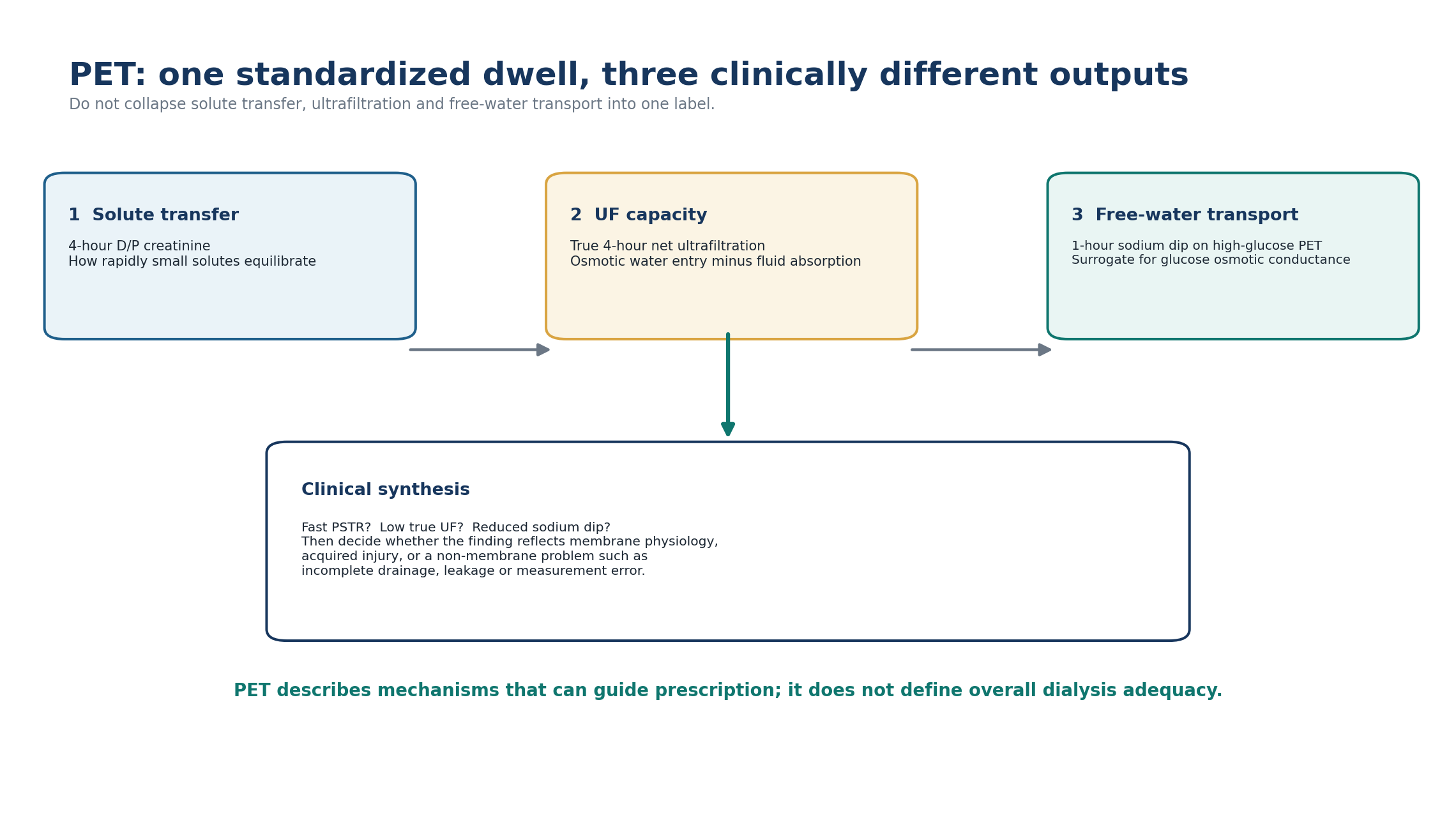
MASTER PRINCIPLE A PET is not a pass/fail test of dialysis adequacy. Interpret it in order: test validity -> 4-hour D/P creatinine -> true 4-hour ultrafiltration -> 1-hour sodium dip when indicated -> clinical context. Then act on the mechanism, not the label.
0. One-page chapter map
Table 3.1 - The eight decisions that govern PET interpretation.
| Decision | Core question | Bedside output |
|---|---|---|
| 1. Question | Is this an early baseline membrane assessment or investigation of a new clearance/UF problem? | Correct indication and timing |
| 2. Run | Was a standardized 4-hour PET performed with a defined glucose concentration and sampling protocol? | Comparable physiology test |
| 3. Validate | Were timing, samples, creatinine assay, true fill volume and complete drainage reliable? | Artefact excluded before interpretation |
| 4. Solute | How fast did creatinine equilibrate relative to the centre/population reference? | PSTR phenotype |
| 5. Water | What was the true 4-hour net UF for the glucose concentration used? | UF-capacity screen |
| 6. Free water | If UF is insufficient, is the 1-hour sodium dip preserved on a high-glucose test? | Osmotic-conductance signal |
| 7. Prescribe | Which dwell architecture or osmotic strategy fits the measured mechanism? | Targeted prescription response |
| 8. Trend | Is this stable baseline physiology, a transient change, or acquired membrane deterioration? | Longitudinal plan and escalation trigger |
Learning outcomes
Explain what a PET measures and why PSTR, ultrafiltration capacity and free-water transport must be interpreted as separate properties.
Calculate and interpret D/P creatinine and D/D0 glucose conceptually without confusing them with clearance or overall dialysis adequacy.
Perform a guideline-concordant 4-hour PET framework and identify technical errors that can invalidate the result.
Use 4-hour D/P creatinine relative to centre/population distributions rather than treating historical transporter bands as universal treatment thresholds.
Recognize guideline screening thresholds for insufficient ultrafiltration and state why they are not, by themselves, proof of membrane failure.
Explain the 1-hour sodium dip and sodium-sieving ratio and use the ISPD diagnostic thresholds when intrinsic low osmotic conductance is suspected.
Distinguish fast PSTR, intrinsic low osmotic conductance and acquired membrane injury from non-membrane causes of low drained volume.
Translate fast or slow solute transfer into dwell-time and osmotic-agent reasoning while protecting residual kidney function and minimizing unnecessary glucose exposure.
Interpret serial PET changes in context, including inflammation, peritonitis, treatment duration and measurement variability.
Know when derivative membrane tests add specialist information and when routine clinical care should remain anchored to the standard PET.
EVIDENCE POSTURE The 2021 ISPD membrane-dysfunction recommendations remain the principal contemporary standard for PET measurement and interpretation. They recommend a 4-hour PET at 6-12 weeks after PD initiation and subsequently when clinically indicated, using creatinine as the index solute with either 2.27%/2.5% or 3.86%/4.25% glucose/dextrose. Modern interpretation emphasizes continua, centre effects, true UF capacity and selective high-glucose sodium-dip testing rather than rigid transporter labels. The original Twardowski PET, contemporary PET review literature, large cohorts, mechanistic AQP1 work and longitudinal studies provide the supporting evidence. [1–8]
1. Core concept: PET is a physiologic stress test, not a transporter score
The peritoneal membrane is not a fixed dialyzer cartridge. Its effective vascular surface area, small-pore solute permeability, free-water pathway, interstitial structure and fluid absorption vary between people and can change over time. The PET imposes a standardized dwell so that these properties can be observed under reproducible conditions. [1–3]
The most common error is to collapse the whole test into one label - “high transporter” or “low transporter.” Four-hour D/P creatinine mainly describes the speed of small-solute equilibration. It does not tell you whether glucose will generate adequate free-water flow, whether the patient is euvolemic, whether the catheter drains completely, or whether the total PD prescription is adequate. [1,2]
Table 3.2 - What each PET output actually tells you.
| Output | Primary physiologic question | What it does NOT prove |
|---|---|---|
| 4-h D/P creatinine | How rapidly creatinine equilibrates between plasma and dialysate | Overall adequacy, sodium balance or UF mechanism |
| 4-h D/D0 glucose | How much of the starting dialysate glucose remains | Exact glucose absorption or free-water conductance by itself |
| 4-h net UF | How much drained volume exceeds true instilled volume | That low UF is caused by the membrane |
| 1-h sodium dip | How much early solute-free water entry dilutes dialysate sodium | That AQP1 expression alone is abnormal |
| Serial PET trend | Whether membrane behavior is changing over time | The structural diagnosis without clinical context |
BEDSIDE TRANSLATION Read PET as three questions: How fast do small solutes move? How much fluid is removed? If fluid removal is poor, is free-water transport preserved? Only after those answers should you decide which prescription lever to change.
2. The two curves: D/P creatinine rises while D/D0 glucose falls
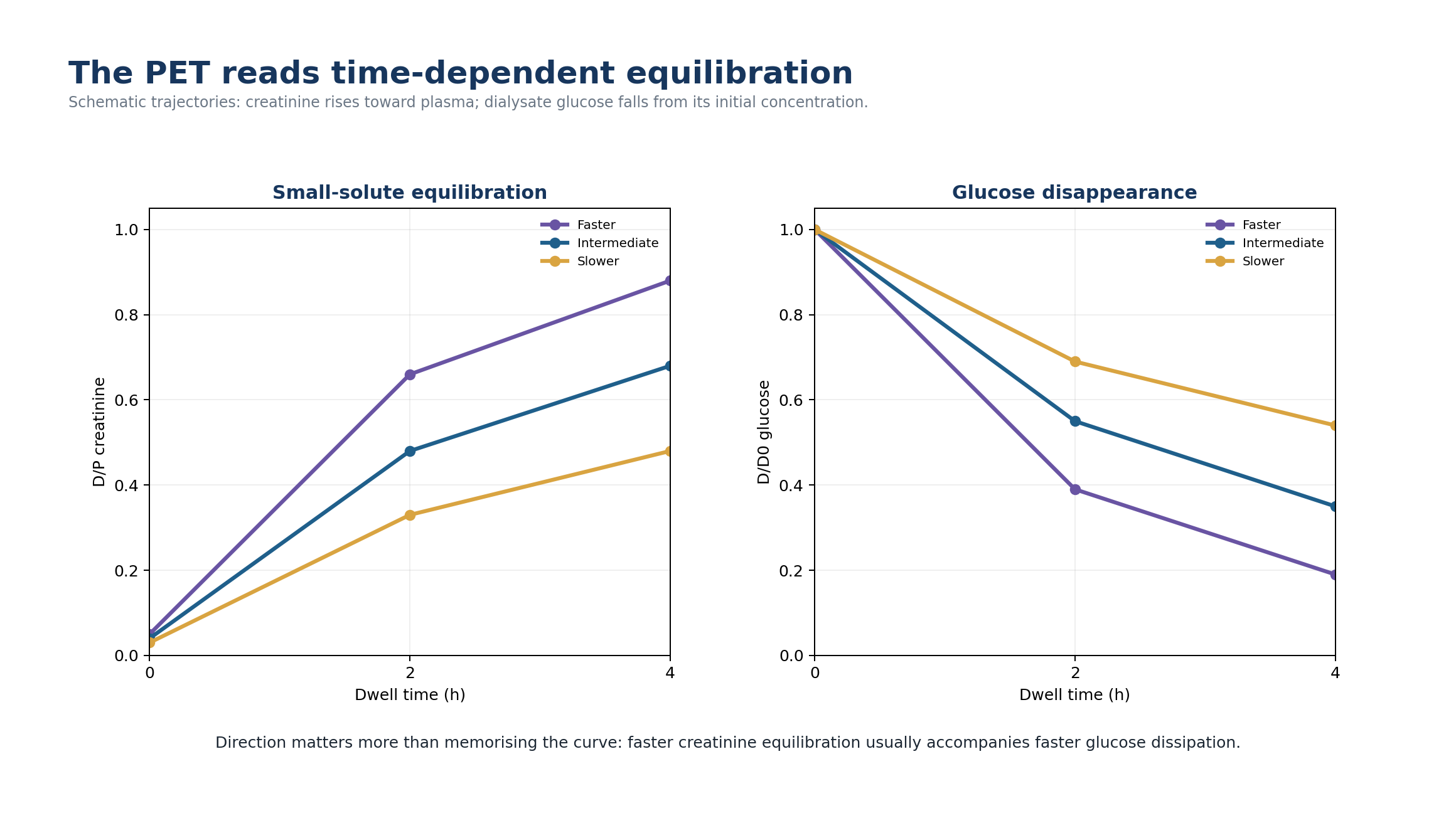
For creatinine, the dialysate-to-plasma ratio (D/P creatinine) starts low and rises toward 1 as equilibration proceeds. A higher 4-hour D/P creatinine means more rapid small-solute transfer. For glucose, the dialysate concentration at time t is divided by its initial concentration (D/D0 glucose). This ratio falls as glucose leaves the cavity; faster transport therefore tends to produce a lower D/D0 at 4 hours. [2,3]
Table 3.3 - D/P and D/D0: opposite directions, one transport story.
| Measure | Numerator / denominator | Direction with faster transport | Clinical use |
|---|---|---|---|
| D/P creatinine | Dialysate creatinine / plasma creatinine | Higher at 4 h | Preferred index of PSTR |
| D/D0 glucose | Dialysate glucose at time t / initial dialysate glucose | Lower at 4 h | Corroborates glucose disappearance / equilibration |
| Net UF | Drained volume - true instilled volume | Often lower during long glucose dwell when PSTR is fast | Screens water-removal capacity |
MEMORY HOOK Creatinine climbs; glucose falls. Fast transport makes both happen sooner.
3. What makes PSTR fast? Think effective vascular surface area, not a “leaky mesothelium”
PSTR is influenced by the effective perfused microvascular surface available for exchange. More perfused capillary area brings blood solutes closer to dialysate and increases diffusion. Local intraperitoneal inflammatory activity is one important determinant of higher PSTR; systemic inflammation and local inflammation are not interchangeable biological signals. [1,8]
Fast PSTR therefore has a paradoxical clinical phenotype: small-solute equilibration is efficient, yet a long glucose dwell may remove less water because glucose also disappears more rapidly and the crystalloid osmotic gradient is lost earlier. This is why “fast transporter” should never be translated as simply “good membrane” or “bad membrane.”
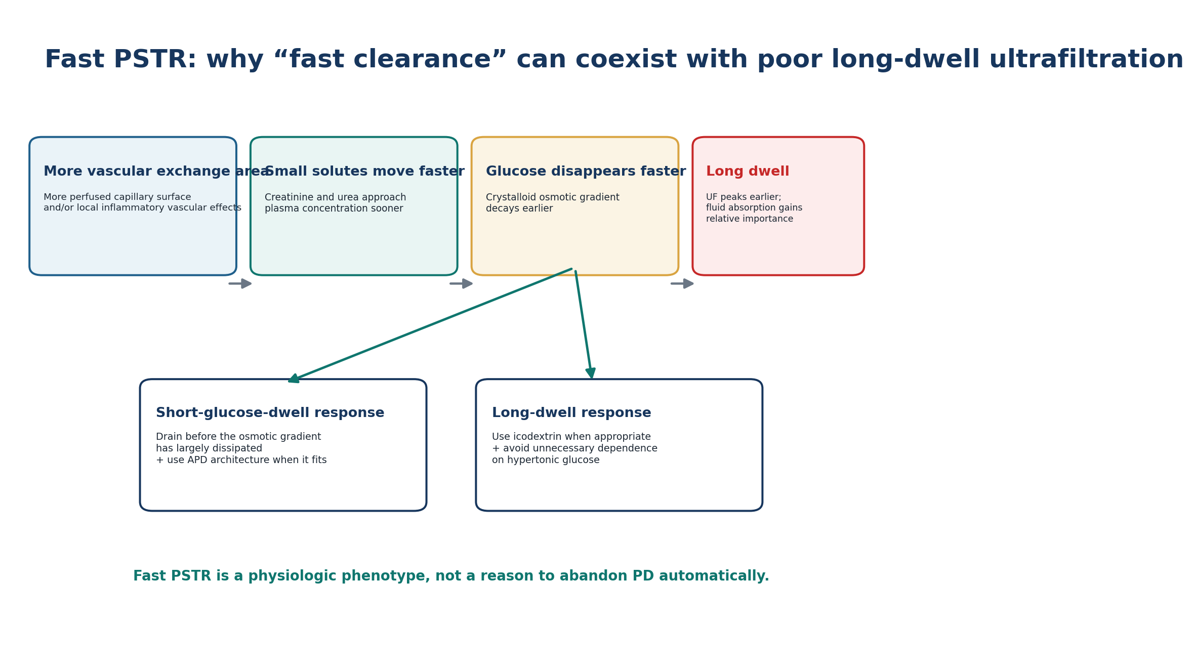
Table 3.4 - Fast PSTR: mechanism -> phenotype -> response.
| Mechanism | Bedside phenotype | Prescription implication |
|---|---|---|
| Larger effective vascular exchange area | Higher 4-h D/P creatinine | Expect faster equilibration |
| Faster glucose diffusion to blood | Lower late D/D0 glucose | Osmotic gradient fades earlier |
| Earlier decline in crystalloid osmotic force | Long glucose dwell may have low net UF | Consider shorter glucose dwells |
| More time for fluid absorption after UF peak | Late dwell may lose previously gained volume | Use a long-dwell strategy that sustains UF, commonly icodextrin when appropriate |
| Associated local inflammation in some patients | PSTR may change around inflammatory events | Interpret serial trend with clinical timing |
4. Historical four-group PET categories: useful language, not universal treatment thresholds
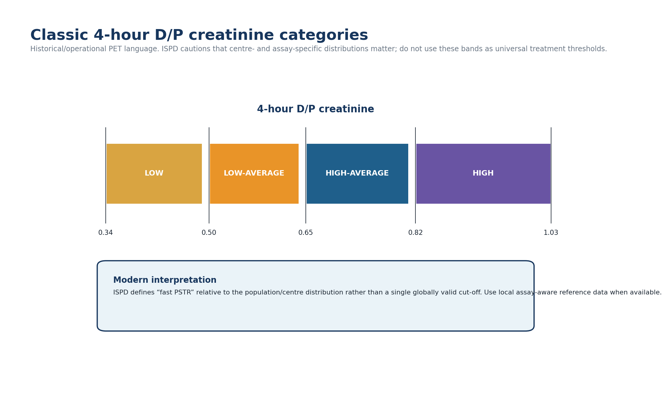
The classic PET language divides 4-hour D/P creatinine into low, low-average, high-average and high groups. These categories remain useful for teaching, local protocols and communication. The Oman Ministry of Health 2025 PD guideline reproduces the commonly used classic bands shown below. [20]
Table 3.5 - Classic operational 4-h D/P creatinine categories.
| Category | 4-h D/P creatinine | How to use it safely |
|---|---|---|
| Low | 0.34-0.49 | Historical/operational reference band; interpret with local assay and clinical context |
| Low-average | 0.50-0.64 | Historical/operational reference band |
| High-average | 0.65-0.81 | Historical/operational reference band |
| High | 0.82-1.03 | Historical/operational reference band |
Modern ISPD guidance deliberately avoids defining fast PSTR by one global number. In a large US cohort of more than 10,000 patients, mean 4-hour D/P creatinine was approximately 0.65 +/- 0.12; other cohorts report different means. ISPD therefore defines fast PSTR relative to the population or centre distribution, ideally using local assay-aware reference data. [1,4]
THRESHOLD DISCIPLINE Do not convert the classic four bands into automatic modality rules. A patient at D/P creatinine 0.82 does not require APD merely because a historical table calls the result “high.” The relevant question is whether the measured physiology creates a clinical clearance or fluid problem for this person.
5. When to perform PET
ISPD recommends determining PSTR early in the dialysis course, between 6 and 12 weeks after starting PD, and repeating the assessment subsequently when clinically indicated. The purpose of the early test is to establish a stable baseline after the immediate start period and provide an individualized membrane phenotype for prescription decisions. [1]
Table 3.6 - PET timing: routine baseline versus problem-driven repetition.
| Situation | Reason for PET | Interpretation safeguard |
|---|---|---|
| 6-12 weeks after PD initiation | Establish early PSTR phenotype | Avoid treating immediate post-start physiology as the long-term baseline |
| New or worsening volume overload | Assess whether membrane transport/UF contributes | Also measure urine, intake, daily UF and mechanical function |
| Unexpected fall in UF | Distinguish fast PSTR from low osmotic conductance or mimic | Use high-glucose PET with 1-h sodium dip if intrinsic UF insufficiency is suspected |
| Unexplained change in clearance / prescription performance | Reassess solute-transfer phenotype | Verify delivery and residual kidney function first |
| Long-term PD with progressive UF deterioration | Assess acquired membrane change | Interpret with serial trajectory and EPS risk context |
| Soon after peritonitis or acute inflammatory illness | Usually defer if clinically safe until stable | Inflammation may transiently alter transport; document timing if testing cannot wait |
DO NOT OVER-TEST There is no evidence that frequent scheduled PETs in every stable patient improve outcomes. Repeat the test when the answer will change management or when longitudinal membrane behavior is clinically important. [1]
6. Run a valid PET: standardization is part of the diagnosis
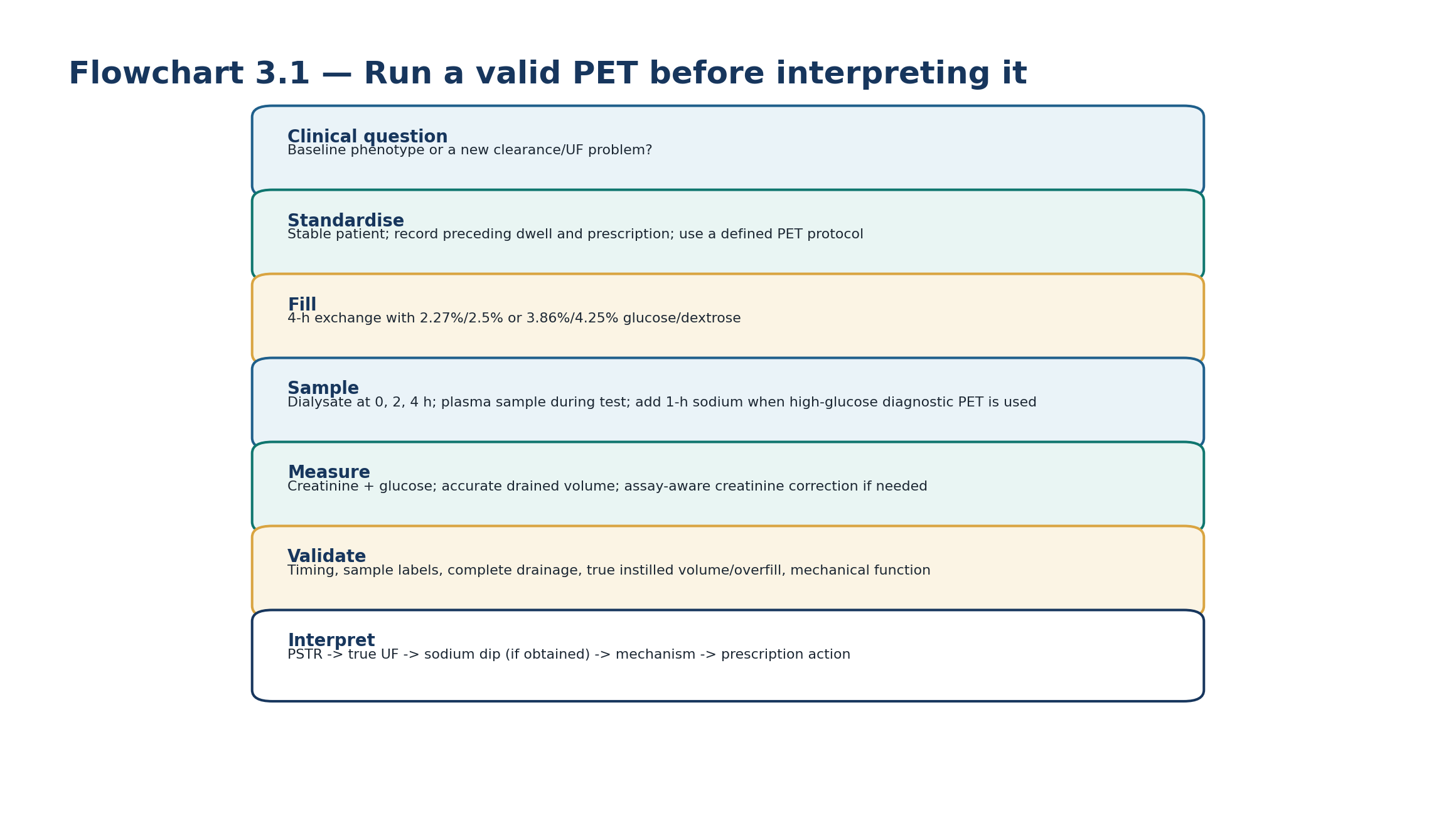
The original PET used a 4-hour dwell with 2 L of 2.5% dextrose, standardized preceding exchange, inflow/drain times, position, sampling and laboratory methods. Contemporary ISPD guidance permits either 2.27%/2.5% or 3.86%/4.25% glucose/dextrose for the 4-hour assessment of PSTR. [1,3]
Table 3.7 - Core PET sequence: what must be standardized.
| Stage | Core action | Why it matters |
|---|---|---|
| Before test | Document indication, current PD prescription, preceding dwell, recent peritonitis/illness and residual urine context | Defines whether the test is comparable to baseline and clinically interpretable |
| Drain preceding dwell | Achieve a complete drain and document unusual residual volume / drain difficulty | Retained fluid alters starting concentration and apparent volume |
| Fill | Use the defined test solution and prescribed test volume; record true instilled volume | Required for valid concentration and UF interpretation |
| Time 0 dialysate | Obtain the initial dialysate sample after standardized mixing according to local protocol | Defines D0 glucose and baseline sodium when measured |
| During dwell | Use standardized sampling times; common standard protocols include dialysate at 2 h and plasma sampling during the test | Prevents timing-dependent misclassification |
| 4 h drain | Complete the drain, measure drained volume and obtain final dialysate sample | Generates D/P creatinine, D/D0 glucose and net UF |
| High-glucose diagnostic PET | Add 1-h dialysate sodium when evaluating intrinsic UF insufficiency | Enables sodium-dip / sieving assessment |
PROTOCOL BOUNDARY The clinical principles are universal; exact fill speed, patient positioning, sample-mixing manoeuvres, aliquot volumes and laboratory transport are local-protocol details. Do not improvise them at the bedside, because a PET only has meaning if its method is reproducible.
7. PET quality assurance: invalidate the artefact before diagnosing the membrane
PET ratios are highly sensitive to timing, sample identity, glucose interference in creatinine assays, residual intraperitoneal volume and the accuracy of fill/drain measurement. Standardization is therefore not administrative detail; it is the analytical validity of the test. [1–3,12–15]
Table 3.8 - High-frequency PET errors and their direction of bias.
| Error | How it distorts the test | Correction |
|---|---|---|
| Wrong sample time | Changes D/P and D/D0 because equilibration is time dependent | Use clocked, documented sampling times |
| Plasma/dialysate sample mislabelling | Can create physiologically impossible ratios | Stop interpretation; repeat if identity cannot be proven |
| Creatinine assay glucose interference | Can falsely elevate dialysate creatinine with some methods | Laboratory must validate method-specific correction or assay approach |
| Incomplete pre-test drain | Dilutes fresh dialysate and changes starting volume | Resolve drainage issue and repeat under standardized conditions if needed |
| Incomplete final drain | Underestimates drained volume and may appear as low UF | Assess catheter/position/constipation; do not diagnose membrane failure |
| Bag overfill ignored | Makes true instilled volume larger than nominal and falsely increases calculated UF if nominal volume is subtracted | Use measured/validated true fill or center-specific overfill correction |
| Dialysate leak | Lowers recovered volume without proving low transcapillary UF | Evaluate for leak before membrane diagnosis |
| Acute peritoneal inflammation | Can increase PSTR transiently | Interpret timing; repeat after recovery when clinically appropriate |
LABORATORY RULE Do not copy a universal Jaffe creatinine “glucose correction formula” from another center. Glucose interference is analyzer- and method-dependent; the laboratory should validate the method used for dialysate creatinine. [14,15]
8. Which glucose concentration should be used? Standard PET versus high-glucose diagnostic PET
PSTR can be determined with either 2.27%/2.5% or 3.86%/4.25% glucose/dextrose. The higher glucose concentration has an additional diagnostic advantage: it creates a strong crystalloid osmotic gradient, allowing simultaneous assessment of 4-hour UF capacity and the 1-hour sodium dip. [1,5,11]
Table 3.9 - Middle-strength versus high-glucose PET.
| Feature | 2.27% glucose / 2.5% dextrose | 3.86% glucose / 4.25% dextrose |
|---|---|---|
| PSTR classification | Validated for 4-h D/P creatinine | Also valid for 4-h D/P creatinine |
| Historical evidence base | Large registry/cohort experience | Large reference cohort also available |
| UF-capacity screening threshold | <100 mL at 4 h suggests insufficient UF | <400 mL at 4 h suggests insufficient UF |
| 1-h sodium dip | Not the preferred diagnostic osmotic-conductance test | Recommended when intrinsic UF insufficiency is suspected |
| Glucose exposure | Lower for the test | Higher acute glucose exposure |
| Best use | Routine local PET protocol where established | Mechanism-focused evaluation when UF insufficiency needs characterization |
CLINICAL CHOICE If the main question is simply PSTR, either glucose strength is acceptable under ISPD guidance. If the question is WHY ultrafiltration is insufficient, the high-glucose PET with 1-hour sodium measurement is the more informative diagnostic experiment. [1]
9. Ultrafiltration capacity: simple to measure, easy to misinterpret
Net PET ultrafiltration is the drained volume minus the true instilled volume. It is a valuable screening signal because it integrates transcapillary osmotic water entry and competing fluid absorption over the test. However, it is not membrane-specific; a low result can arise from drainage failure, leak, variable residual volume or excessive fluid absorption unrelated to fast PSTR. [1,12,13]
Table 3.10 - ISPD screening thresholds for insufficient 4-hour UF.
| Test solution | Insufficient UF should be suspected when net 4-h UF is | Interpretation |
|---|---|---|
| 3.86% glucose / 4.25% dextrose | <400 mL | Screen for membrane dysfunction AND non-membrane causes |
| 2.27% glucose / 2.5% dextrose | <100 mL | Screen for membrane dysfunction AND non-membrane causes |
These are guideline screening thresholds, not a complete diagnosis. ISPD explicitly notes that low UF capacity may result from mechanical problems, leaks or increased fluid absorption. A clinically important volume problem may also be present even when the PET number is above the screening threshold; daily sodium/water balance and residual kidney function remain essential. [1]
SCREEN, THEN DIAGNOSE A 4-hour high-glucose PET with 320 mL net UF tells you that UF is insufficient. It does not yet tell you whether the mechanism is fast PSTR, low osmotic conductance, catheter dysfunction, leak or measurement error.
10. Sodium sieving: the diagnostic window into free-water transport
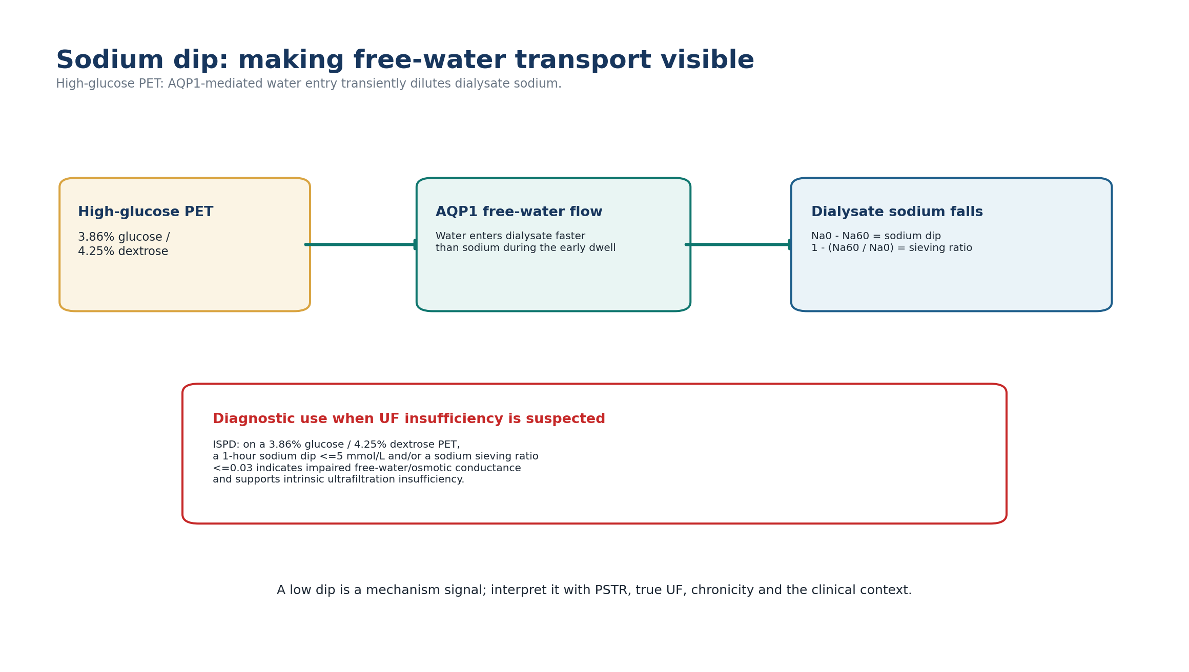
During the early phase of a 3.86% glucose/4.25% dextrose exchange, water crosses through AQP1 channels without accompanying sodium. Dialysate sodium therefore falls transiently. The 1-hour dip is used to minimize the confounding effect of later small-pore sodium diffusion, although faster PSTR can still blunt the apparent dip. [1]
Table 3.11 - Sodium-dip equations and ISPD interpretation.
| Measure | Equation | Interpretation |
|---|---|---|
| Absolute sodium dip | Dialysate Na at time 0 - dialysate Na at 60 min | Larger positive dip = more early solute-free water entry |
| Sodium-sieving ratio | 1 - (Na60 / Na0) | Dimensionless expression of the same early dilution |
| ISPD abnormal threshold | Dip <=5 mmol/L and/or ratio <=0.03 at 1 h | Indicates UF insufficiency due to impaired free-water/osmotic conductance when tested with 3.86%/4.25% solution |
In a multicenter cohort of 758 incident patients tested with 3.86% glucose, mean sodium sieving was about 8.4 mmol/L, while corrected 4-hour UF averaged 675 mL; the broad distribution illustrates why individual variation in osmotic conductance cannot be inferred from PSTR alone. [5]
MEASUREMENT DETAIL ISPD recommends measuring dialysate sodium by the indirect electrode method for this diagnostic approach. Initial dialysate sodium differs among products, which is why the change from the patient’s own time-0 value is used. [1]
11. PSTR and osmotic conductance are separate axes
PSTR explains only a minority of the between-patient variation in UF capacity. Two patients with the same D/P creatinine can have very different glucose-driven water transport. Mechanistic and genetic studies of AQP1 reinforce this distinction: variation in AQP1 expression can alter ultrafiltration without requiring a corresponding change in small-solute transfer. [1,7]
Table 3.12 - The two-axis membrane model.
| PSTR | Sodium dip / osmotic conductance | Likely clinical phenotype |
|---|---|---|
| Fast | Preserved | Fast solute transfer with potentially early glucose-gradient loss; free-water pathway intact |
| Fast | Reduced | Combined fast transfer + low osmotic conductance; higher risk of poor glucose UF |
| Average/slower | Preserved | Solute equilibration slower but glucose osmotic water pathway intact |
| Average/slower | Reduced | Intrinsic UF limitation despite non-fast PSTR; do not miss this phenotype |
CLINICAL PEARL A normal-looking D/P creatinine does not exclude intrinsic ultrafiltration insufficiency. If the patient has a credible UF problem, examine true UF and, when indicated, the sodium dip.
12. Mechanism-based taxonomy of membrane dysfunction
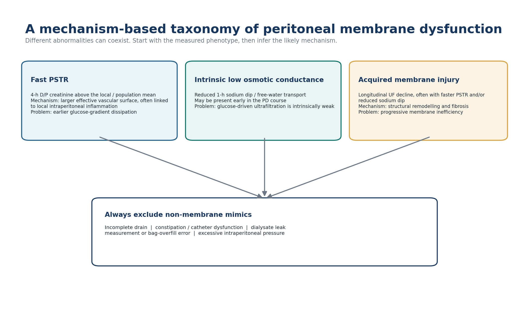
Table 3.13 - Membrane dysfunction as three syndromes.
| Syndrome | Dominant test signal | Mechanism / implication |
|---|---|---|
| Fast PSTR | 4-h D/P creatinine above centre/population average | Earlier glucose-gradient loss; lower long-dwell UF; outcome association partly mediated by fluid balance |
| Intrinsic low osmotic conductance | Reduced 1-h sodium dip on high-glucose PET with UF insufficiency | Reduced free-water response to glucose; may be present early |
| Acquired membrane insufficiency | Progressive longitudinal fall in UF and/or sodium dip, often with faster PSTR | Structural remodeling/fibrosis; raises concern about long-term membrane health and modality sustainability |
The categories are a reasoning framework rather than mutually exclusive diseases. A patient may have fast PSTR and low osmotic conductance simultaneously. Conversely, a low drained volume can be entirely non-membrane. The clinician must therefore name the measured phenotype before inferring mechanism. [1]
13. Clinical syndrome: fast PSTR
Fast PSTR matters most when it creates a clinical mismatch between dwell time and osmotic-gradient duration. The classic patient has rapid solute equilibration but disappointing net UF during a long glucose dwell, especially after residual urine has declined. [1,4]
Table 3.14 - Fast PSTR: bedside recognition.
| Finding | Interpretation | Next question |
|---|---|---|
| 4-h D/P creatinine above local/population mean | Fast solute transfer phenotype | Is volume control actually impaired? |
| Low late D/D0 glucose | Glucose gradient dissipates rapidly | Is the glucose dwell too long? |
| Low 4-h UF with preserved sodium dip | Fast PSTR may dominate rather than intrinsic low conductance | Can dwell architecture be shortened? |
| Good small-solute clearance but edema | Clearance and water removal have separated | What are sodium intake, RKF, daily UF and long-dwell strategy? |
| Increasing PSTR after inflammatory events | Possible acquired/local inflammatory effect | Is the change persistent after recovery? |
PATTERN RECOGNITION Fast PSTR is not synonymous with “under-dialysis.” In fact, small-solute equilibration may be excellent. The clinical problem is often loss of the glucose osmotic gradient before the dwell ends.
14. Managing fast PSTR: shorten the mismatch, not simply the patient’s life on PD
ISPD recommends mitigation strategies that preserve net UF by reducing exposure to late ineffective glucose dwell time and using osmotic agents strategically. Shorter glucose-based exchanges, icodextrin for a suitable long dwell, and selected use of higher glucose concentrations can improve fluid removal. Compared with glucose, icodextrin improves fluid status and reduces episodes of fluid overload in randomized evidence. [1,9]
Table 3.15 - Fast PSTR prescription levers.
| Lever | Why it works | Trade-off / safeguard |
|---|---|---|
| Shorten glucose dwell | Drain before glucose gradient is largely dissipated and fluid absorption dominates | Too-short dwell may reduce per-exchange solute equilibration and increase treatment complexity |
| Use APD architecture when it fits the patient | Creates multiple shorter nocturnal glucose dwells | APD is not mandatory; lost dwell time and treatment burden matter |
| Use icodextrin for appropriate long dwell | Provides sustained colloid osmotic effect despite fast glucose diffusion | Follow product-specific safety/monitoring; not a substitute for dietary sodium control |
| Increase glucose concentration selectively | Raises crystalloid osmotic driving force | More glucose exposure; do not normalize recurrent hypertonic dependence |
| Preserve residual kidney function | Maintains water/sodium and solute removal outside the peritoneum | Requires broader clinical strategy beyond PET |
DO NOT AUTOMATE “Fast transporter -> APD” is an oversimplification. Choose CAPD or APD according to the needed dwell architecture, residual kidney function, patient goals, treatment burden and actual delivered therapy. The PET informs the prescription; it does not choose the modality by itself.
15. Slower PSTR: do not make every dwell short
When PSTR is slower, small solutes need more time to equilibrate. A prescription dominated by very short APD cycles may therefore produce good machine completion yet insufficient solute transfer, particularly after residual kidney function falls. The physiologic response is to increase effective contact time or dialysate volume in a way the patient can tolerate, not to label the membrane “poor.”
Table 3.16 - Slower PSTR: clinical reasoning.
| Problem | Mechanism | Potential response |
|---|---|---|
| Low solute clearance on very short cycles | Insufficient time for equilibration | Lengthen effective dwell, reduce unnecessary cycling, or add daytime contact as appropriate |
| Good UF but biochemical underdialysis | Water movement adequate; solute exposure insufficient | Target clearance architecture, not stronger glucose |
| Large treatment burden from many cycles | Repeated gradient renewal may not compensate for short contact time efficiently | Simplify prescription while preserving total effective dwell |
| Preserved RKF | Kidney clearance may mask peritoneal limitations | Reassess when RKF declines |
MECHANISM RULE For slow solute transfer, the prescription lever is TIME and effective dialysate contact. For poor water removal, the lever may instead be osmotic strategy. Do not use glucose concentration to solve a solute-time problem.
16. PET does not decide CAPD versus APD
Older teaching often mapped transporter category directly to modality: fast transport to APD and slow transport to CAPD. Modern goal-directed practice is more nuanced. APD is useful when short glucose dwells are required, but the same patient may need a long icodextrin daytime dwell. A slower transporter may still prefer and succeed with APD if dwell duration and total contact are adequate. Observational studies suggest APD can mitigate some risk associated with fast transport, but modality choice remains individualized. [1,10,18]
Table 3.17 - Use PET to design dwells, not to assign a machine.
| PET / clinical signal | CAPD logic | APD logic |
|---|---|---|
| Fast PSTR + poor long-glucose UF | Shorter manual glucose dwell and/or long icodextrin dwell may work | Multiple shorter glucose cycles often physiologically attractive |
| Slower PSTR + clearance problem | Long manual dwells can provide contact time | Avoid excessively short cycles; consider longer cycles/daytime dwell |
| Intrinsic low osmotic conductance | Neither modality fixes weak glucose conductance by itself | Neither modality fixes weak glucose conductance by itself |
| High treatment burden / sleep disruption | May favor fewer daytime exchanges for some | May favor nocturnal automation for others |
| Strong patient preference | Can be decisive if physiology can be safely accommodated | Can be decisive if physiology can be safely accommodated |
17. Discordant PET results are a diagnostic opportunity
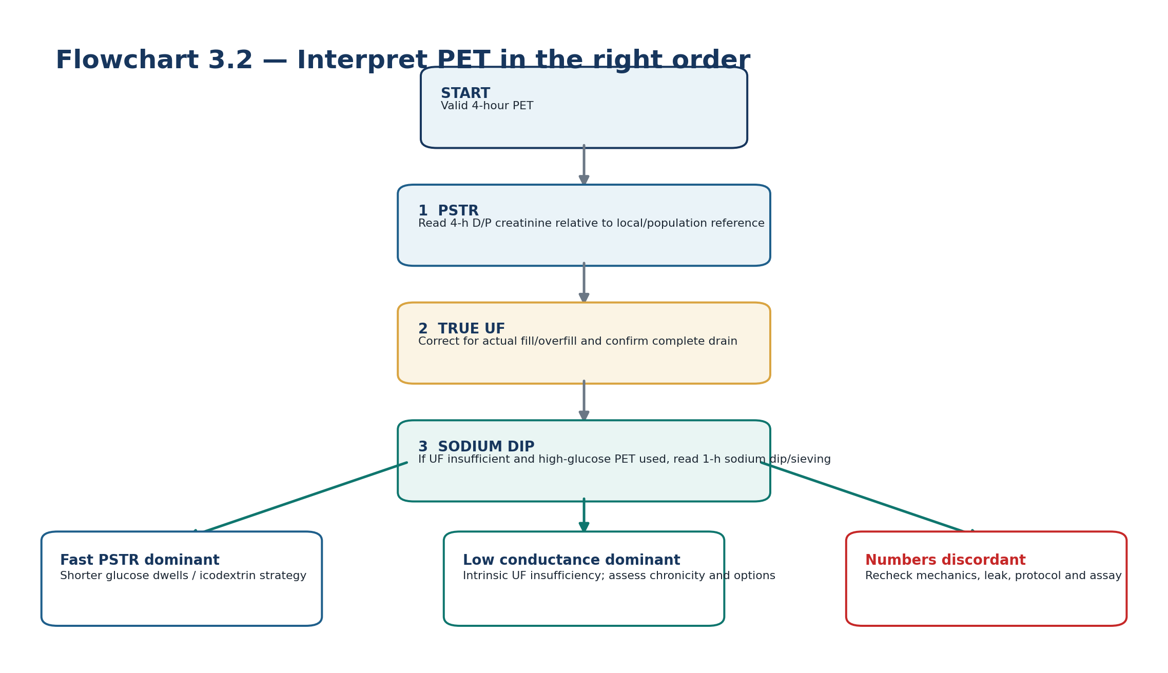
Table 3.18 - Discordant patterns and what they suggest.
| Pattern | Think | Do next |
|---|---|---|
| Fast D/P Cr + adequate high-glucose UF + preserved sodium dip | Fast solute transfer without major intrinsic water-transport defect | Prescribe to clinical volume/clearance needs; no “failure” diagnosis |
| Fast D/P Cr + low UF + preserved sodium dip | Early glucose-gradient dissipation / increased absorption may dominate | Check mechanics, then shorten glucose dwell / long-dwell strategy |
| Not-fast D/P Cr + low UF + reduced sodium dip | Intrinsic low osmotic conductance | Confirm validity and chronicity; assess clinical volume consequences |
| Low UF + normal-looking transport ratios + poor drain | Mechanical mimic | Correct catheter/constipation/position issue before repeating PET |
| Implausible D/P or D/D0 values | Timing, sample or assay error | Verify laboratory and sampling before clinical action |
DIAGNOSTIC DISCIPLINE When the numbers do not fit one transporter label, do not average them into a story. Ask which physiological property each number measures and whether the test itself was valid.
18. Low PET ultrafiltration: membrane dysfunction versus mimic
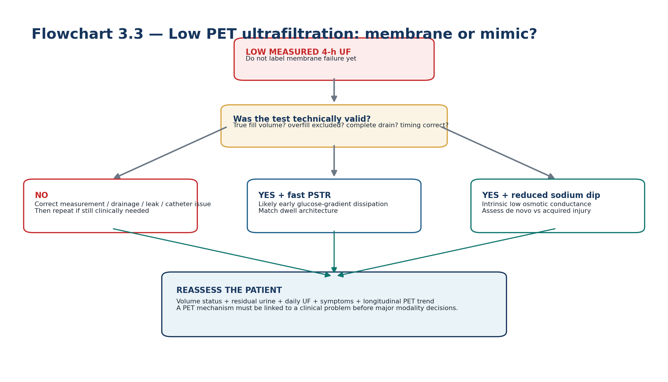
Table 3.19 - Rapid differential for low 4-hour PET UF.
| Cause | Clue | Discriminator |
|---|---|---|
| Fast PSTR | High D/P Cr, rapid glucose disappearance | Sodium dip may remain relatively preserved; dwell mismatch evident |
| Intrinsic low osmotic conductance | Low high-glucose UF with reduced 1-h sodium dip | Free-water transport impaired |
| Incomplete drainage | Slow/positional drain, constipation, catheter history | Drain troubleshooting / imaging as indicated |
| Dialysate leak | Edema or swelling pattern, unexpectedly low recovery | Clinical/imaging leak assessment |
| Bag overfill / fill-volume error | Nominal rather than true fill used in UF calculation | Measured/validated fill volume changes result |
| Variable residual intraperitoneal volume | Large/variable sump volume | Repeat standardized complete drain; interpret cautiously |
| Increased fluid absorption | Low net UF not fully explained by PSTR | Specialist interpretation; not readily quantified routinely |
STOP RULE Do not diagnose acquired membrane failure from one low drain volume. A membrane diagnosis requires a valid physiologic test plus a clinical phenotype, and often a longitudinal pattern.
19. Longitudinal change: the trend may matter more than the category
PSTR commonly rises over time on PD. ISPD summarizes longitudinal evidence showing that roughly one in three patients may increase 4-hour D/P creatinine by at least 0.1 after one year. Drivers include peritoneal inflammation, glucose/GDP exposure and episodes of peritonitis, although individual trajectories vary widely. [1]
A 2025 observational study of patients treated for at least four years found increasing PSTR over time among those exposed to standard glucose solutions, while PSTR appeared more stable among patients receiving low-GDP solutions. The result is hypothesis-supporting rather than proof of causality because treatment allocation was not randomized. [17]
Table 3.20 - Serial PET: what changes deserve attention?
| Change | Possible meaning | Response |
|---|---|---|
| Small isolated D/P change | Biological / analytical variability | Compare protocol, assay, clinical state and prior values |
| Persistent faster PSTR | Increased effective vascular area / inflammation / acquired change | Review fluid strategy and clinical volume status |
| Progressive fall in sodium dip | Worsening osmotic conductance; may precede severe UF insufficiency | Assess chronicity, daily UF and long-term membrane sustainability |
| Progressive decline in UF capacity | Acquired membrane injury OR evolving non-membrane problem | Repeat mechanism-based evaluation |
| Change after peritonitis | Inflammation-related functional change may be transient or persistent | Repeat after recovery if result will change management |
TREND PRINCIPLE A patient moving from D/P 0.62 to 0.70 is clinically more interesting than a stable patient at 0.70 if the change is reproducible and accompanied by worsening UF. Serial physiology is a story, not a sequence of labels.
20. Acquired membrane injury and the EPS boundary
Progressive fibrosis can reduce osmotic conductance and UF capacity over years, often in association with faster PSTR. A falling sodium dip may be an earlier functional marker of severe acquired UF insufficiency than PSTR alone. ISPD advises that acquired membrane injury, particularly after prolonged PD, should prompt discussion of the risks of continuing PD, including encapsulating peritoneal sclerosis (EPS), within shared decision-making. [1]
This is not the same as saying that an abnormal PET diagnoses EPS. Most long-term PD patients do not develop severe UF insufficiency, and EPS remains uncommon. Clinical diagnosis, risk assessment and management of long-term membrane failure and EPS belong to Chapter 14.
Table 3.21 - PET findings that should trigger a long-term membrane review.
| Signal | Why it matters | What it does NOT mean |
|---|---|---|
| Progressive UF decline | May reflect acquired structural membrane change | Not diagnostic of EPS |
| Progressive loss of sodium dip | Suggests worsening osmotic conductance | Not proof of absent AQP1 expression |
| Increasing PSTR + low UF | Consistent with acquired vascular/interstitial change | Not an automatic indication to stop PD |
| Dependence on repeated hypertonic glucose | Signals shrinking fluid-management reserve | Not solved by PET alone |
| Long PD vintage + obstructive GI symptoms | Raises clinical concern beyond membrane testing | Requires dedicated EPS diagnostic pathway |
SAFETY BOUNDARY PET can reveal declining membrane reserve; it cannot diagnose EPS by itself. Chapter 14 develops the long-term membrane failure and EPS pathway.
21. Additional membrane tests: know what exists, but do not overcomplicate routine care
Several derivative tests have been developed to separate water pathways or model transport more precisely. They are valuable in research and selected specialist evaluations, but ISPD does not recommend complex measurements of protein loss, intraperitoneal pressure, osmotic conductance or lymphatic reabsorption for routine clinical practice. [1,2,6]
Table 3.22 - Common derivative membrane tests.
| Test | What it adds | Routine role |
|---|---|---|
| High-glucose / modified PET | PSTR + stronger UF-capacity signal + 1-h sodium dip | Useful when UF insufficiency needs mechanistic assessment |
| Mini-PET | Short high-glucose study focused on free-water/small-pore transport | Specialist/research use; not a replacement for all standard PET information |
| Double mini-PET | Uses different glucose strengths to estimate osmotic conductance | Research/specialist physiology |
| Standard permeability analysis (SPA) | More detailed solute/water transport modeling | Research/specialist centers |
| Personal Dialysis Capacity / modeling systems | Model-based transport parameters from multiple exchanges | Center/device specific; not universal standard |
| Peritoneal protein clearance | Large-pore/macromolecule information | Not recommended as routine membrane-function test |
PRACTICAL RULE For most bedside decisions, the high-value core remains: 4-hour D/P creatinine + accurate 4-hour UF + 1-hour sodium dip when UF insufficiency is being characterized.
22. Clinical problem framework: from patient phenotype to PET action
Table 3.23 - Clinical question -> data required -> interpretation -> next action.
| Clinical question | Data required | Interpretation | Next action |
|---|---|---|---|
| Why is the patient fluid overloaded? | Weight/edema/BP, sodium intake, urine, daily UF, drain function, PET | Separate total-volume problem from membrane mechanism | Correct intake/RKF/mechanics; PET-targeted prescription only if membrane contributes |
| Why is long-dwell UF poor? | D/P Cr, high-glucose UF, 1-h sodium dip | Fast gradient loss vs low conductance vs mimic | Shorten glucose dwell / icodextrin / address mechanics according to mechanism |
| Why is clearance falling? | Delivered prescription, RKF, D/P Cr, dwell time | Loss of RKF vs inadequate contact vs delivery problem | Adjust contact time/volume only after delivery/RKF review |
| Has the membrane changed over years? | Serial PETs done by comparable method, UF trend, peritonitis history | Persistent PSTR/UF/sodium-dip trajectory | Long-term membrane review and shared modality planning |
| Is this one bad drain “UF failure”? | Exchange log, catheter function, constipation, leak clues | Mechanical or measurement cause may dominate | Fix mimic before diagnostic PET interpretation |
23. Clinical pearls
Table 3.24 - High-value PET pearls.
| Pearl | Why it matters |
|---|---|
| PET describes membrane behavior, not total dialysis adequacy. | A perfect PET cannot tell you whether the patient is symptomatically or volumetrically well dialyzed. |
| Fast transport is good for equilibration and can be bad for long glucose UF. | The same vascular surface area accelerates solute transfer and glucose loss. |
| The 4-hour D/P creatinine should be interpreted against a local/population distribution. | Centre and creatinine-assay effects make universal cut-offs unreliable. |
| Low PET UF is a screening signal, not a membrane diagnosis. | Drainage, leak, overfill and residual volume can produce the same number. |
| A low 1-hour sodium dip is a free-water transport signal. | It separates osmotic-conductance dysfunction from PSTR alone. |
| Serial decline in sodium dip can be more informative than a stable transporter label. | Acquired membrane injury is longitudinal physiology. |
| Do not use stronger glucose to solve a slow-solute-transfer problem. | Clearance requires contact time; osmotic strength primarily targets water. |
| The most useful PET is one that changes a named clinical decision. | Testing without a question generates labels rather than care. |
24. Mini-cases: decisions, not transporter trivia
Case 1 - Fast transport, poor long-dwell UF
A patient on CAPD has a 4-h D/P creatinine above the centre mean, adequate small-solute clearance, preserved 1-hour sodium dip on a high-glucose PET, but poor UF from the long daytime glucose dwell and progressive edema.
REASONING This is a classic dwell-mismatch phenotype. The preserved sodium dip argues against dominant intrinsic low osmotic conductance. Review sodium intake and residual urine, then shorten ineffective glucose exposure and use an appropriate sustained long-dwell strategy such as icodextrin rather than simply escalating glucose.
Case 2 - Low UF with a reduced sodium dip
A patient with little residual urine has persistent volume overload. A valid high-glucose PET yields <400 mL net UF at 4 h and a 1-hour sodium dip of 3 mmol/L; D/P creatinine is not especially fast.
REASONING The UF screening threshold is crossed and free-water transport is impaired. This is intrinsic low osmotic conductance rather than a fast-PSTR-only problem. Confirm no mechanical/leak explanation, assess chronicity and redesign volume strategy; if acquired and progressive, discuss membrane sustainability and modality options.
Case 3 - “Ultrafiltration failure” after constipation
A previously stable patient has one PET with only 150 mL net UF using high-glucose solution. The final drain was slow and positional, and the patient has not opened the bowel for four days.
REASONING Do not diagnose membrane failure. The test is mechanically compromised. Treat constipation and catheter/drain factors, then repeat the PET only if a membrane question remains clinically important.
Case 4 - Slow PSTR on very short APD cycles
An anuric patient on APD has low-average/slow D/P creatinine and progressive biochemical underdialysis despite acceptable nightly UF. The cycler uses multiple very short dwells.
REASONING The problem is likely insufficient solute contact time, not inadequate osmotic strength. Rebuild effective dwell time and total dialysate contact; do not “fix” the clearance problem by increasing glucose concentration.
Case 5 - Serial change after years on PD
A patient’s D/P creatinine has risen gradually over several years, daily UF has fallen, and the high-glucose sodium dip has progressively decreased. Hypertonic glucose use is increasing.
REASONING The pattern is more concerning than any single category. Exclude delivery and mechanical problems, then consider acquired membrane injury. Review long-term PD risks, residual kidney function, treatment burden and modality alternatives. PET does not diagnose EPS; Chapter 14 addresses that pathway.
Case 6 - Classic “high” category without clinical problem
A clinically well patient has a 4-hour D/P creatinine of 0.83 on the centre’s standard PET, good daily UF, preserved urine output, no congestion and an acceptable prescription burden.
REASONING Do not intervene because the classic table says “high.” Confirm that the result is valid and establish it as the patient’s baseline. Continue goal-directed care and respond only if the physiology creates a clinical problem or changes longitudinally.
25. Common pitfalls - and the correction
Table 3.25 - High-frequency PET errors.
| Pitfall | Why it fails | Correction |
|---|---|---|
| Calling PET an adequacy test | It measures membrane transport, not total clinical dialysis quality | Integrate symptoms, volume, RKF and delivered prescription |
| Treating 0.82 as a universal high-transport threshold | Centre/assay distributions differ | Use local/population reference; classic bands are operational language |
| Diagnosing UF failure from drain volume alone | Mechanical and measurement errors are common | Validate true fill, complete drain, leak and catheter function |
| Using a 2.5% PET to diagnose osmotic conductance without sodium testing | It cannot provide the recommended high-glucose sodium-dip signal | Use high-glucose PET with 1-h sodium when intrinsic UF insufficiency is suspected |
| Applying a universal creatinine glucose correction | Assay interference is method-specific | Use laboratory-validated approach |
| Automatically moving fast transporters to APD | Modality is patient- and prescription-specific | Design dwell architecture around physiology and goals |
| Giving stronger glucose for poor solute clearance | Osmotic strength targets water, not contact time | Adjust dwell/time/volume for solute transfer |
| Ignoring a changing serial PET because category is unchanged | Important physiology can change within one band | Compare absolute trajectory with UF and sodium dip |
| Calling progressive PET change EPS | Functional decline is not a structural diagnosis | Use dedicated long-term membrane/EPS clinical pathway |
26. Active recall
MUST MEMORIZE
1. Q: When should baseline PSTR be measured after starting PD? A: ISPD recommends between 6 and 12 weeks, then subsequently when clinically indicated.
2. Q: What is the index solute for PSTR in current ISPD guidance? A: Creatinine, measured as the 4-hour dialysate-to-plasma ratio.
3. Q: Which glucose strengths are acceptable for the 4-hour PSTR PET? A: Either 2.27% glucose/2.5% dextrose or 3.86% glucose/4.25% dextrose.
4. Q: What 4-hour UF values should trigger suspicion of insufficient UF? A: <400 mL with 3.86%/4.25% or <100 mL with 2.27%/2.5%; these are screening thresholds.
5. Q: When insufficient UF is suspected, what additional test does ISPD recommend? A: A 1-hour sodium dip during a 3.86% glucose/4.25% dextrose PET.
6. Q: What sodium-dip threshold indicates UF insufficiency? A: A dip <=5 mmol/L and/or sodium-sieving ratio <=0.03 at 1 hour.
7. Q: What is the sodium-dip equation? A: Dialysate sodium at time 0 minus dialysate sodium at 60 minutes.
8. Q: What is the sodium-sieving ratio? A: 1 - (Na60/Na0).
9. Q: What are the three ISPD membrane-dysfunction phenotypes? A: Fast PSTR, intrinsic poor UF/low osmotic conductance, and acquired membrane insufficiency over time.
10. Q: What must be excluded before a low UF result is called membrane dysfunction? A: Mechanical problems, leak, measurement/overfill errors and other causes of excess fluid absorption.
MUST REASON
1. Q: Why can a fast PSTR produce poor long-dwell UF? A: Glucose leaves dialysate faster, so the crystalloid osmotic gradient dissipates early and later fluid absorption reduces net UF.
2. Q: Why can a normal D/P creatinine coexist with severe UF insufficiency? A: PSTR and osmotic conductance are partly independent; free-water transport can be impaired despite non-fast solute transfer.
3. Q: Why is one low PET drain volume insufficient to diagnose membrane failure? A: The recovered volume depends on true fill, residual volume, complete drainage, leak and catheter function as well as membrane water transport.
4. Q: Why can APD underperform in a slow transporter despite many cycles? A: If individual dwells are too short, small solutes do not have enough time to equilibrate; repeated cycling does not eliminate the time requirement.
5. Q: Why is serial sodium-dip decline concerning? A: It suggests progressive loss of osmotic conductance and may precede severe acquired UF insufficiency.
6. Q: Why should classic transporter bands not dictate modality? A: They are historical categories; clinical response depends on actual volume/clearance goals, local reference, RKF and patient preference.
7. Q: Why might peritonitis temporarily change PET? A: Local intraperitoneal inflammation can increase effective solute transport; testing during acute inflammation may not represent stable baseline physiology.
8. Q: Why does a high-glucose PET provide more diagnostic information when UF failure is suspected? A: It stresses glucose osmotic conductance strongly enough to assess both UF capacity and the 1-hour sodium dip.
USE AS REFERENCE
Classic four-group D/P creatinine bands and local Oman operational PET table.
Centre-specific D/P creatinine reference distribution and laboratory creatinine method.
Exact local PET sampling, mixing, fill/drain and specimen-handling protocol.
Product-specific dialysate sodium concentration and laboratory sodium methodology.
Derivative PET/mini-PET/double mini-PET procedures used only in specialist settings.
27. Flashcards: active recall
1. Q: PET in one sentence? A: A standardized 4-hour dwell used to characterize peritoneal solute and water transport.
2. Q: Fast PSTR marker? A: Higher 4-hour D/P creatinine relative to the relevant centre/population distribution.
3. Q: D/D0 glucose moves which direction with time? A: Down.
4. Q: Creatinine D/P moves which direction with time? A: Up.
5. Q: Fast PSTR means the membrane clears solute poorly? A: No - small-solute equilibration is faster.
6. Q: Why can fast PSTR reduce long-dwell glucose UF? A: The glucose gradient dissipates earlier.
7. Q: Low 4-h UF on 4.25% PET threshold? A: <400 mL suggests insufficient UF.
8. Q: Low 4-h UF on 2.5% PET threshold? A: <100 mL suggests insufficient UF.
9. Q: 1-hour sodium dip of 3 mmol/L means? A: Abnormally reduced free-water/osmotic conductance when obtained on the recommended high-glucose diagnostic PET.
10. Q: Normal D/P creatinine excludes low osmotic conductance? A: No.
11. Q: PET says “high transporter”; first clinical question? A: Is there actually a clearance or volume problem caused by this physiology?
12. Q: One bad drain = membrane failure? A: No; check mechanics, leak and measurement validity.
13. Q: Best long-dwell osmotic strategy for many fast transporters? A: Icodextrin when clinically appropriate.
14. Q: Slow transporter + underclearance on short APD cycles: first physiologic lever? A: Increase effective dwell/contact time.
15. Q: Should every stable patient have frequent routine PETs? A: No; repeat when clinically indicated and when the result can change management.
16. Q: Serial PET after peritonitis: interpret how? A: In relation to timing and recovery from local inflammation.
17. Q: Can PET diagnose EPS? A: No.
18. Q: What defines successful PET use? A: A valid test that identifies a clinically relevant mechanism, leads to a targeted action when needed and is followed by reassessment.
28. Final revision sheet
TEN TAKE-HOME RULES 1) PET is a physiology test, not an adequacy score. 2) Use 4-h D/P creatinine for PSTR. 3) Baseline PET is recommended at 6-12 weeks. 4) Interpret PSTR against centre/population data. 5) Low 4-h UF is a screen, not a membrane diagnosis. 6) High-glucose PET + 1-h sodium dip diagnoses impaired osmotic conductance when UF insufficiency is suspected. 7) Fast PSTR can clear solute well and remove long-dwell fluid poorly. 8) Exclude drainage, leak and overfill errors before membrane failure. 9) Serial physiology matters. 10) Match the prescription to the measured mechanism and the patient’s clinical goal.
Table 3.26 - Key numbers and definitions.
| Item | Value / definition | Use |
|---|---|---|
| Baseline PET timing | 6-12 weeks after starting PD | Establish early PSTR phenotype |
| PSTR index | 4-h D/P creatinine | Primary solute-transfer measure |
| Acceptable PET glucose strengths | 2.27%/2.5% or 3.86%/4.25% | Both valid for PSTR |
| High-glucose UF screen | <400 mL at 4 h | Suspect insufficient UF |
| Middle-strength UF screen | <100 mL at 4 h | Suspect insufficient UF |
| Sodium dip | Na0 - Na60 | Free-water transport surrogate |
| Sodium-sieving ratio | 1 - (Na60/Na0) | Alternative free-water expression |
| Low sodium dip | <=5 mmol/L at 1 h | Intrinsic UF insufficiency signal |
| Low sieving ratio | <=0.03 at 1 h | Intrinsic UF insufficiency signal |
Table 3.27 - One-minute bedside synthesis.
| If you see... | Think... | Do now... |
|---|---|---|
| Fast D/P Cr + poor long glucose UF | Gradient dissipates early | Shorten glucose dwell; review icodextrin and total volume strategy |
| Low high-glucose UF + low sodium dip | Low osmotic conductance | Confirm validity/chronicity; review membrane sustainability |
| Low UF + poor drain | Mechanical mimic | Fix constipation/catheter/position before membrane diagnosis |
| Slow D/P Cr + low clearance on short APD | Insufficient contact time | Lengthen effective dwell / add contact |
| Serial UF + sodium-dip decline | Acquired membrane injury possible | Longitudinal membrane review; shared modality planning |
| Classic high category but patient clinically well | Phenotype without current problem | Document baseline; do not treat the label |
Table 3.28 - Post-PET success checklist.
| Check | Pass criterion |
|---|---|
| Question | The clinical reason for PET is explicit. |
| Protocol | Glucose strength, volume, timing and sampling method are documented. |
| Laboratory | Creatinine assay handling and sodium method are appropriate. |
| Volume | True instilled and completely drained volumes are credible. |
| PSTR | 4-h D/P creatinine is interpreted against relevant reference data. |
| UF | 4-h net UF is interpreted as a screen, not a diagnosis. |
| Free water | 1-h sodium dip is obtained when intrinsic UF insufficiency is suspected. |
| Mechanics | Catheter, constipation, leak and measurement error have been considered. |
| Action | Any prescription change is tied to a named mechanism. |
| Reassessment | Clinical response, daily UF, volume status and treatment burden will be reviewed. |
FINAL MENTAL MODEL Clinical question -> valid standardized PET -> 4-h D/P creatinine -> true 4-h UF -> 1-h sodium dip when indicated -> exclude mimics -> identify mechanism -> change the relevant prescription lever -> reassess the patient. The membrane is a continuum; the patient is the outcome.
Rapid oral viva
Explain why a fast PSTR can simultaneously improve creatinine equilibration and worsen long-dwell glucose ultrafiltration.
Walk through the quality checks that must be passed before interpreting a low PET UF volume.
Defend why 0.82 should not be taught as a universal treatment threshold for “high transport.”
A patient has low high-glucose UF and a sodium dip of 3 mmol/L. Explain the mechanism and the next clinical questions.
Contrast the prescription response to fast PSTR with the response to slow PSTR and insufficient solute clearance.
Explain how serial PET findings can raise concern for acquired membrane injury without diagnosing EPS.
SCOPE BOUNDARY This chapter teaches membrane-function measurement and its translation into physiology-based prescription reasoning. Detailed initial prescription construction is developed in Chapter 6; adequacy and residual kidney function in Chapter 7; volume, sodium balance and formal ultrafiltration-failure management in Chapter 8; CAPD/APD optimization in Chapter 9; and long-term membrane failure/EPS in Chapter 14.
29. Selected authoritative references
1. Morelle J, Stachowska-Pietka J, Öberg C, Gadola L, La Milia V, Yu Z, et al. ISPD recommendations for the evaluation of peritoneal membrane dysfunction in adults: classification, measurement, interpretation and rationale for intervention. Perit Dial Int. 2021;41(4):352–372. https://doi.org/10.1177/0896860820982218. PMID: 33563110.
2. Gu J, Bai E, Ge C, Winograd J, Shah AD. Peritoneal equilibration testing: your questions answered. Perit Dial Int. 2023;43(5):361–373. https://doi.org/10.1177/08968608221133629. PMID: 36350033.
3. Twardowski ZJ, Nolph KD, Khanna R, Prowant BF, Ryan LP, Moore HL, Nielsen MP. Peritoneal Equilibration Test. Perit Dial Int. 1987;7(3):138–148. https://doi.org/10.1177/089686088700700306.
4. Mehrotra R, Ravel V, Streja E, Kuttykrishnan S, Adams SV, Katz R, et al. Peritoneal equilibration test and patient outcomes. Clin J Am Soc Nephrol. 2015;10(11):1990–2001. https://doi.org/10.2215/CJN.03470315. PMID: 26463882.
5. La Milia V, Cabiddu G, Virga G, Vizzardi V, Giuliani A, Finato V, et al. Peritoneal Equilibration Test Reference Values Using a 3.86% Glucose Solution During the First Year of Peritoneal Dialysis: Results of a Multicenter Study of a Large Patient Population. Perit Dial Int. 2017;37(6):633–638. https://doi.org/10.3747/pdi.2017.00004. PMID: 28698252.
6. Smit W, Langedijk MJ, Schouten N, van den Berg N, Struijk DG, Krediet RT. Quantification of free water transport in peritoneal dialysis. Kidney Int. 2004;66(2):849–854. https://doi.org/10.1111/j.1523-1755.2004.00815.x.
7. Morelle J, Marechal C, Yu Z, Debaix H, Corre T, Lambie M, et al. AQP1 Promoter Variant, Water Transport, and Outcomes in Peritoneal Dialysis. N Engl J Med. 2021;385(17):1570–1580. https://doi.org/10.1056/NEJMoa2034279. PMID: 34670044.
8. Lambie M, Chess J, Donovan KL, Kim YL, Do JY, Lee HB, et al.; Global Fluid Study Investigators. Independent effects of systemic and peritoneal inflammation on peritoneal dialysis survival. J Am Soc Nephrol. 2013;24(12):2071–2080. https://doi.org/10.1681/ASN.2013030314. PMID: 24009237.
9. Goossen K, Becker M, Marshall MR, Bühn S, Breuing J, Firanek CA, et al. Icodextrin versus glucose solutions for the once-daily long dwell in peritoneal dialysis: an enriched systematic review and meta-analysis of randomized controlled trials. Am J Kidney Dis. 2020;75(6):830–846. https://doi.org/10.1053/j.ajkd.2019.10.004. PMID: 32033860.
10. Johnson DW, Hawley CM, McDonald SP, Brown FG, Rosman JB, Wiggins KJ, et al. Superior survival of high transporters treated with automated versus continuous ambulatory peritoneal dialysis. Nephrol Dial Transplant. 2010;25(6):1973–1979. https://doi.org/10.1093/ndt/gfp780. PMID: 20097847.
11. Pride ET, Gustafson J, Graham A, Spainhour L, Mauck V, Brown P, et al. Comparison of a 2.5% and a 4.25% dextrose peritoneal equilibration test. Perit Dial Int. 2002;22(3):365–370. https://doi.org/10.1177/089686080202200311.
12. McCafferty K, Fan SLS. Are we underestimating the problem of ultrafiltration in peritoneal dialysis? An observational study of overfill of dialysis bags. Perit Dial Int. 2006;26(3):349–352. https://doi.org/10.1177/089686080602600312. PMID: 16722028.
13. La Milia V, Pozzoni P, Crepaldi M, Locatelli F. Overfill of peritoneal dialysis bags as a cause of underestimation of ultrafiltration failure. Perit Dial Int. 2006;26(4):503–505. https://doi.org/10.1177/089686080602600416. PMID: 16881347.
14. Da Rin G, Amici G, Virga G, Bardin C, Calzavara P, Bocci C. Correction of glucose concentration interference on Jaffe kinetic creatinine assay in peritoneal dialysis. Am J Nephrol. 1995;15(6):480–487. https://doi.org/10.1159/000168890. PMID: 8546169.
15. Valentine RM, Sharrod-Cole H, Ford C, Gama R. Peritoneal equilibration test: does glucose interfere with enzymatic creatinine measurement? Ann Clin Biochem. 2019;56(2):302–303. https://doi.org/10.1177/0004563218806559.
16. Krediet RT. Physiology of peritoneal dialysis; pathophysiology in long-term patients. Front Physiol. 2024;15:1322493. https://doi.org/10.3389/fphys.2024.1322493.
17. Davenport A. Longitudinal changes in peritoneal solute transport rate and the impact of lower glucose degradation product glucose dialysates. Ther Apher Dial. 2025;29(3):471–478. https://doi.org/10.1111/1744-9987.70012. PMID: 40129079.
18. Brown EA, Blake PG, Boudville N, Davies S, de Arteaga J, Dong J, et al. International Society for Peritoneal Dialysis practice recommendations: Prescribing high-quality goal-directed peritoneal dialysis. Perit Dial Int. 2020;40(3):244–253. https://doi.org/10.1177/0896860819895364.
19. Boudville N, de Moraes TP. 2005 Guidelines on targets for solute and fluid removal in adults being treated with chronic peritoneal dialysis: 2019 update of the literature and revision of recommendations. Perit Dial Int. 2020;40(3):254–260. https://doi.org/10.1177/0896860819898307.
20. Ministry of Health, Sultanate of Oman. Guideline on Running the Peritoneal Dialysis Service. MoH/DGHS&P/GUD/007/Vers.02. April 2025. Appendix 7: Peritoneal Equilibration Test.
21. Van Biesen W, Heimbürger O, Krediet R, Rippe B, La Milia V, Covic A, et al. Evaluation of peritoneal membrane characteristics: clinical advice for prescription management by the ERBP working group. Nephrol Dial Transplant. 2010;25(7):2052–2062. https://doi.org/10.1093/ndt/gfq100.
SOURCE NOTE ISPD membrane recommendations, PET evidence and contemporary longitudinal literature were checked through 1 September 2026. The 2021 ISPD membrane-dysfunction guideline remains the principal PET-specific international standard identified. Exact PET sample handling, creatinine assay correction, local reference distribution and operational steps remain governed by the validated laboratory and PD-unit protocol; local Oman guidance is shown where it provides an operational PET reference, not as a replacement for ISPD interpretation principles.
CHAPTER 3 COMPLETE
NEXT:
CHAPTER 4 - PATIENT SELECTION, MODALITY CHOICE AND SHARED DECISION-MAKING