Peritoneal Dialysis Catheters:
Placement, Function and Access Care
Planning | Device | Mapping | Insertion | Healing | Function | Exit Site | Salvage
CHAPTER MISSION Build a bedside-to-procedure mental model that converts a decision for PD into a safe, durable and comfortable peritoneal access: map the patient before choosing the catheter, match insertion technique to anatomy and expertise, protect healing, prove function, maintain a healthy exit site, and localise mechanical failure before the access is unnecessarily replaced.
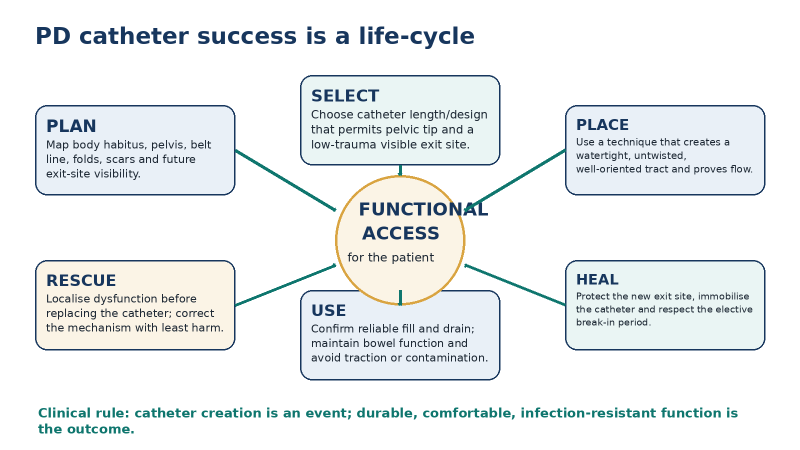
MASTER PRINCIPLE A PD catheter is successful only when it repeatedly fills and drains with acceptable comfort, has a visible low-trauma exit site, resists infection, and can be maintained or rescued without unnecessary interruption of PD. Catheter creation is an event; usable access is the outcome.
0. One-page chapter map
Table 5.1 - The eight decisions that govern PD catheter care.
| Decision | Core question | Bedside output |
|---|---|---|
| 1. Map | Where must the tip and exit site be for this patient’s body and daily life? | Preoperative body map |
| 2. Device | Which catheter length/design can achieve both pelvic position and a maintainable exit site? | Standard or extended catheter plan |
| 3. Technique | Which insertion approach best matches anatomy, urgency, anaesthetic risk and local expertise? | Named placement strategy |
| 4. Prepare | Have bowel, bladder, infection prophylaxis and surgical details been addressed? | Safe procedural readiness |
| 5. Prove | Did the catheter lie correctly, flow freely and leave a watertight tract? | Function documented at placement |
| 6. Heal | Can the access mature without traction, contamination or excessive intraperitoneal pressure? | Protected break-in / urgent-start plan |
| 7. Maintain | Is the exit site healthy, immobilised and cared for consistently? | Daily/regular preservation plan |
| 8. Rescue | If flow or the exit site changes, what is the mechanism and least harmful correction? | Cause-based salvage pathway |
Learning outcomes
Use preoperative mapping to select an insertion point and exit site that jointly permit pelvic catheter-tip position, visibility, accessibility and low mechanical stress.
Explain the functional anatomy of the intraperitoneal segment, deep cuff, intercuff tunnel, superficial cuff, exit site, external limb and transfer set.
Choose among standard and extended catheter configurations without treating straight versus coiled tips or any proprietary design as universally superior.
Compare open surgical, advanced laparoscopic, peritoneoscopic and image-guided percutaneous insertion approaches using patient anatomy, anaesthetic risk, urgency and operator expertise.
Apply the peri-insertion safety bundle: multidisciplinary planning, bowel/bladder preparation, antimicrobial prophylaxis, sterile technique, watertight placement and immediate catheter immobilisation.
Use the elective break-in period correctly and understand how urgent-start PD modifies fill volume and posture when PD must begin earlier.
Recognise normal healing, mechanical irritation, definitive exit-site infection and tunnel-infection warning signs without overcalling erythema alone.
Localise poor flow into outflow-predominant, two-way obstruction, positional pain or leakage phenotypes and troubleshoot in least-invasive order.
Distinguish constipation, bladder distension, catheter migration, kink, intraluminal fibrin, omental wrapping and adhesions by mechanism and bedside clues.
Choose nonoperative versus operative catheter salvage according to urgency, likely mechanism, local capability and the goal of preserving PD continuity.
Protect long-term access by preventing traction, maintaining bowel function, monitoring the exit site, and acting on change from the patient’s baseline.
EVIDENCE POSTURE The 2019 ISPD adult peritoneal-access guideline remains the principal comprehensive adult access guideline, complemented by the 2023 ISPD catheter-related infection recommendations, the 2024 SAGES PD-access guideline update, the 2023 Cochrane review of insertion techniques, and newer comparative evidence through 2026. Where guidelines diverge, this chapter shows the certainty and clinical context rather than forcing a single universal technique. [1–8]
1. Core concept: the catheter is an access system, not a tube
The catheter is the only permanent interface between the peritoneal cavity and the outside world. Its intraperitoneal tip must sit where dialysate can move freely; its abdominal-wall passage must resist leakage and migration; its cuffs must support tissue ingrowth; and the external segment must exit through skin that the patient can see, clean and secure. A defect in any one of these domains can present later as slow drainage, pain, leak, cuff extrusion, infection or technique failure. [1]
This explains why a “successful operation” is not the same as successful access. A catheter can be patent on the operating table yet be poorly positioned for the patient’s body habitus, hidden under a pannus, repeatedly compressed by a belt, or so mobile that the exit sinus is continually traumatized. Conversely, a patient with prior abdominal surgery or obesity can have excellent long-term PD when the access is deliberately mapped and placed by an experienced team. [1,3]
BEDSIDE TRANSLATION When a catheter problem appears, identify which component failed: intraperitoneal tip, lumen, abdominal-wall tract, cuff/tunnel, exit site, external limb or connection system. “Catheter dysfunction” is a syndrome, not a diagnosis.
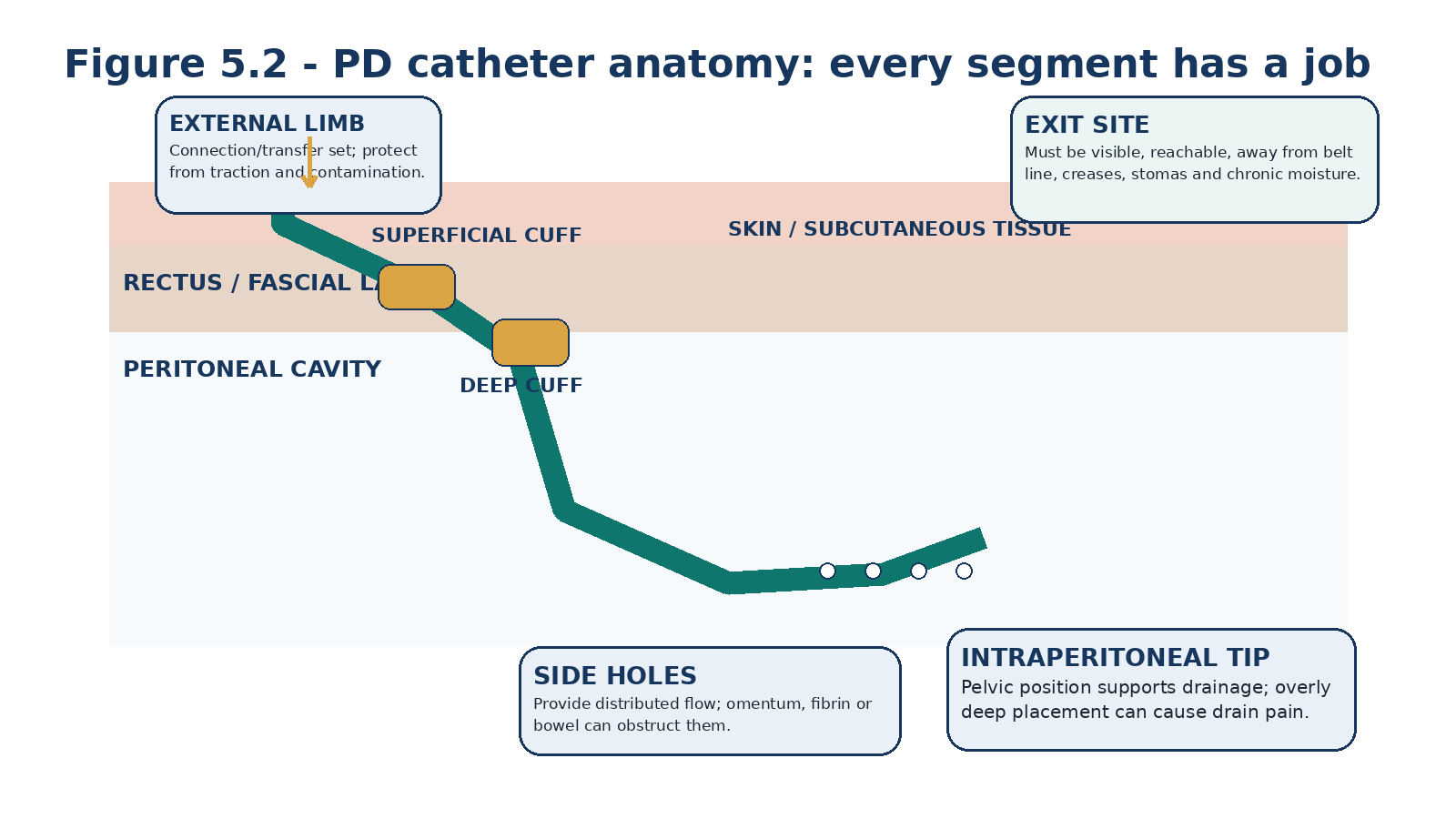
Table 5.2 - Catheter segment -> function -> failure clue.
| Segment | Normal role | Failure clue |
|---|---|---|
| External limb / transfer set | Connects dialysis system while allowing disconnection between treatments. | Crack, contamination, loose adapter, traction, external leak. |
| Exit site | Stable epithelialized skin interface around catheter. | Discharge, erythema, granuloma, trauma, cuff exposure. |
| Superficial cuff | Tissue ingrowth and tunnel stability. | Extrusion, chronic infection around cuff, local pain. |
| Intercuff tunnel | Subcutaneous route that directs and stabilises catheter. | Tenderness, swelling, induration, tunnel fluid on ultrasound. |
| Deep cuff / rectus tract | Anchors abdominal-wall passage and helps create a watertight seal. | Early leak, tract movement, deep infection. |
| Intraperitoneal segment | Provides distributed fill/drain flow. | Migration, omental wrap, adhesion, tip pain. |
| Side holes / lumen | Permit inflow and outflow. | Fibrin plug, debris, tissue occlusion, two-way resistance. |
2. Catheter design: keep the device simple and make the geometry right
ISPD recommends silicone-rubber catheters for chronic PD, generally with double polyester cuffs. Standard adult catheters may have straight or coiled intraperitoneal tips and straight or preformed-arc intercuff segments. The guideline does not support a universal functional superiority of one standard geometry over all others. The practical priority is choosing a device whose length and shape fit the patient’s anatomy. [1]
Randomized trials have sometimes favoured straight intraperitoneal tips over coiled designs for migration or dysfunction, including a 2020 multicentre trial, but results across eras, insertion methods and catheter families are not sufficiently uniform to turn tip shape into the dominant access decision. Proper insertion site, catheter length, tunnel geometry and operator technique matter at least as much. [1,9,10]
Table 5.3 - Catheter design: what matters clinically.
| Feature | What the evidence supports | Practical use |
|---|---|---|
| Material | Silicone rubber remains the standard chronic catheter material. | Use validated chronic PD devices; avoid improvised substitutes. |
| Number of cuffs | ISPD recommends standard double-cuff catheters. | Deep and superficial cuffs serve different stabilising/infection-barrier functions. |
| Straight vs coiled tip | No universally decisive advantage across the total evidence. | Use local experience and a device that fits the planned pelvic geometry. |
| Straight vs preformed intercuff bend | No convincing universal functional superiority. | Match tunnel shape without forcing a straight catheter into an excessive bend. |
| Extended catheter | Recommended when a standard catheter cannot provide both pelvic tip and satisfactory exit-site location. | Useful for obesity, folds, stomas, incontinence or remote upper-abdominal/presternal exits. |
DEVICE RULE Do not let a familiar catheter force an unfamiliar body into bad geometry. Keep a small inventory that allows the access team to choose length and exit-site reach deliberately. [1]
3. Preoperative mapping: the tip and exit site are one decision
Preoperative mapping is one of the highest-yield opportunities to prevent later problems because catheter design, insertion site and exit-site location are mechanically linked. ISPD recommends a protocol that maps the patient before the procedure, using fixed anatomical landmarks rather than the umbilicus alone. [1]
The sequence is important. First determine an insertion site and catheter length that place the intraperitoneal tip in the true pelvis without excessive depth. Then, with the patient sitting, verify that the reachable exit site is visible and accessible and does not fall under the belt line, within a skin crease, on the blind side or apex of a pannus, near a stoma, or in an area of chronic moisture or friction. If both goals cannot be achieved with a standard catheter, an extended catheter may solve the problem. [1]
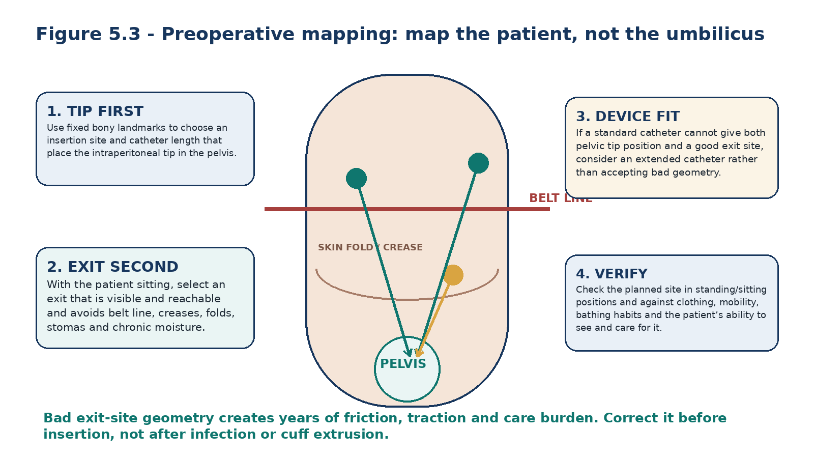
Table 5.4 - Mapping findings that change the plan.
| Finding | Why it matters | Planning response |
|---|---|---|
| High or low belt line | Chronic compression/friction can traumatize the sinus. | Move exit away from belt; re-check while seated. |
| Obesity / pannus | Lower exits may be invisible, moist or mobile. | Consider upper-abdominal or extended/presternal exit if needed. |
| Deep skin crease | Moisture and movement increase care difficulty. | Avoid placing exit within the crease. |
| Stoma / gastrostomy / suprapubic catheter | Contamination risk and equipment interference. | Maximise separation; extended catheter may be useful. |
| Previous scars | May imply adhesions and alter tunnel options. | Map around scars; choose insertion technique accordingly. |
| Unilateral mobility limitation | Patient may not reach/see one side. | Place where the patient or caregiver can inspect and secure it. |
| Preferred sleep side / occupation | Pressure or repetitive traction may occur. | Select opposite or protected location where feasible. |
MAPPING RULE The best exit site is not the cosmetically easiest place for the operator; it is the place the patient can maintain for years with minimal friction, traction and contamination.
4. Pre-procedure assessment: identify the anatomy that changes technique
The access history should actively search for previous abdominal or pelvic surgery, peritonitis, hernia repair, ostomy, mesh, chronic constipation, urinary retention, organomegaly, severe obesity, inflammatory bowel or abdominal-wall disease, and prior PD access. These findings rarely function as simple yes/no contraindications; they determine whether direct visualization, adhesiolysis, hernia planning, remote exit sites or a different anaesthetic strategy is needed. [1,3]
Physical examination should be done in more than one posture. Look for hernias, scars, folds, ostomies, skin disease, belt position and locations the patient can see. If a hernia is clinically important, coordinate repair strategy with the access surgeon; the 2024 SAGES update conditionally favours staged hernia repair before catheter insertion in adults when both are required, but certainty is very low and local expertise/patient timing matter. [3]
Table 5.5 - Pre-procedure anatomy -> access consequence.
| Finding | Potential problem | Access strategy |
|---|---|---|
| Multiple prior abdominal operations | Adhesions, compartmentalization, difficult blind entry. | Favour an approach with safe entry and ability to diagnose/treat adhesions when necessary. |
| Known hernia | Leak/hernia enlargement with intraperitoneal pressure. | Coordinate repair timing and postoperative low-pressure plan. |
| Obesity / thick pannus | Hidden exit, long subcutaneous route, catheter mobility. | Anthropometric mapping; consider extended catheter. |
| Ostomy / chronic incontinence | Higher contamination exposure. | Remote exit location and meticulous site separation. |
| Severe cardiopulmonary risk | General anaesthesia/pneumoperitoneum may be undesirable. | Consider image-guided percutaneous or open/local-anaesthetic approach when appropriate. |
| Need for immediate dialysis | Healing time is limited. | Coordinate urgent-start capable access pathway; detailed prescription in Chapter 17. |
5. Peri-insertion preparation: prevent avoidable failure before the first incision
The 2019 ISPD access guideline frames catheter placement as a multidisciplinary process rather than a stand-alone procedure. Its best-practice checklist includes bowel management to prevent perioperative constipation, emptying the bladder, surgical-site antisepsis, and a single preoperative antibiotic dose. The 2023 ISPD catheter-infection update more strongly states that prophylactic antibiotics should be administered immediately before catheter insertion to prevent peritonitis (1A); known nasal Staphylococcus aureus carriers should receive nasal antibiotic prophylaxis according to local protocol (1C). [1,2]
The exact antibiotic and surgical antisepsis protocol should follow local microbiology, allergies, resistance patterns and institutional policy. This chapter deliberately avoids inventing a universal drug or dose. The principle is temporal and mechanistic: antimicrobial protection must be active at the time the peritoneal cavity and new catheter tract are created. [2,8]
Table 5.6 - Pre-insertion safety bundle.
| Domain | Action | Why it matters |
|---|---|---|
| Access plan | Confirm catheter, insertion site, exit-site mark and backup technique. | Prevents intraoperative improvisation. |
| Bowel | Prevent/treat constipation before procedure. | Reduces immediate outflow obstruction and tip displacement. |
| Bladder | Have patient void; catheterise if clinically required. | Avoids bladder injury/compression and poor pelvic space. |
| Infection prevention | Administer prophylactic antibiotic immediately before insertion per local protocol. | Reduces early peritonitis risk. |
| Surgical preparation | Use validated skin antisepsis, sterile attire and draping. | Protects a permanent foreign-body tract. |
| Equipment | Have correct adapter/transfer set and rescue equipment available. | Avoids unplanned manipulation after placement. |
| Urgency | State whether elective or urgent-start PD is anticipated. | Changes wound closure and postoperative pressure strategy. |
SAFETY BOUNDARY Exact antibiotic selection, anaesthesia, skin-preparation agent, bowel regimen and operative details remain governed by local protocol and the procedural team. This chapter teaches the access logic, not an unsupervised procedure recipe.
6. Insertion approaches: four routes to the same physiological goal
Chronic PD catheters can be placed by open surgical dissection, image-guided percutaneous techniques, peritoneoscopy or laparoscopy. The appropriate question is not which specialty “owns” PD access; it is which approach can safely deliver the required catheter geometry for this patient, at the needed time, with a team able to rescue complications. [1,3,4]
Evidence does not support a single universal winner. The 2023 Cochrane review found low-certainty evidence and insufficient high-quality data to show one insertion method consistently reduces catheter dysfunction. SAGES 2024 conditionally favours advanced laparoscopy over basic laparoscopy or open insertion and considers either advanced laparoscopy or image-guided percutaneous insertion reasonable in adults, but the certainty is very low. More recent meta-analyses continue to show broadly comparable outcomes between contemporary percutaneous and laparoscopic strategies. [3–6]
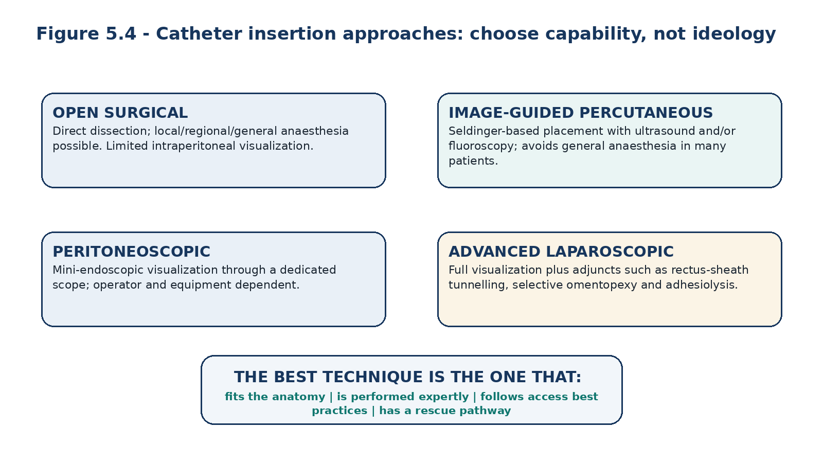
Table 5.7 - Insertion approaches: operational trade-offs.
| Approach | Strengths | Limitations / when to think twice |
|---|---|---|
| Open surgical | Direct abdominal-wall dissection; can be performed without pneumoperitoneum and sometimes under local/regional anaesthesia. | Limited intraperitoneal visualization; cannot easily perform omentopexy/adhesiolysis. |
| Image-guided percutaneous | Less invasive; often avoids general anaesthesia; ultrasound/fluoroscopy can improve safe entry and pelvic placement. | Limited ability to treat dense adhesions or omental pathology; requires imaging expertise. |
| Peritoneoscopic | Direct limited intraperitoneal view through dedicated scope. | Equipment/operator availability; adjunctive surgery limited. |
| Basic laparoscopy | Full visualization and pelvic placement. | Visualization alone does not prevent migration/omental problems. |
| Advanced laparoscopy | Adds rectus-sheath tunnelling, selective omentopexy, adhesiolysis and hernia-related procedures when indicated. | General anaesthesia/pneumoperitoneum; resource and surgeon expertise requirements. |
EVIDENCE CALIBRATION Do not misread a conditional guideline recommendation as proof of superiority. Advanced laparoscopy has mechanistic advantages and favourable observational data, but RCT evidence comparing modern techniques remains limited. [3–6]
7. Advanced laparoscopy: use adjuncts only when they solve a failure mechanism
The value of “advanced” laparoscopy is not the camera itself. It is the opportunity to prevent or correct specific mechanical causes of failure. Rectus-sheath tunnelling fixes the craniocaudal direction of the catheter through the abdominal wall; omentopexy can move redundant omentum away from the side holes when it lies near the tip; adhesiolysis can open a compartmentalized peritoneal cavity; and hernias can be identified and managed in a planned fashion. [1,3]
These adjuncts should be selective rather than ritual. An omentopexy is useful when redundant omentum threatens the catheter, not because every patient has an omentum. Adhesiolysis is useful when adhesions would compromise placement or drainage, not as a search for “perfect” anatomy. The operative goal is reliable flow with the least additional intervention. [1]
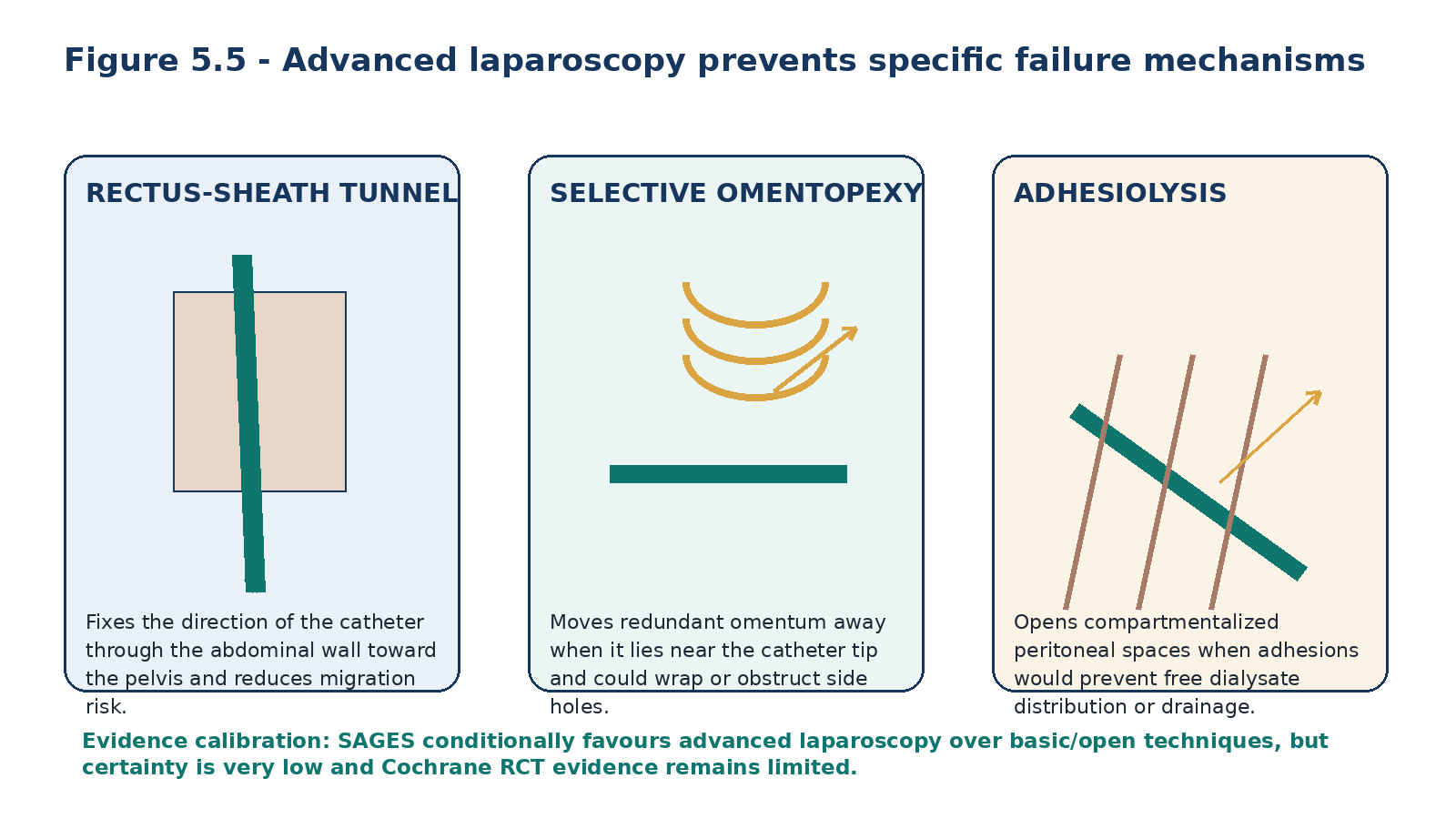
Table 5.8 - Advanced laparoscopic adjunct -> mechanism -> endpoint.
| Adjunct | Mechanism addressed | What success should look like |
|---|---|---|
| Rectus-sheath tunnelling | Catheter migration / poor directional fixation. | Catheter enters peritoneum in a stable craniocaudal direction toward pelvis. |
| Selective omentopexy | Redundant omentum near tip/side holes. | Omentum held away from catheter without unnecessary resection. |
| Adhesiolysis | Compartmentalization or obstructed pelvic path. | Free communication of dialysate and unobstructed placement. |
| Hernia identification/repair | Abdominal-wall pressure defect. | Access plan accounts for leak/hernia risk and postoperative pressure. |
| Intraoperative flow test | Unrecognized kink, malposition or obstruction. | Prompt fill and drain before closing access procedure. |
8. Choosing the insertion technique: a patient-specific access conference
The access team should make an explicit technique decision before the day of insertion. For a patient with no prior surgery, favourable anatomy and high anaesthetic risk, an image-guided percutaneous approach may be ideal. A patient with multiple prior laparotomies, suspected adhesions and a hernia may gain more from advanced laparoscopy. An urgent inpatient with limited theatre access may require a different pathway again. [1,3]
Table 5.9 - Patient feature -> technique discussion.
| Patient/context | Technique consideration | Do not assume |
|---|---|---|
| No prior abdominal surgery; high anaesthetic risk | Image-guided percutaneous or open/local-anaesthetic pathway may reduce anaesthetic burden. | Laparoscopy is mandatory for every patient. |
| Multiple prior operations / suspected adhesions | Approach with safe visual entry and ability to lyse clinically important adhesions. | Prior surgery makes PD impossible. |
| Severe obesity / remote exit needed | Technique must reproduce mapped catheter length and tunnel; extended catheter may be key. | A standard lower-abdominal exit is acceptable because it is familiar. |
| Urgent dialysis requirement | Insertion method and team must support early low-pressure use and rapid troubleshooting. | Urgency means temporary HD is the only option. |
| Local high-volume expert pathway | Experience may outweigh theoretical technique differences. | Published technique labels guarantee local outcomes. |
PROGRAMME RULE A PD programme without timely catheter insertion and rescue capacity does not truly offer PD. Access expertise is part of modality availability.
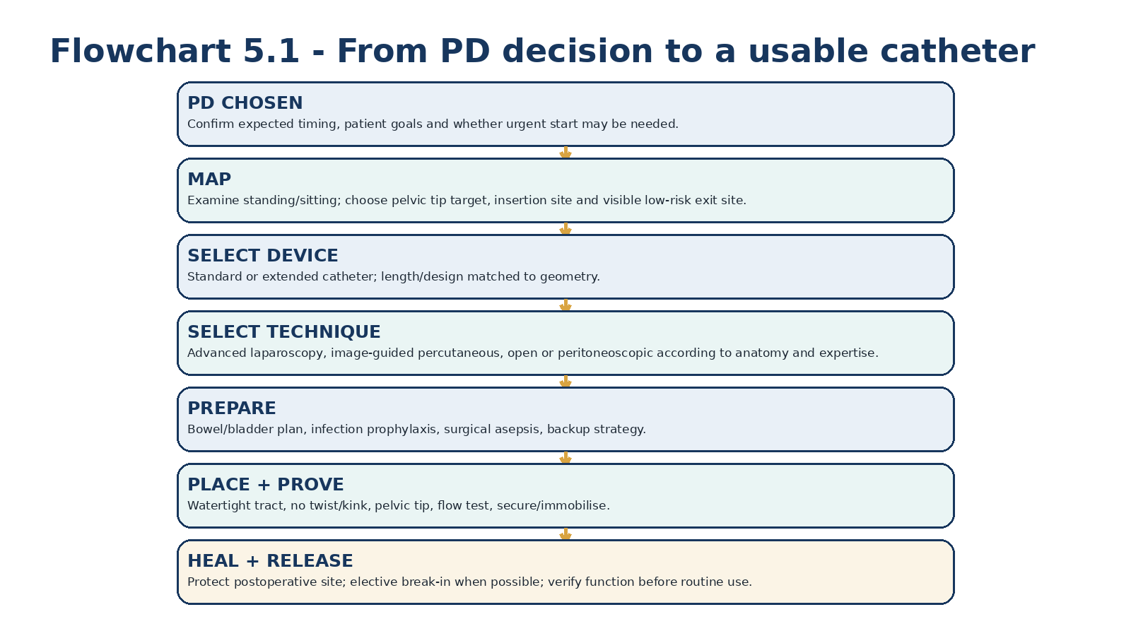
9. Placement endpoints: what must be true before the procedure is considered finished
Regardless of insertion method, several end-state principles are shared: the catheter should not be twisted or kinked; the deep cuff should sit in an appropriate muscular/fascial position; the intraperitoneal tip should be directed to the pelvis; the abdominal-wall passage should be watertight; the exit site should be small, appropriately directed and separated from the superficial cuff; and flow should be tested before completion. [1]
The exit site should not be secured by sutures through or immediately beside the skin-catheter interface because local trauma can impair healing. The catheter and extension should instead be immobilized in a way that prevents torque on the new tract. [1,2]
Table 5.10 - Insertion completion checklist.
| Check | Pass criterion | If not |
|---|---|---|
| Orientation | No twist or acute bend along radiopaque stripe/tract. | Correct before closure. |
| Pelvic position | Tip directed into true pelvis without excessive depth. | Reposition; reassess insertion level/length. |
| Abdominal wall | Deep cuff/tract stable and closure watertight. | Correct tract/closure before use. |
| Flow test | Prompt inflow and gravity drainage appropriate to test conditions. | Investigate kink, obstruction, malposition. |
| Exit site | Visible, reachable, small, not on belt/crease, no anchoring suture at exit. | Re-site if geometry is clearly poor. |
| External limb | Correct adapter/transfer set attached; catheter securely immobilised. | Fix before postoperative movement. |
10. Immediate postoperative care: protect the tract while tissue ingrowth develops
The newly created exit site and tunnel are mechanically vulnerable. Early handling should be minimized, the catheter should be immobilized, and the dressing should remain clean and dry according to the procedural and PD-unit protocol. The operative team should document catheter type, insertion technique, exit-site location, any adhesiolysis/omentopexy/hernia repair, intraoperative flow findings, complications and the intended earliest-use plan. [1,2]
The catheter should not simply disappear under a dressing until the first dialysis. The PD team needs a defined postoperative pathway: who reviews the wound, when the catheter is flushed if local practice requires it, how constipation is prevented, what symptoms trigger urgent contact, and whether the patient will start electively or urgently. [1]
Table 5.11 - First postoperative days: what to protect.
| Risk | Why early period is vulnerable | Prevention direction |
|---|---|---|
| Traction | Cuffs and sinus have not matured. | Immobilise catheter/extension; avoid pulling. |
| Leak | Abdominal-wall seals are fresh. | Respect break-in; use low intraperitoneal pressure if urgent start is unavoidable. |
| Constipation | May immediately impair drainage or move tip. | Maintain bowel plan from day 1. |
| Contamination | Fresh wound plus permanent foreign body. | Minimal handling; sterile unit protocol. |
| Unrecognized dysfunction | Catheter may not be used for days/weeks. | Document flow at insertion and planned verification before routine use. |
HANDOFF RULE Every catheter insertion should end with a written handoff to the PD team: device, technique, flow test, wound/exit plan, break-in plan, complications and who owns the first problem.
11. Break-in and early use: healing is part of access function
ISPD recommends a break-in period of at least 2 weeks before elective PD start (1B). The 2024 SAGES guideline similarly conditionally favours traditional over urgent start in adults when the patient can safely wait, although the evidence certainty is very low. Earlier use is associated particularly with more leak and early mechanical events. [1,3]
When dialysis cannot safely be deferred, ISPD recommends a modified prescription using low-volume exchanges with the patient supine when PD is started before 2 weeks (1C). Urgent-start PD is therefore not “normal PD sooner”; it is deliberately low-pressure PD designed to protect a fresh abdominal-wall tract. Detailed urgent-start prescriptions, escalation and inpatient scenarios belong to Chapter 17. [1,3]
Table 5.12 - Elective versus urgent catheter use.
| Situation | Access principle | Trade-off |
|---|---|---|
| Elective start feasible | Allow at least 2 weeks of healing before routine PD. | Lower early leak/mechanical risk; requires timely planning. |
| PD needed before 2 weeks | Use urgent-start pathway with low-volume supine exchanges and careful escalation. | Avoids/limits temporary HD but increases early mechanical risk. |
| Major leak or wound concern | Reduce intraperitoneal pressure or rest per access team. | May require temporary alternative KRT depending on clinical urgency. |
THRESHOLD DISCIPLINE The 2-week interval is a guideline-supported break-in standard for elective start, not a biological switch at day 14. Healing, concomitant surgery, leak risk and clinical urgency still matter. [1,3]
12. Mature exit-site care: clean, inspect, immobilise
The 2023 ISPD catheter-related infection update provides the current preventive framework. It recommends topical antibiotic cream or ointment at the catheter exit site daily (mupirocin or gentamicin; 1C), notes that no cleansing agent has proved superior to another (2B), recommends cleansing at least twice weekly and after showering or vigorous exercise (1C), and recommends catheter immobilisation to prevent traction injury (1C). [2]
A dressing cover is not mandatory after routine exit-site care and topical antibiotic application once beyond the immediate postoperative context (2D); evidence comparing dressing systems is low certainty, and a 2024 network meta-analysis found no basis for a strong universal dressing recommendation. Local climate, skin sensitivity, dust/pet exposure, sweating and patient preference can legitimately modify the dressing plan. [2,11]
Exit-site care must continue even if PD is temporarily interrupted or discontinued while the catheter remains in place. “Not currently using the catheter” does not mean the skin tunnel is no longer a foreign-body interface. [2]
Table 5.13 - Mature exit-site preservation bundle.
| Action | Evidence-based principle | Bedside interpretation |
|---|---|---|
| Inspect | Compare with the patient’s normal exit site at every care episode. | Change from baseline matters. |
| Clean | At least twice weekly and after shower/vigorous exercise; no cleanser proven universally superior. | Use a consistent tolerated local protocol. |
| Topical prophylaxis | Daily mupirocin or gentamicin per ISPD/local programme. | Prevention strategy; monitor local resistance/skin tolerance. |
| Immobilise | Prevent traction/friction at the skin-catheter interface. | Secure external catheter/extension without creating pressure injury. |
| Dress or not | Cover is not mandatory for a mature site after care; evidence is low certainty. | Individualise to environment and patient needs. |
| Continue care off PD | Maintain exit-site care as long as catheter remains. | Catheter risk persists even when not exchanging. |
ACCESS-CARE RULE The exit site should be boring: stable colour, no purulent drainage, minimal trauma, no tension and a routine the patient can perform consistently.
13. Read the exit site correctly: irritation, infection and tunnel disease are different
A newly placed catheter can have mild erythema, and a mature site can become red after traction, dressing allergy or a change in cleansing materials. ISPD therefore does not define exit-site infection by redness alone. Definitive exit-site infection is purulent discharge at the catheter-epidermal interface, with or without surrounding erythema. Tunnel infection is inflammation such as erythema, swelling, tenderness or induration along the subcutaneous catheter pathway, with or without an ultrasound fluid collection. [2]
This distinction prevents two opposite errors: giving repeated antibiotics for mechanical dermatitis, and dismissing deep tunnel disease because the skin opening looks modest. This chapter teaches recognition and immediate access protection; organism-specific treatment, antibiotic duration, cuff shaving/exit relocation and catheter-removal decisions are developed in Chapter 11. [2]
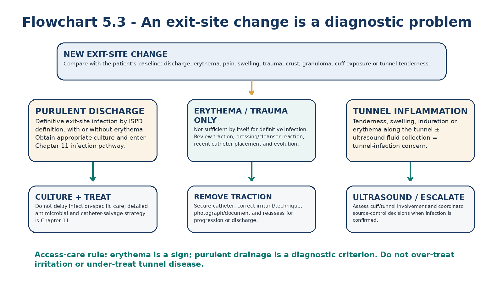
Table 5.14 - Exit-site finding -> interpretation.
| Finding | Interpretation | Immediate response |
|---|---|---|
| Purulent discharge ± erythema | Definitive exit-site infection by ISPD definition. | Culture appropriately and enter infection pathway. |
| Erythema alone after traction/new dressing | May be irritation or early infection; not definitive by itself. | Remove trauma/irritant; document and reassess closely. |
| Tenderness/induration along tunnel | Tunnel infection concern. | Examine entire tunnel; ultrasound can assess peri-catheter fluid. |
| Granuloma/crust without purulence | Local tissue problem; may coexist with infection. | Assess cause, trauma and evolving discharge; manage per trained PD team. |
| Extruded superficial cuff | Mechanical exposure that can become a bacterial reservoir. | Protect site and arrange access review; do not ignore. |
| Positive swab with normal-looking site | Colonisation does not equal infection. | Do not diagnose ESI from culture alone. |
14. Catheter function: separate fill, dwell and drain
Catheter function should be described rather than labelled. Is inflow slow? Is outflow slow? Are both impaired? Is drainage positional? Does the patient have end-of-drain pain? Is the cycler producing repeated low-drain alarms? These phenotypes localise the likely mechanism more effectively than the phrase “poor catheter.” [1]
Outflow dysfunction is more common than isolated inflow failure. A catheter may fill because positive hydrostatic pressure pushes dialysate past a partial obstruction, yet fail to drain by gravity. Conversely, an intraluminal fibrin plug or tubing kink often produces two-way resistance. The exact pattern should be documented before any salvage procedure. [1]
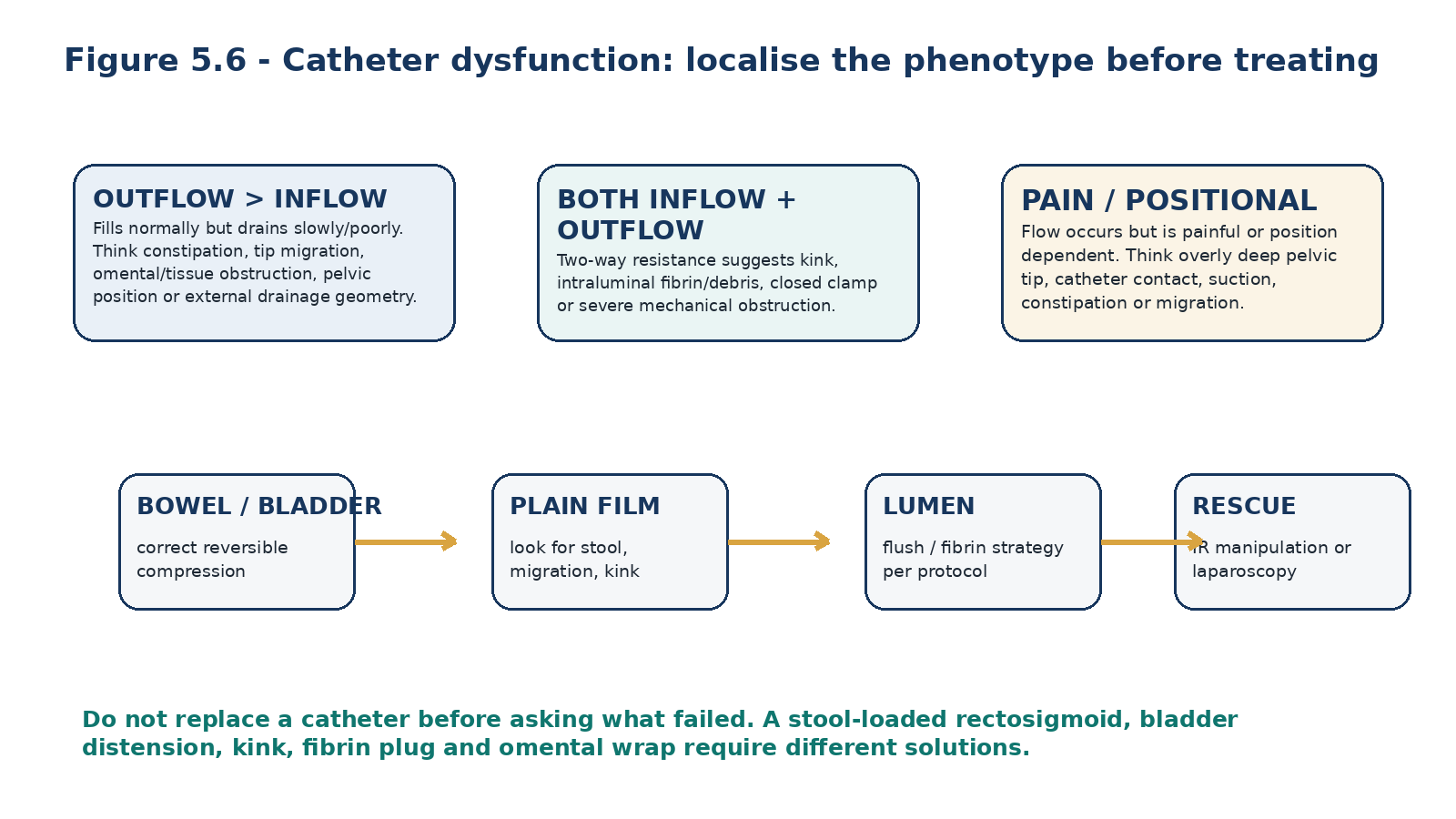
Table 5.15 - Flow phenotype -> first hypotheses.
| Phenotype | Think first | Important alternatives |
|---|---|---|
| Normal fill, poor drain | Constipation; tip migration; tissue/omental obstruction. | Patient position, pelvic anatomy, excessive suction. |
| Poor fill + poor drain | Kink; clamp/transfer-set issue; fibrin/debris. | Severe tissue entrapment or external tubing damage. |
| Position-dependent drain | Migration, tip contact, bowel/bladder relationship. | External line geometry or cycler setup. |
| Drain pain with otherwise adequate flow | Tip too deep/contact with pelvic structures; suction. | Constipation, inflammation, prescription-related negative pressure. |
| Sudden dysfunction after prior good function | Constipation, fibrin, migration, omental wrap. | New hernia/leak or external mechanical damage. |
15. Constipation and bladder distension: fix the common reversible causes first
ISPD identifies constipation as the most common cause of PD catheter outflow dysfunction. A stool-filled rectosigmoid colon can directly block side holes or displace the catheter tip away from a favourable drainage position. Urinary retention can compress the catheter less commonly. These diagnoses matter because they are both common and reversible without access intervention. [1]
Before ordering catheter manipulation for an outflow problem, ask about stool frequency/consistency, recent opioid or iron use, abdominal fullness and urinary symptoms. Examine the abdomen and review a plain film when appropriate. A catheter that drains normally after bowel decompression did not need a catheter procedure. [1]
Table 5.16 - Bowel/bladder clues.
| Clue | Mechanism | Response direction |
|---|---|---|
| New constipation + low drain | Rectosigmoid compression or tip displacement. | Treat constipation; reassess flow. |
| Fecal loading on film | Mechanical crowding around catheter. | Bowel programme before access escalation. |
| Suprapubic fullness / retention symptoms | Distended bladder competing for pelvic space. | Assess/relieve urinary retention appropriately. |
| No change after bowel/bladder correction | Another catheter mechanism is likely. | Proceed to imaging/lumen assessment. |
COMMON-CAUSE RULE Constipation should be actively prevented, not rediscovered after the catheter stops draining.
16. Imaging: ask a specific mechanical question
A plain abdominal radiograph is often the first useful test after simple bedside causes have been excluded because it can show fecal loading, catheter migration and obvious kinking. It does not diagnose omental wrapping or every adhesion, and radiographic pelvic position does not guarantee freedom from tissue obstruction. [1,3]
Catheterography can assess contrast flow and sometimes enables guidewire manipulation. Ultrasound is particularly useful for exit-site/tunnel assessment and selected abdominal-wall or fluid-collection questions. CT peritoneography is reserved for leak, hernia or complex anatomic questions rather than routine poor drainage. Imaging should answer a hypothesis—not substitute for one. [1,2]
Table 5.17 - Test -> question -> limitation.
| Test | Best question | Limitation |
|---|---|---|
| Plain abdominal film | Where is the tip? Is there fecal loading or a visible kink? | Cannot reliably identify omental wrap or all adhesions. |
| Catheterography / fluoroscopy | Is lumen patent? Can the catheter be repositioned/recanalized? | Technical success may not persist if underlying pathology remains. |
| Ultrasound | Is there tunnel/cuff fluid? Is there a superficial collection? | Negative tunnel ultrasound does not completely exclude infection. |
| CT / CT peritoneography | Is there complex leak, hernia or anatomic complication? | Radiation/contrast; not first-line for simple flow dysfunction. |
| Laparoscopy | What is the intraperitoneal mechanical cause, and can it be corrected now? | Invasive, anaesthesia/resource burden. |
17. Intraluminal fibrin and tubing kinks: two-way resistance has a different logic
When both inflow and outflow are restricted, first trace the external circuit for closed clamps, transfer-set problems and obvious compression. If external causes are excluded, an intraluminal fibrin plug or catheter kink becomes more likely. Blood in the effluent after insertion, peritonitis or inflammation can promote fibrin deposition. [1]
ISPD supports attempting fibrinolytic therapy when imaging has excluded displacement/kink, bladder distension and constipation and intraluminal debris is suspected. Exact flush volumes, drug concentration and dwell technique are device- and programme-specific and should follow a validated local protocol; they should not be improvised from a textbook. Persistent two-way obstruction after appropriate lumen treatment warrants structural reassessment. [1]
Table 5.18 - Two-way obstruction sequence.
| Step | Question | Action direction |
|---|---|---|
| 1. External | Are clamps, transfer set and tubing open/intact? | Correct external cause. |
| 2. Geometry | Is there kink or major migration on film? | Structural rescue pathway. |
| 3. Reversible compression | Constipation or bladder distension? | Correct and retest. |
| 4. Lumen | Fibrin/debris likely? | Use approved flush/fibrinolytic protocol. |
| 5. Persistent | Still poor after above? | Catheterography/operative assessment depending urgency and resources. |
18. Salvage: preserve a working access when the cause is correctable
Catheter salvage should progress from low-risk reversible causes to interventions that can correct the suspected mechanism. The 2019 ISPD guideline recommends a logical sequence from conservative/noninvasive approaches toward more aggressive intervention. SAGES 2024 conditionally supports either nonoperative or operative salvage, acknowledging very low certainty evidence. [1,3]
Fluoroscopic guidewire manipulation is attractive because it is minimally invasive and can restore patency or reposition the catheter without general anaesthesia. Contemporary series show useful success in selected patients, but recurrence is substantial when the underlying problem is omental wrap or adhesions. Laparoscopic rescue is more invasive but can diagnose and directly treat migration, omental entrapment and adhesions. The urgency of dialysis and local availability should determine how long to persist with low-success repeated manipulations. [1,3,12,13]
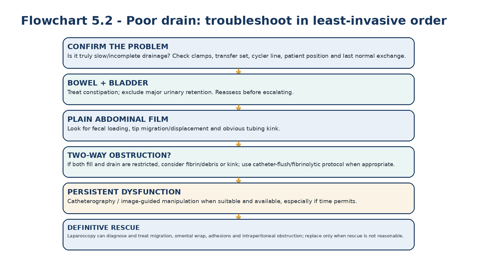
Table 5.19 - Rescue option -> strength -> limitation.
| Option | Strength | Limitation |
|---|---|---|
| Bowel/bladder correction | Low risk; treats common causes. | Only works if compression is the mechanism. |
| Fibrinolytic lumen strategy | Minimally invasive for fibrin/debris. | Does not fix migration, kink or omental wrap. |
| Fluoroscopic manipulation | Can recanalize/reposition without surgery; useful when time permits. | Recurrent dysfunction if underlying pathology persists. |
| Laparoscopic rescue | Diagnoses and treats omentum, adhesions, migration and kink directly. | Anaesthesia/invasive resource burden. |
| Simultaneous replacement | Provides new access when salvage is not reasonable. | Exposes new catheter to all initial-placement risks; should not be reflex first choice. |
SALVAGE RULE Do not perform repeated interventions just because they are available. Each intervention should have a named mechanism, expected benefit and stopping rule.
19. Pericatheter and other dialysate leaks: access failure can occur outside the lumen
A catheter can fill and drain yet still fail as an access because dialysate escapes from the peritoneal cavity. Early pericatheter leak is promoted by a fresh tract, high intraperitoneal pressure and early routine-volume use. It may present as clear fluid at the exit, abdominal-wall or genital swelling, unexpected weight gain or apparent loss of instilled volume. [1]
The 2-week elective break-in is partly intended to reduce this risk. When urgent-start PD is necessary, low-volume supine exchanges reduce pressure on the new tract. Detailed diagnosis and management of abdominal-wall, pleuroperitoneal, genital and occult leaks are deferred to Chapter 12. [1,3]
Table 5.20 - Leak clues that should trigger an access pathway.
| Finding | Possible leak pattern | Next question |
|---|---|---|
| Clear fluid at exit site | Pericatheter leak. | Is glucose-rich dialysate present and was PD started early/high pressure? |
| New genital/scrotal/labial oedema | Tracking dialysate through patent processus/hernia pathway. | Is there inguinal defect or recent pressure increase? |
| Focal abdominal-wall swelling | Subcutaneous/abdominal-wall leak or hernia. | Relation to fill volume and posture? |
| Dyspnoea + unilateral pleural effusion | Pleuroperitoneal communication. | Does pleural fluid profile support dialysate leak? |
| Low apparent UF with unexplained weight gain | Occult leak. | Is dialysate leaving cavity without returning to drain bag? |
20. Fill and drain pain: function includes comfort
A catheter that technically flows but produces severe pain is not a successful access. End-of-drain pain can occur when a tip lies too deep in the pelvis or when hydraulic suction brings sensitive peritoneal structures against side holes during APD. Constipation, inflammation, migration and local anatomy can amplify the symptom. [1]
Describe when pain occurs: during inflow, throughout dwell, or only at final drainage; whether it is positional; whether it began with a new cycler prescription; and whether effluent or abdominal findings suggest infection. Prescription strategies such as tidal drainage may reduce suction-related pain, but detailed APD optimization is deferred to Chapter 9. Persistent anatomic pain may require catheter reassessment or repositioning. [1]
Table 5.21 - Flow pain phenotype.
| Pain timing | Think | Cross-reference |
|---|---|---|
| During inflow | Jet/contact irritation, cold solution, local pathology. | Solutions/system issues Chapter 2; abdominal pain differential Chapter 12. |
| During drain only | Tip contact or suction, excessive negative pressure, constipation. | APD/tidal options Chapter 9; access position in this chapter. |
| Continuous abdominal pain | Peritonitis, surgical abdomen, hernia/leak or other pathology. | Peritonitis Chapter 10; complications Chapter 12. |
| Position-dependent pain | Tip relationship to pelvis/bowel/abdominal wall. | Check constipation and catheter position. |
21. Cuff extrusion and external catheter damage: small defects can become access emergencies
Superficial cuff extrusion is often a mechanical problem before it becomes an infectious one. Excessive bend stress in the subcutaneous tract, a superficial cuff placed too close to the exit, or chronic piston-like catheter motion can push the cuff outward. Once exposed, the porous cuff becomes difficult to keep clean and can act as a bacterial reservoir. [1]
External tubing can also crack, separate or be cut. A damaged catheter is both a leak and contamination event. Depending on the location and device, validated repair kits or splicing may preserve the access, but this requires trained staff, manufacturer-compatible components, contamination assessment and peritonitis prophylaxis as indicated. Do not tape a leaking catheter and continue exchanges. [1,8]
Table 5.22 - External access defect -> response.
| Defect | Risk | Immediate direction |
|---|---|---|
| Superficial cuff visible/extruding | Chronic contamination, infection, unstable sinus. | Protect and refer for access review; infection assessment if discharge. |
| External catheter crack/leak | Open contamination pathway and fluid leak. | Clamp/stop unsafe use; urgent PD-unit evaluation. |
| Loose adapter/transfer set | Contamination and disconnection. | Stop, secure using approved technique and follow contamination protocol. |
| Traction injury with bleeding | Sinus trauma and increased ESI risk. | Immobilise; reassess exit site over subsequent days. |
| Accidental catheter cut | Potential loss of sterile integrity. | Do not improvise; assess repair versus replacement with trained team. |
22. Long-term access preservation: make good function the default
Catheter maintenance is mostly a system of small repeated actions: stable bowel habits, immobilisation, consistent exit-site care, protection of the external limb, rapid response to new drainage changes and periodic competency review. The 2026 ASN core interventions for PD-related infection prevention reinforce programme-level surveillance, standardized staff and patient training, routine infection-prevention assessments and antimicrobial prophylaxis. [7]
Patients should know their own baseline: how the exit site normally looks, how quickly bags usually drain, which positions are normal, what the catheter feels like under clothing, and which changes require same-day contact. A photo of a mature healthy site and a written “call now” list can make subtle change more visible than generic instructions. [2,7]
Table 5.23 - Patient self-check: five things to know.
| Domain | Normal baseline | Call the PD team for |
|---|---|---|
| Exit site | No purulent drainage; stable appearance. | Purulent discharge, spreading redness, pain, swelling or tunnel tenderness. |
| Catheter position | Secure external limb with no pulling. | New traction injury, exposed cuff or damaged tubing. |
| Drain | Predictable time/volume for that prescription. | Repeated slow/incomplete drain or new cycler low-drain alarms. |
| Bowel | Regular pattern for that patient. | Constipation accompanying drainage change. |
| Abdominal wall | No new fluid swelling or leak. | Exit-site fluid, hernia swelling, genital edema or unexplained dialysate loss. |
23. Access programme quality: measure what happens after insertion
An access service should audit more than the number of catheters inserted. ISPD recommends monitoring catheter outcomes, mechanical events and insertion-related complications. The 2023 infection guideline recommends annual monitoring of catheter-related infection incidence and suggests that insertion-related exit-site/tunnel infection within 30 days should remain below 5% of inserted catheters (2C). The overall exit-site infection rate target is no more than 0.40 episodes per patient-year at risk (2C). [1,2]
These are programme quality targets, not patient-level treatment thresholds. A centre with frequent early leaks, poor primary function or emergency catheter replacements should investigate mapping, insertion technique, constipation pathways, postoperative handoff and rescue availability rather than attributing every event to “bad catheters.” [1,2]
Table 5.24 - Access programme dashboard.
| Metric | Why track it | What a poor result should trigger |
|---|---|---|
| Catheter functional at intended start | Measures usable access, not insertion count. | Review mapping, placement and flow testing. |
| Early leak / mechanical dysfunction | Detects tract/healing and placement problems. | Review break-in, closure, technique and urgent-start practice. |
| Insertion-related ESI/tunnel infection | Links infection to procedure/early wound period. | Review prophylaxis, dressing/handling and handoff. |
| Exit-site infection rate | Monitors long-term access care. | Training, prophylaxis, immobilisation and CQI review. |
| Time to rescue after dysfunction | Measures ability to preserve PD continuity. | Improve imaging/IR/surgical access pathway. |
| Catheter removal for mechanical failure | High-impact access loss outcome. | Root-cause review of avoidable failures. |
QUALITY RULE A programme that cannot tell why its catheters fail cannot improve its catheter outcomes.
24. Clinical pearls
PEARL 1 The insertion site determines the pelvic position; the exit site determines whether the patient can care for the catheter.
PEARL 2 Map the patient sitting before the patient is anaesthetized and supine.
PEARL 3 If a standard catheter cannot give both pelvic tip and good exit site, change the catheter - not the anatomy.
PEARL 4 Constipation is the commonest outflow problem and belongs in access prevention, not only troubleshooting.
PEARL 5 A normal fill with poor drain localises differently from two-way obstruction.
PEARL 6 A low-drain alarm is a symptom; the cycler cannot diagnose constipation, migration or omental wrap.
PEARL 7 Exit-site erythema alone is not definitive infection; purulent discharge is the key ISPD diagnostic criterion.
PEARL 8 Traction injury is preventable. Immobilisation is an infection-prevention intervention as well as a comfort measure.
PEARL 9 Image-guided manipulation is valuable when the mechanism is amenable; repeated recurrence should prompt definitive pathology-directed rescue.
PEARL 10 A catheter that flows but causes severe pain is not a satisfactory access.
25. Mini-cases: decisions, not hardware trivia
Case 1 - Obesity and the invisible exit site
A 52-year-old with obesity chooses PD. A standard catheter placed from the lower abdomen would leave the exit site under a deep pannus where the patient cannot see it while seated.
REASONING QUESTION Is this patient unsuitable for PD?
INTERPRETATION No. The problem is exit-site geometry, not PD eligibility. A lower hidden exit would increase care difficulty, moisture and traction risk.
MANAGEMENT DECISION Perform formal preoperative mapping and choose a catheter/route that preserves pelvic tip position while relocating the exit to a visible upper-abdominal or remote site if needed.
TEACHING POINT Use catheter design to fit the body; do not use the body as a reason to deny the modality.
Case 2 - Prior laparotomy and a request for “blind percutaneous” placement
A 64-year-old with two prior midline laparotomies has chosen PD. The proposed service has immediate access to blind bedside percutaneous insertion but no ability to manage adhesions if encountered.
REASONING QUESTION What should change?
INTERPRETATION Prior surgery does not exclude PD, but it raises the probability that intraperitoneal visualization or adhesiolysis may be useful and makes blind entry less attractive.
MANAGEMENT DECISION Refer to an experienced access pathway capable of image-guided or laparoscopic placement and rescue rather than proceeding for convenience alone.
TEACHING POINT Technique choice is an anatomy-and-capability decision, not a waiting-time contest.
Case 3 - The catheter fills but will not drain
Three months after starting PD, a patient has normal inflow but progressively incomplete drainage. He has not opened his bowels for four days and recently began opioid analgesia.
REASONING QUESTION What is the best first hypothesis?
INTERPRETATION Constipation-related rectosigmoid compression or catheter displacement is more likely than sudden catheter failure.
MANAGEMENT DECISION Correct constipation, review the external circuit and reassess drainage before requesting invasive catheter manipulation; obtain a plain film if dysfunction persists.
TEACHING POINT The common reversible cause comes before the catheter laboratory.
Case 4 - Two-way obstruction with fibrin
After a bloody postoperative flush, a new catheter becomes difficult both to fill and drain. External clamps and transfer set are normal; film shows no migration or kink.
REASONING QUESTION What mechanism fits the phenotype?
INTERPRETATION Intraluminal fibrin/debris is plausible because resistance is two-way and structural displacement has not been demonstrated.
MANAGEMENT DECISION Use the centre’s approved catheter-flush/fibrinolytic protocol and reassess. Persistent obstruction should progress to structural imaging/rescue rather than repeated blind flushing.
TEACHING POINT Two-way resistance points to lumen or kink before it points to constipation alone.
Case 5 - Red exit site after a new dressing
A stable PD patient develops a 1-cm area of erythema after changing adhesive dressings. There is no purulent discharge, tenderness, swelling or tunnel pain.
REASONING QUESTION Is this definitive exit-site infection?
INTERPRETATION No. ISPD does not define ESI by erythema alone; contact irritation or traction are plausible.
MANAGEMENT DECISION Remove the likely irritant, ensure catheter immobilisation, document the site and reassess closely for purulent drainage or progression. Escalate if infection features develop.
TEACHING POINT Avoid both antibiotic reflexes and complacency: redness requires context and follow-up.
Case 6 - Recurrent guidewire salvage
A catheter has required two fluoroscopic repositioning procedures in six weeks and again migrates with poor outflow. The patient needs reliable dialysis now.
REASONING QUESTION Should a third identical manipulation be the default?
INTERPRETATION Repeated recurrence suggests an unresolved mechanical driver such as omental entrapment, unstable trajectory or adhesions.
MANAGEMENT DECISION Discuss timely laparoscopic diagnosis and pathology-directed rescue versus replacement rather than repeating a low-durability intervention without a new hypothesis.
TEACHING POINT Salvage has a stopping rule: recurrence is information about mechanism.
26. Common pitfalls - and the correction
Table 5.25 - High-frequency errors in PD catheter care.
| Pitfall | Why it fails | Correction |
|---|---|---|
| Using the umbilicus as the main landmark | Body proportions vary; tip may be too deep or too high. | Use fixed bony landmarks and catheter-specific mapping. |
| Choosing the exit site after insertion | The insertion site limits reachable exit geometry. | Plan tip + insertion + exit as one system. |
| Standard catheter for every body | May hide exit under pannus or create excessive bend stress. | Use extended catheter when geometry requires it. |
| Calling laparoscopy universally superior | Evidence certainty is low and expertise matters. | Match technique to anatomy, anaesthetic risk and programme skill. |
| Ignoring constipation until dysfunction | Common reversible cause becomes recurrent access failure. | Maintain bowel prevention from perioperative period onward. |
| Treating low-drain alarm as catheter failure | Alarm reports outcome, not cause. | Localise phenotype and troubleshoot sequentially. |
| Antibiotics for redness alone | Irritation/trauma can mimic ESI. | Use ISPD definition and serial assessment. |
| Leaving catheter mobile at exit | Traction increases sinus injury/infection risk. | Immobilise catheter/extension consistently. |
| Continuing routine-volume PD immediately after insertion | Raises fresh-tract pressure and leak risk. | Respect break-in or use urgent-start low-volume supine pathway. |
| Replacing before attempting cause-based rescue | Consumes access and exposes patient to new insertion risks. | Correct reversible causes; use IR/laparoscopy when appropriate. |
| Repeated fluoroscopic manipulation without new hypothesis | Underlying omentum/adhesion/geometry may persist. | Escalate to definitive diagnosis when recurrence indicates it. |
| Stopping exit care when PD is temporarily held | Foreign-body tunnel remains at risk. | Continue exit care while catheter remains in place. |
27. Active recall: MUST MEMORIZE / MUST REASON / USE AS REFERENCE
MUST MEMORIZE
Table 5.26 - Must memorize.
| Prompt | Answer |
|---|---|
| Elective catheter break-in? | ISPD: at least 2 weeks before elective PD start. |
| Urgent start before 2 weeks? | Low-volume exchanges with patient supine; detailed prescription in Chapter 17. |
| Most common cause of outflow dysfunction? | Constipation. |
| Definitive exit-site infection criterion? | Purulent discharge at catheter-epidermal interface, with or without erythema. |
| Tunnel infection definition? | Clinical inflammation along tunnel with or without ultrasound fluid collection. |
| Mature exit-site cleansing frequency? | At least twice weekly and after shower/vigorous exercise per ISPD 2023. |
| Catheter immobilisation? | Recommended to prevent traction injury. |
| Dressing over mature site mandatory? | No; ISPD says not mandatory after care/topical antibiotic, evidence low certainty. |
| When standard catheter cannot achieve pelvic tip + good exit? | Use an extended catheter rather than accept poor geometry. |
| Outflow-only vs two-way resistance? | Outflow-only often bowel/migration/tissue; two-way suggests kink/lumen/debris/external obstruction. |
MUST REASON
Table 5.27 - Must reason.
| Clinical problem | Reasoning chain |
|---|---|
| Obese patient cannot see lower exit | Not a contraindication -> map body -> remote/extended catheter -> preserve pelvic tip + accessible exit. |
| Poor drain after opioid use | Medication -> constipation -> rectosigmoid crowding -> correct bowel first -> reassess catheter. |
| Both fill and drain are difficult | External line/clamp -> kink -> fibrin/debris -> imaging/lumen strategy -> rescue if persistent. |
| Repeated migration after guidewire rescue | Recurrence -> unresolved anatomy/mechanics -> definitive laparoscopic diagnosis more valuable. |
| Red exit after adhesive change | Irritant/trauma vs infection -> purulence? tunnel signs? -> remove irritant + reassess rather than reflex antibiotics. |
| Urgent dialysis after new insertion | Need KRT now -> avoid routine fill pressures -> low-volume supine urgent-start protocol -> monitor leak/mechanics. |
USE AS REFERENCE
Table 5.28 - Use as reference, not rote memory.
| Item | Why reference it |
|---|---|
| Specific catheter lengths and stencil measurements | Device-specific and dependent on patient anatomy. |
| Exact antibiotic agent/dose before insertion | Local microbiology, allergy and policy determine regimen. |
| Fibrinolytic drug concentration/flush volume | Programme/device-specific protocol. |
| Advanced laparoscopic operative steps | Requires procedural training and local technique. |
| Urgent-start fill volumes/escalation | Prescription belongs to Chapter 17 and individual patient mechanics. |
| Repair kit/splice technique | Manufacturer- and catheter-specific; trained staff only. |
28. Flashcards: active recall
1. Q: What is the patient-important catheter outcome? A: Reliable, comfortable fill/drain with a maintainable low-risk exit site and durable access.
2. Q: What does preoperative mapping link? A: Pelvic tip target, insertion site, catheter length/tunnel and exit-site location.
3. Q: Why is the umbilicus a poor sole landmark? A: It varies with body habitus and does not reliably predict pelvic catheter position.
4. Q: When should an extended catheter be considered? A: When a standard catheter cannot provide both correct pelvic tip and satisfactory exit site.
5. Q: What does SAGES 2024 say about advanced laparoscopy? A: Conditionally favours it over basic/open insertion, but certainty is very low.
6. Q: What does Cochrane 2023 say about insertion technique? A: Evidence is insufficient to identify a universally superior technique for dysfunction/outcomes.
7. Q: Peri-insertion antibiotic timing? A: Immediately before catheter insertion per ISPD.
8. Q: Elective break-in? A: At least 2 weeks.
9. Q: Urgent-start posture? A: Supine with low-volume exchanges.
10. Q: Most common outflow problem? A: Constipation.
11. Q: Normal fill, poor drain suggests? A: Outflow mechanism such as constipation, migration or tissue/omental obstruction.
12. Q: Both inflow and outflow poor suggests? A: External obstruction, kink or intraluminal fibrin/debris.
13. Q: First imaging for persistent mechanical dysfunction? A: Often a plain abdominal film after bedside causes are addressed.
14. Q: Role of fluoroscopic manipulation? A: Minimally invasive recanalization/repositioning in selected dysfunction; recurrence can occur.
15. Q: Role of laparoscopy in salvage? A: Direct diagnosis and correction of omentum, adhesions, migration and other intraperitoneal causes.
16. Q: Definitive ESI? A: Purulent discharge ± erythema.
17. Q: Erythema alone? A: Not definitive ESI; assess trauma, allergy, recent placement and evolution.
18. Q: Catheter immobilisation? A: Prevents traction/friction injury and supports infection prevention.
19. Q: Mature dressing always required? A: No; cover is not mandatory after routine care in ISPD 2023.
20. Q: Exit care when PD is paused? A: Continue while catheter remains in place.
29. Rapid troubleshooting: catheter problem -> mechanism -> next step
Table 5.29 - One-minute catheter troubleshooting.
| If you see... | Think... | Do now... |
|---|---|---|
| Slow drain after constipation | Rectosigmoid compression / tip displacement. | Correct bowel; re-test; film if persistent. |
| Normal fill, poor drain, normal bowel | Migration / tissue or omental obstruction. | Plain film -> targeted salvage pathway. |
| Poor fill + poor drain | Kink / fibrin / external line issue. | Trace external system; film; approved lumen protocol. |
| New drain pain on cycler | Tip contact / suction / constipation. | Check bowel/position; prescription and access review. |
| Clear fluid at exit after early start | Pericatheter leak. | Stop escalating fill pressure; urgent access/Chapter 12 pathway. |
| Purulent exit-site discharge | Exit-site infection. | Culture and Chapter 11 infection pathway. |
| Tunnel tenderness/induration | Tunnel infection concern. | Ultrasound/clinical escalation; infection pathway. |
| Exposed superficial cuff | Mechanical extrusion with infection risk. | Protect; access review; culture if purulence. |
| External catheter crack | Leak + contamination. | Clamp/stop unsafe use; urgent repair/replacement assessment. |
| Recurrent failure after IR manipulation | Unresolved intraperitoneal mechanism. | Timely laparoscopic diagnosis/rescue discussion. |
30. Final revision sheet
TEN TAKE-HOME RULES 1) Map the patient before choosing the catheter. 2) Plan pelvic tip and exit site as one mechanical system. 3) Use an extended catheter when standard geometry forces a bad exit. 4) Technique choice depends on anatomy, urgency, anaesthetic risk and expertise; evidence for a universal winner is low certainty. 5) Give antimicrobial prophylaxis immediately before insertion. 6) Respect a 2-week elective break-in; urgent start is low-volume and supine. 7) Immobilise the catheter and maintain a consistent exit-site care routine. 8) Purulent discharge defines ESI; erythema alone does not. 9) Constipation is the commonest outflow problem. 10) Salvage the mechanism before replacing the access.
Table 5.30 - One-minute bedside synthesis.
| Clinical question | Core answer |
|---|---|
| Where should the catheter be? | Tip in pelvis; tract untwisted/watertight; exit visible, accessible, low-trauma. |
| Which catheter? | Standard silicone double-cuff unless geometry requires a different validated/extended design. |
| Which insertion technique? | The expert technique that fits anatomy/urgency and can reproduce best-practice geometry. |
| How to protect after insertion? | Immobilise, prevent constipation, protect wound and respect break-in. |
| How to read poor flow? | Describe inflow/outflow/pain/position first, then localise mechanism. |
| How to read exit-site redness? | Look for purulence and tunnel signs; irritation is common. |
| When to escalate salvage? | When reversible causes and appropriate low-risk strategies fail or dialysis urgency demands definitive rescue. |
Table 5.31 - Catheter-access verification checklist.
| Domain | Pass criterion |
|---|---|
| Body map | Pelvic tip target and exit site verified in relevant postures. |
| Device | Catheter length/design matches anatomy; extended option considered if needed. |
| Technique | Insertion strategy explicitly matched to anatomy, urgency and expertise. |
| Peri-procedure | Bowel/bladder plan, antibiotic prophylaxis and sterile protocol completed. |
| Placement | No twist/kink; watertight tract; flow tested; catheter immobilised. |
| Break-in | Elective ≥2-week plan or documented urgent-start strategy. |
| Exit site | Care, prophylaxis, cleaning frequency and securement taught with teach-back. |
| Function | Baseline fill/drain performance documented. |
| Rescue | Patient knows who to contact; programme has imaging/IR/surgical pathway. |
| Continuity | Access care continues during PD interruption while catheter remains. |
FINAL MENTAL MODEL Map -> choose device -> choose technique -> prepare -> place and prove -> heal -> maintain -> localise failure -> rescue mechanism -> verify durable function.
Rapid oral viva
Explain why preoperative catheter mapping should use bony landmarks and sitting examination rather than the umbilicus alone.
Compare advanced laparoscopic and image-guided percutaneous insertion using evidence certainty, anaesthetic burden and anatomy.
Give the least-invasive differential for normal inflow with poor outflow.
Explain why constipation can stop a catheter draining and why it must be prevented from the perioperative period.
Distinguish a fibrin plug from catheter migration using the inflow/outflow phenotype and imaging.
Walk through the response to purulent exit-site discharge versus erythema after traction.
Explain the rationale for the 2-week break-in period and how urgent-start PD changes the prescription.
Defend catheter salvage versus replacement in a patient with recurrent malfunction.
SCOPE BOUNDARY This chapter teaches catheter selection, placement logic, access preservation and first-line mechanical reasoning. Detailed PD initiation is Chapter 6; adequacy/RKF Chapter 7; volume/UF failure Chapter 8; CAPD/APD prescription optimization Chapter 9; peritonitis Chapter 10; exit-site/tunnel infection treatment Chapter 11; and comprehensive mechanical complications/leaks Chapter 12.
31. Selected authoritative references
1. Crabtree JH, Shrestha BM, Chow KM, et al. Creating and Maintaining Optimal Peritoneal Dialysis Access in the Adult Patient: 2019 Update. Perit Dial Int. 2019;39(5):414–436. https://doi.org/10.3747/pdi.2018.00232. PMID: 31028108.
2. Chow KM, Li PKT, Cho Y, et al. ISPD Catheter-related Infection Recommendations: 2023 Update. Perit Dial Int. 2023;43(3):201–219. https://doi.org/10.1177/08968608231172740. PMID: 37232412.
3. Haggerty SP, Kumar SS, Collings AT, et al. SAGES peritoneal dialysis access guideline update 2023. Surg Endosc. 2024;38(1):1–23. https://doi.org/10.1007/s00464-023-10550-8. PMID: 37989887.
4. Briggs VR, Jacques RM, Fotheringham J, Maheswaran R, Campbell M, Wilkie ME. Catheter insertion techniques for improving catheter function and clinical outcomes in peritoneal dialysis patients. Cochrane Database Syst Rev. 2023;2:CD012478. https://doi.org/10.1002/14651858.CD012478.pub2. PMID: 36810986; PMCID:PMC9946371.
5. Chui JN, Kim P, Cooper TE, et al. Percutaneous versus surgical catheter insertion for peritoneal dialysis: a systematic review and meta-analysis of randomized controlled trials. ANZ J Surg. 2025;95(4):664–674. https://doi.org/10.1111/ans.19315. PMID: 39641197.
6. Wang X, Jin W, Li X, Ji W, Ren T. Percutaneous versus laparoscopic catheter placement for peritoneal dialysis: a meta-analysis. BMC Nephrol. 2025;26(1):687. https://doi.org/10.1186/s12882-025-04597-z. PMID: 41339808; PMCID:PMC12676859.
7. Perl J, Teitelbaum I, Warady BA, et al. Core Interventions for the Prevention of Peritoneal Dialysis-Related Infections. Clin J Am Soc Nephrol. 2026;21(8):1439–1444. https://doi.org/10.2215/CJN.0000000976. PMID: 41348491.
8. Li PKT, Chow KM, Cho Y, et al. ISPD peritonitis guideline recommendations: 2022 update on prevention and treatment. Perit Dial Int. 2022;42(2):110–153. https://doi.org/10.1177/08968608221080586. PMID: 35264029.
9. Xie J, Kiryluk K, Ren H, et al. Coiled versus straight peritoneal dialysis catheters: a randomized controlled trial and meta-analysis. Am J Kidney Dis. 2011;58(6):946–955. https://doi.org/10.1053/j.ajkd.2011.06.026. PMID: 21872978.
10. Chow KM, Wong SSM, Ng JKC, et al. Straight Versus Coiled Peritoneal Dialysis Catheters: A Randomized Controlled Trial. Am J Kidney Dis. 2020;75(1):39–44. https://doi.org/10.1053/j.ajkd.2019.05.024. PMID: 31445925.
11. Feng C, Liu Y, Jin W, Lu M, Su CY. Effect of different exit-site care dressings on preventing peritoneal dialysis related infection from nontropical area: a systematic review and network meta-analysis. Ren Fail. 2024;46(2):2376331. https://doi.org/10.1080/0886022X.2024.2376331. PMID: 39011577.
12. Li P, Choo D, Deved V, et al. Salvage of Malfunctioning Peritoneal Dialysis Catheters: An Algorithm for Recanalization and Repositioning. J Vasc Interv Radiol. 2021;32(6):902–906. https://doi.org/10.1016/j.jvir.2021.03.522. PMID: 33771711.
13. Wong RCL, Cheung EWP, Wong A, Lee BKH, Cheng KK, Cho DHY. Efficacy and safety of repositioning malfunctioning peritoneal dialysis catheters with fluoroscopically guided guidewire manipulation. Clin Nephrol. 2025;104(3):159–169. https://doi.org/10.5414/CN111605. PMID: 40356439.
14. Crabtree JH, Chow KM. Peritoneal dialysis catheter insertion. Semin Nephrol. 2017;37(1):17–29. https://doi.org/10.1016/j.semnephrol.2016.10.004. PMID: 28153191.
15. Ouyang CJ, Huang FX, Yang QQ, et al. Comparing the incidence of catheter-related complications with straight and coiled Tenckhoff catheters in peritoneal dialysis patients: a single-center prospective randomized trial. Perit Dial Int. 2015;35(4):443–449. https://doi.org/10.3747/pdi.2013.00016. PMID: 24584608; PMCID:PMC4520727.
16. Johnson DW, Wong J, Wiggins KJ, et al. A randomized controlled trial of coiled versus straight swan-neck Tenckhoff catheters in peritoneal dialysis patients. Am J Kidney Dis. 2006;48(5):812–821. https://doi.org/10.1053/j.ajkd.2006.08.010. PMID: 17060001.
17. Wallace EL, Fissell RB, Golper TA, et al. Catheter insertion and perioperative practices within the ISPD North American Research Consortium. Perit Dial Int. 2016;36(4):382–386. https://doi.org/10.3747/pdi.2015.00089. PMID: 26493754; PMCID:PMC4934430.
SOURCE NOTE ISPD, SAGES, Cochrane and bibliographic sources were checked 1 September 2026. The 2019 ISPD document remains the principal comprehensive adult PD-access guideline identified in the current evidence search. Technique-specific procedural details, antibiotic choice/dose, fibrinolytic protocols, device repair and urgent-start prescriptions remain subject to current local policy, manufacturer instructions and trained specialist practice.