Timing | Baseline Assessment | CAPD | APD | Incremental PD | Urgent Start | Reassessment
| CHAPTER MISSION Build the first PD prescription from clinical goals and physiology: decide when dialysis is truly needed, measure what the kidneys still contribute, select CAPD or APD deliberately, choose fill volume/dwell/exchange number/osmotic strategy, protect a new catheter during urgent start, and reassess early enough that an initial prescription never becomes an unexamined permanent prescription. |
|---|
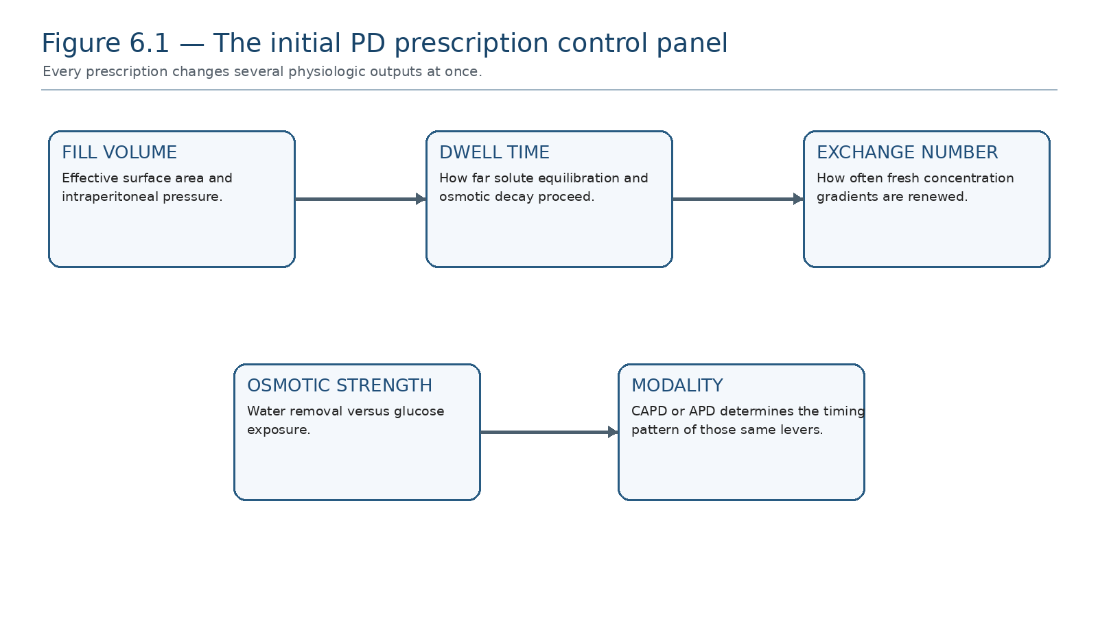
Every prescription combines fill volume, dwell time, exchange frequency, osmotic strength and modality. Changing one lever can improve one target while worsening glucose exposure, intraperitoneal pressure or treatment burden.
| MASTER PRINCIPLE The correct initial prescription is not the largest prescription that achieves a clearance number. It is the least burdensome prescription expected to meet the person’s current clinical goals safely, with an explicit plan for escalation as residual kidney function, membrane behavior and life circumstances change. |
|---|
0. One-page chapter map
Table 6.1 — The eight decisions that govern a safe PD start.
| Decision | Core question | Bedside output |
|---|---|---|
| 1. Need | Why does this person need dialysis now? | Clinical indication, not eGFR alone |
| 2. Readiness | Is the catheter, patient, home and training pathway ready? | Safe start pathway |
| 3. Kidney contribution | What urine volume and residual clearance remain? | Incremental vs fuller starting dose |
| 4. Modality | Does CAPD or APD fit physiology and life better? | Named modality architecture |
| 5. Dose architecture | What fill, dwell, exchanges and total volume are required now? | Initial prescription |
| 6. Volume plan | How will sodium and water balance be achieved? | Osmotic + diet/diuretic strategy |
| 7. Urgency | Is this a traditional start or urgent start? | Access-protection protocol |
| 8. Verify | Did the delivered treatment achieve the goals without excess burden? | Early reassessment + escalation trigger |
Learning outcomes
Initiate dialysis using a composite clinical assessment rather than an isolated eGFR threshold.
Build an initial PD prescription from patient goals, body size, residual kidney function, volume status and expected membrane physiology.
Distinguish the physiologic strengths and limitations of CAPD and APD at the start of therapy.
Choose fill volume, dwell duration, exchange number and osmotic strategy by mechanism and trade-off.
Use incremental PD deliberately when residual kidney function and clinical goals permit, with an explicit escalation plan.
Use weekly Kt/V urea as a small-solute metric without confusing it with the total definition of high-quality dialysis.
Start PD urgently with a fresh catheter using a low-pressure strategy and recognise when urgent HD or another modality is safer.
Reassess the first prescription using symptoms, volume/sodium balance, urine, laboratory trends, treatment delivery and burden.
| EVIDENCE POSTURE KDIGO 2024 recommends starting dialysis from a composite assessment of symptoms, signs, quality of life, preferences, GFR and laboratory abnormalities. ISPD 2020 reframes PD as high-quality goal-directed care, while the 2019 solute/fluid update retains weekly Kt/V ≥1.7 mainly as a low-certainty practice point, especially for anuric adults. Incremental PD is supported as a person-centred strategy when residual kidney function is meaningful, but comparative evidence remains heterogeneous. Urgent-start PD can avoid temporary HD in selected patients but carries increased mechanical-risk concerns and requires a protected low-pressure start. [1–8] |
|---|
1. Starting dialysis is a clinical transition—not an eGFR event
The decision to start PD begins before a bag is chosen. KDIGO 2024 states that dialysis initiation should be based on the combined pattern of symptoms, signs, quality of life, patient preferences, kidney function and laboratory abnormalities. The relevant clinical question is therefore not “Has the eGFR fallen below X?” but “Has kidney failure created a problem that cannot be acceptably controlled without kidney replacement therapy?” [1]
Table 6.2 — Findings that can make dialysis clinically necessary.
| Domain | Examples | Why it matters |
|---|---|---|
| Uremic syndrome | Anorexia, nausea, fatigue, cognitive/neurologic symptoms, serositis/pruritus in context | Signals toxin/physiologic burden not controlled conservatively |
| Volume/BP | Persistent congestion or hypertension despite optimized medical therapy | Dialysis may be required for sodium/water control |
| Electrolyte/acid-base | Medically resistant hyperkalaemia or acidosis | Immediate safety issue |
| Nutrition | Progressive nutritional deterioration despite appropriate intervention | Kidney failure may be driving catabolism/intake failure |
| Quality of life / goals | Symptoms or treatment burden unacceptable to the person | Patient-centred indication |
| THRESHOLD DISCIPLINE KDIGO notes that dialysis often—but not invariably—begins when GFR is approximately 5–10 mL/min/1.73 m². This is descriptive context, not a stand-alone trigger. [1] |
|---|
2. Before the first exchange: define the baseline
Table 6.3 — Pre-start baseline dataset.
| Domain | What to document | Prescription consequence |
|---|---|---|
| Clinical indication | Symptoms, volume, BP, potassium, bicarbonate, nutrition | Defines the problem the prescription must solve |
| Urine/RKF | 24-h urine volume and residual clearances when feasible | Determines how much support kidneys still provide |
| Body size | Weight, body surface area context, abdominal tolerance | Influences fill and clearance requirement |
| Access | Catheter function, wound, leak risk, break-in interval | Traditional vs urgent-start pathway |
| Home/training | Technique competence, caregiver support, supplies, cycler readiness | CAPD/APD feasibility and safety |
| Lifestyle goals | Work, sleep, travel, treatment burden priorities | Modality architecture |
| Volume inputs | Dietary sodium, fluid intake, diuretic use if responsive | Avoids solving intake problems only with glucose |
| BEDSIDE TRANSLATION Write the initial prescription only after writing the clinical targets. A prescription without named targets cannot be judged successful or unsuccessful. |
|---|
3. The prescription is a set of physiologic levers
Chapter 1 established the transport physiology. At initiation, the same five levers are converted into a practical order: fill volume, dwell time, number of exchanges, osmotic agent/strength and modality timing. Each lever changes more than one outcome.
Table 6.4 — Lever → intended effect → cost.
| Lever | Potential benefit | Trade-off / failure mode |
|---|---|---|
| Fill volume ↑ | More effective contact area and per-exchange clearance | Higher intraperitoneal pressure, discomfort, leak/hernia risk |
| Dwell longer | More small-solute equilibration | Glucose gradient decays; long dwell may lose UF in rapid transport |
| More exchanges | Renews gradients; increases daily clearance | More burden, connections and glucose exposure |
| Stronger glucose | More crystalloid osmotic force | More glucose absorption/exposure |
| Icodextrin long dwell | Sustained long-dwell UF | Specific monitoring/safety issues; not a short-dwell substitute |
| APD | Shorter repeated nocturnal cycles; scheduling flexibility | Short dwells may limit slower-solute equilibration; alarms/sleep burden |
| CAPD | Longer stable dwells; simple technology | Daytime exchanges and connection burden |
4. CAPD or APD for the initial architecture
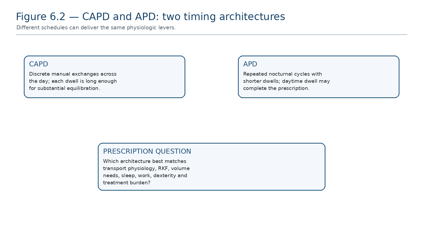
CAPD and APD use the same membrane and osmotic principles. The choice changes when and how often gradients are created, not the fundamental physics.
Table 6.5 — Starting architecture: CAPD versus APD.
| Question | CAPD may fit when… | APD may fit when… |
|---|---|---|
| Daily-life pattern | Daytime exchanges are acceptable | Night-time treatment better preserves daytime activity |
| Dwell physiology | Longer dwells likely to work well | Shorter cycles may better fit rapid transport or nocturnal schedule |
| Technology | Simple gravity system preferred | Cycler competence/support available |
| Sleep | Night free of machine preferred | Machine sleep burden acceptable |
| Assistance | Manual exchange support available | Caregiver/remote support favors cycler workflow |
| DO NOT AUTOMATE APD is not intrinsically “stronger” dialysis and CAPD is not an inferior fallback. Match timing architecture to transport, RKF, volume target and the person’s life. |
|---|
5. Fill volume: enough contact, not unnecessary pressure
Initial fill volume should be individualized. Larger volumes can recruit effective peritoneal surface area and increase per-exchange clearance, but also increase intraperitoneal pressure. Body habitus, comfort, hernia/leak risk, respiratory tolerance and a fresh catheter matter. A new urgent-start catheter is a different pressure environment from a healed chronic catheter.
Table 6.6 — Fill-volume reasoning.
| Finding | Interpretation | Action direction |
|---|---|---|
| Comfortable, healed access, larger body size | Potential to tolerate standard/larger fills | Titrate to clearance/volume need |
| Pain, dyspnoea, reflux, leak or hernia concern | Pressure burden may be excessive | Reduce fill and reassess cause |
| Urgent start / fresh catheter | Leak risk is amplified by pressure | Use lower volumes and recumbent position |
| Low clearance with small fills | Surface-area recruitment may be inadequate | Increase volume if anatomically tolerated |
6. Dwell time: decide what you want the dwell to accomplish
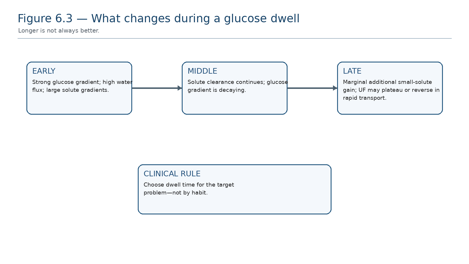
Dwell time is a trade between solute equilibration and preservation of osmotic force.
Table 6.7 — Dwell-time mismatch patterns.
| Pattern | Mechanism | Prescription implication |
|---|---|---|
| Short dwell + poor creatinine/urea equilibration | Insufficient time for diffusion | Lengthen dwell or increase exchange strategy when clinically needed |
| Long glucose dwell + poor UF in rapid transport | Glucose absorbed before drain | Shorter glucose dwell and/or long-dwell icodextrin strategy |
| Long dwell + good clearance + good UF | Appropriate match | Do not change because another patient uses a different schedule |
| APD many very short cycles + biochemical shortfall | Too little effective dwell time | Reassess cycle number, fill, total time and daytime component |
7. Exchange number and total dialysate volume
Increasing exchanges renews concentration gradients and usually increases peritoneal small-solute clearance. The correct starting number is therefore not a ritual four exchanges for every CAPD patient or a fixed cycler program for every APD patient. It is the number required to achieve current goals at acceptable burden, taking RKF into account.
Table 6.8 — Before adding exchanges, ask why.
| Problem | First question | Why blindly adding exchanges may fail |
|---|---|---|
| High urea/creatinine burden | Is delivered treatment complete and RKF falling? | Missed treatments or RKF loss may be the actual event |
| Edema | Is sodium/volume removal the dominant problem? | More low-osmotic exchanges may add clearance without enough net sodium/water removal |
| Poor APD clearance | Are dwells too short? | More cycles can make each dwell even shorter |
| High burden | Could a smaller incremental schedule meet goals? | Full-dose treatment may be unnecessary while RKF is substantial |
8. Osmotic prescription: water removal is a separate target
The osmotic component of the prescription should be designed from sodium and volume needs, not from small-solute clearance alone. Glucose concentration should be the minimum needed to achieve the intended UF pattern, alongside dietary sodium management, fluid intake assessment and diuretics when residual urine is responsive. Icodextrin is physiologically suited to a long dwell when sustained UF is needed. [2,3]
Table 6.9 — Osmotic strategy by clinical problem.
| Clinical problem | Physiologic direction | Trade-off |
|---|---|---|
| Euvolemic with useful urine | Avoid unnecessary hypertonicity | Preserves lower glucose exposure |
| Mild chronic positive balance | Review sodium/intake and diuretic response first | May avoid escalating glucose |
| Long-dwell UF failure from rapid glucose absorption | Consider sustained long-dwell osmotic strategy | Requires correct agent use and monitoring |
| Acute congestion | Increase water/sodium removal safely | Avoid chronic hypertonic rescue becoming permanent default |
9. Residual kidney function changes the starting dose
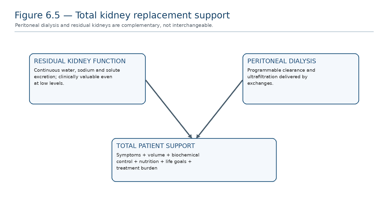
A person starting PD often receives substantial continuous clearance and sodium/water excretion from their own kidneys in addition to PD.
Residual kidney function is not simply an extra line in the adequacy calculation. It contributes continuous solute excretion, sodium and water removal and has a strong association with outcomes. Therefore a patient with substantial RKF may require less peritoneal dialysis initially than an otherwise similar anuric patient.
Table 6.10 — What RKF changes at the bedside.
| If RKF is… | Initial implication | What must be monitored |
|---|---|---|
| Substantial and stable | Incremental regimen may meet goals | Urine, residual clearance, symptoms, volume, labs |
| Falling | The same PD prescription becomes a smaller total therapy | Escalation trigger and repeat total assessment |
| Minimal/absent | PD carries nearly all clearance/volume responsibility | Fuller prescription; Kt/V and UF context become more important |
| Uncertain | Do not guess | Measure urine volume and residual clearance when feasible |
10. Incremental PD: start with what is needed now
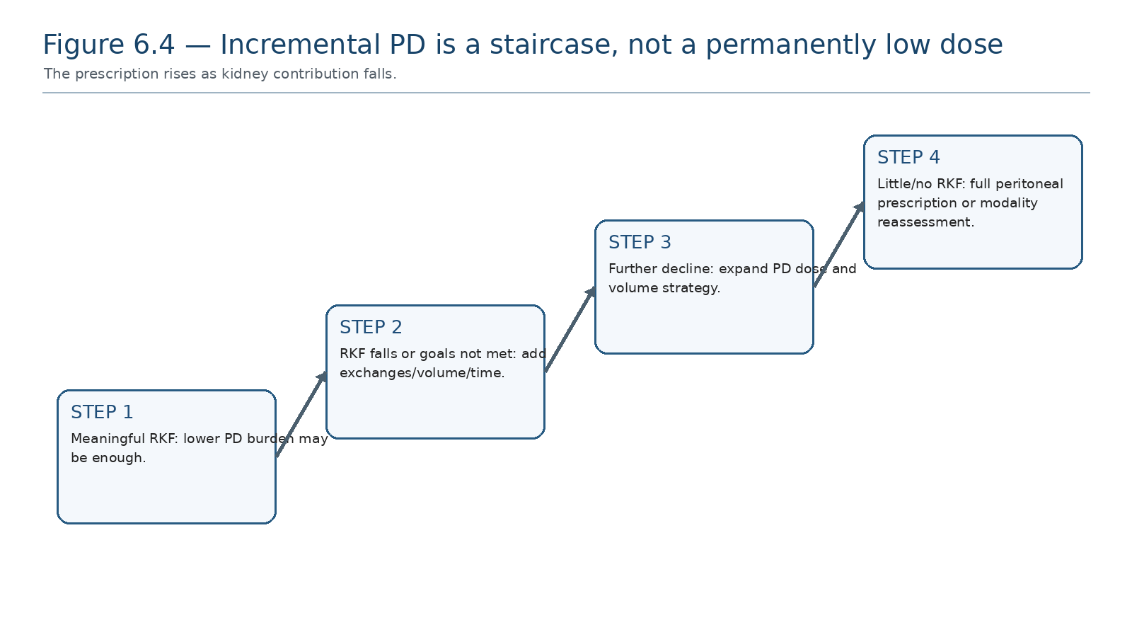
Incremental treatment requires planned escalation. Without monitoring, “incremental” becomes inadvertent underdialysis.
ISPD goal-directed prescribing supports incremental PD as a way to reduce treatment burden when residual kidney function and clinical state make a lower peritoneal dose sufficient. Evidence comparing incremental with standard-dose PD suggests broadly similar survival and technique outcomes in selected incident patients, but definitions vary and evidence for better RKF preservation is inconsistent. A balANZ analysis found similar slopes of RKF and urine decline with incremental and full-dose starts. [2,6,7]
Table 6.11 — Incremental PD is appropriate only if all four elements exist.
| Element | Requirement |
|---|---|
| Clinical suitability | No uncontrolled uremic, electrolyte, nutritional or volume indication requiring greater dose |
| Kidney contribution | Meaningful RKF/urine documented |
| Monitoring | Scheduled reassessment of urine, residual clearance and clinical goals |
| Escalation plan | Predefined triggers to add exchanges, days, volume or modality intensity |
| COMMON TRAP “Incremental” describes a monitored strategy. A low prescription without measurement and escalation is simply an unverified prescription. |
|---|
11. Small-solute clearance: useful metric, incomplete definition
The 2019 ISPD solute/fluid update concluded that available studies support weekly Kt/V urea of at least 1.7, especially in anuric adults, but explicitly graded this as a low-certainty practice point. The 2020 goal-directed recommendations emphasize that this number should not replace assessment of symptoms, volume, nutrition, biochemical control, RKF and treatment burden. [2,3]
Table 6.12 — How to use Kt/V without misusing it.
| Use it to… | Do not use it to… |
|---|---|
| Quantify small-solute removal | Declare the whole patient “adequately dialyzed” |
| Detect change after RKF loss or prescription change | Ignore congestion or symptoms because target is met |
| Support dose adjustment in anuric/low-RKF states | Force unnecessary exchanges in a clinically well person with significant RKF |
| Verify delivered rather than merely prescribed dialysis | Assume peritoneal and renal clearance have identical biological value |
| KEY NUMBER Weekly Kt/V urea ≥1.7 remains a commonly used small-solute practice point, especially for anuric adults; it is not a complete definition of high-quality PD. [3] |
|---|
12. Volume and sodium: prescribe the patient, not the drain bag
Table 6.13 — Initial volume assessment.
| Data | Question | Meaning |
|---|---|---|
| Weight trend | Is there a credible baseline/dry-weight trajectory? | Detects cumulative balance |
| Blood pressure | Is BP rising with congestion or driven by another mechanism? | Supports but does not prove volume excess |
| Edema / lungs / symptoms | Is there clinical congestion? | Patient-important target |
| Net PD UF | What does the peritoneum remove? | Only one part of total output |
| Urine volume | What do kidneys still remove? | Critical early in PD |
| Sodium/fluid intake | Is input overwhelming a reasonable prescription? | Prevents glucose-only escalation |
13. Traditional start versus urgent-start PD
A conventional break-in period after catheter insertion reduces mechanical stress on the new access. Urgent-start PD generally refers to commencing within 14 days of catheter placement. SAGES 2023/2024 suggests traditional start over urgent start in adults when clinically feasible, based on very low-certainty evidence; however, urgent-start PD remains an important strategy when dialysis cannot wait and avoidance of a temporary HD catheter is desirable. [4,5,8]
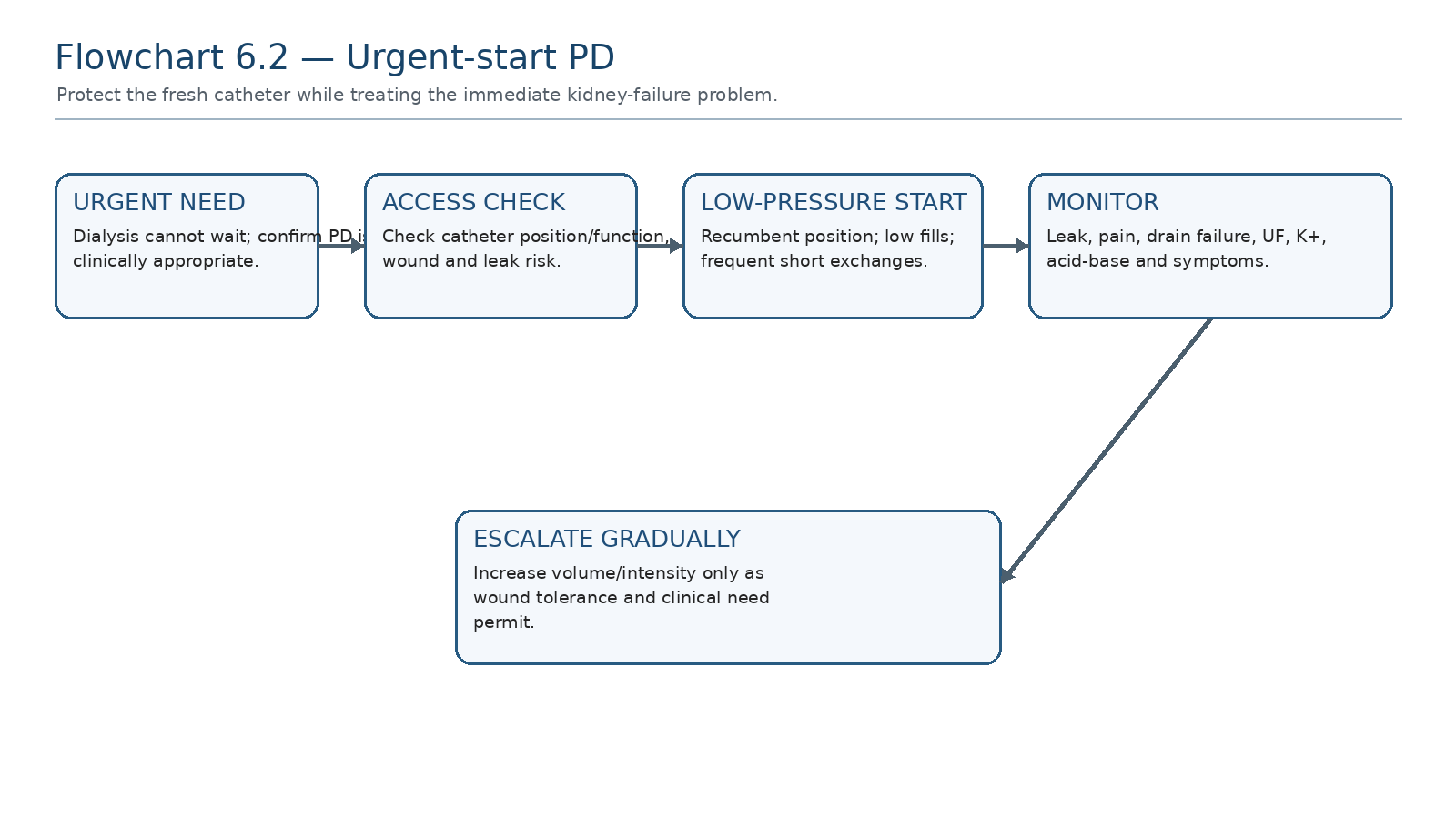
A fresh catheter should be treated as a vulnerable wound-access system. Lower fill volumes and recumbent exchanges reduce intraperitoneal pressure while dialysis intensity is built according to clinical need.
Table 6.14 — Urgent-start priorities.
| Priority | Bedside principle |
|---|---|
| Patient safety | Do not force PD if severe instability or an indication requires a faster/more controllable modality |
| Catheter protection | Recumbent position and low fill volumes initially |
| Clearance | Use repeated exchanges according to biochemical/uremic need |
| Volume | Select osmotic strength according to congestion while limiting pressure burden |
| Leak surveillance | Inspect wound and drain pattern; abdominal wall/genital swelling may indicate leak |
| Escalation | Increase fill/intensity as catheter/wound tolerance permits under local protocol |
| SAFETY BOUNDARY Exact urgent-start fill volumes, exchange schedules and escalation rates are protocol- and catheter-specific. This chapter teaches the low-pressure physiologic strategy; the treating unit’s urgent-start pathway and procedural team govern the exact regimen. |
|---|
14. The elective start pathway
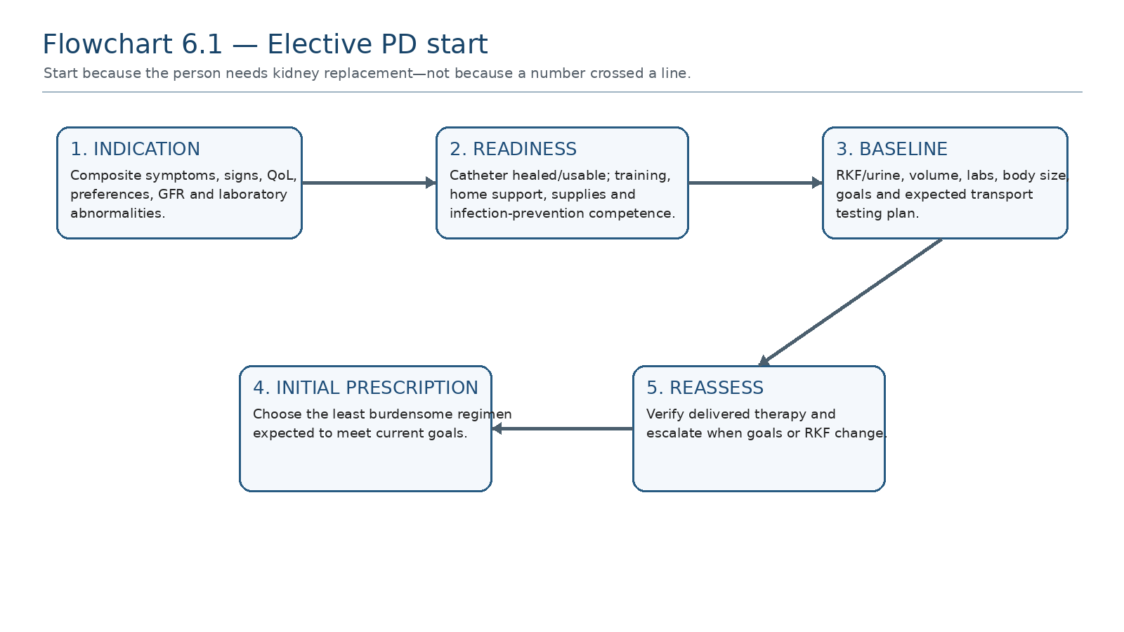
A safe elective start links the clinical indication, healed/functional access, training readiness, baseline RKF/volume assessment, initial dose and an early verification plan.
Table 6.15 — First-prescription sign-off checklist.
| Check | Pass criterion |
|---|---|
| Indication | Reason for dialysis start documented |
| Access | Catheter usable; wound status and break-in pathway known |
| Technique | Patient/caregiver demonstrates safe exchange/cycler workflow |
| RKF | Urine and residual contribution documented or measurement planned |
| Solute target | Clinical/laboratory goal and clearance measurement plan documented |
| Volume target | Weight/BP/UF/urine strategy documented |
| Burden | Regimen compatible with life goals where possible |
| Review | Early reassessment date and escalation triggers named |
15. The first days and weeks: verify what was delivered
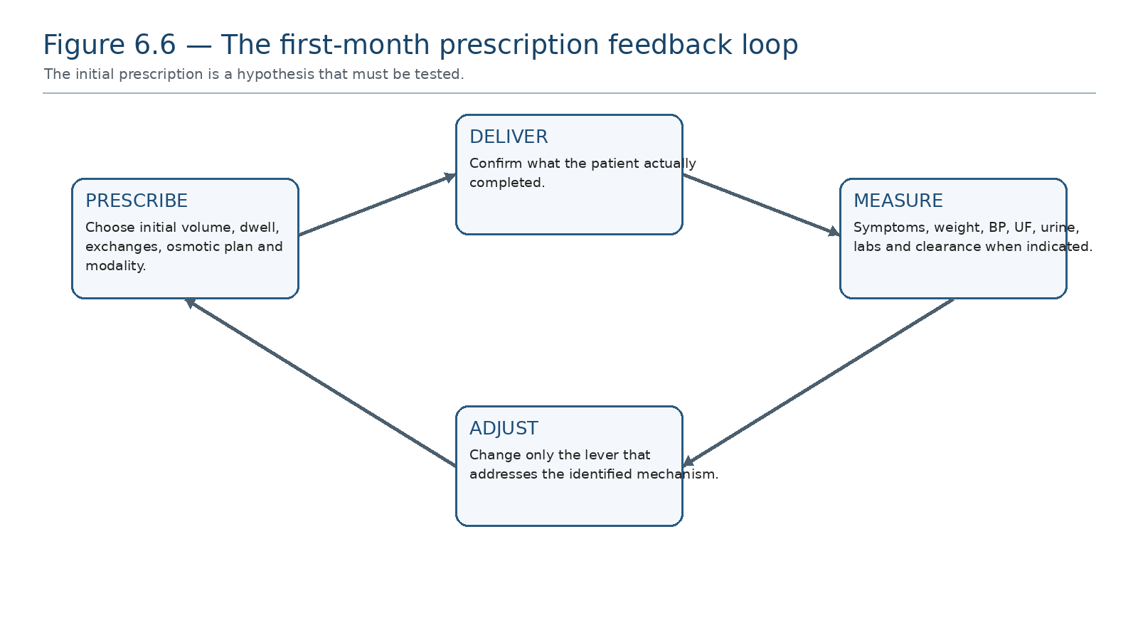
The initial prescription is a testable hypothesis. Delivered treatment and patient response must close the loop.
Table 6.16 — Early follow-up data that change the prescription.
| Finding | What it may mean | Next action |
|---|---|---|
| Missed exchanges / cycler bypass | Delivered dose lower than ordered | Solve workflow, alarm, burden or training issue before increasing prescription |
| Persistent edema | Insufficient total sodium/water removal or high intake | Assess urine, UF, dwell/osmotic strategy and intake |
| Weight falling + dizziness | Excess fluid removal / low intake | Reduce osmotic intensity; reassess target weight |
| Hyperkalaemia/acidosis persists | Insufficient total clearance or ongoing catabolic problem | Verify delivery/RKF and adjust dose as needed |
| Good clinical control + high burden | Possible opportunity for simplification if RKF/targets permit | Reassess incremental strategy |
16. Clinical problem framework: “the initial prescription is not working”
Table 6.17 — Name the phenotype before changing therapy.
| Phenotype | Think first | Typical next data |
|---|---|---|
| Uremic/biochemical | Delivered dose, RKF, dwell/volume, catabolism | Treatment log, urine, clearance, labs |
| Volume overloaded | Sodium/input, urine, UF, glucose-dwell match | Weight/BP, intake, urine, UF records |
| Too dry/hypotensive | Excess osmotic removal, low intake, diuretics | Weight, orthostasis, urine, UF |
| Pain/leak | Fill pressure, catheter/wound, hernia | Exam, volume relationship, imaging if needed |
| Drain alarms | Constipation, catheter position, kink, fibrin | Mechanical troubleshooting |
| Excess burden | Over-prescription, poor timing fit, cycler issues | Goal review + delivered-data review |
17. Diagnostic reasoning framework
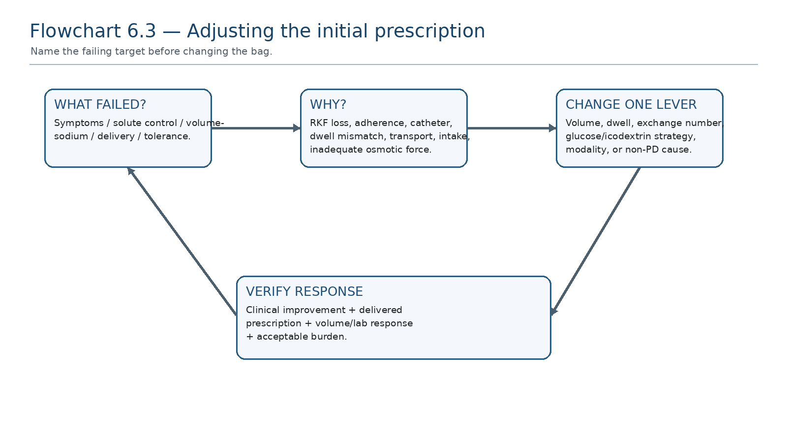
Start with the failing clinical target, identify the responsible mechanism, change the relevant lever and verify the response.
Table 6.18 — Clinical question → data → interpretation → action.
| Clinical question | Data required | Interpretation | Action |
|---|---|---|---|
| Why is clearance poor? | Delivered therapy, RKF, dwell/volume, catheter | Dose vs delivery vs kidney loss vs mechanical issue | Correct mechanism; then increase dose if required |
| Why is volume poor? | Intake, urine, UF, dwell/agent, transport | Input/RKF/osmotic mismatch | Sodium strategy + osmotic/dwell adjustment |
| Why is patient intolerant? | Fill symptoms, pressure-related features, schedule burden | Volume/geometry vs lifestyle problem | Reduce fill/change schedule/modality architecture |
| Why did status change suddenly? | Adherence, catheter, constipation, intercurrent illness | Acute delivery/mechanical/clinical event | Do not assume gradual membrane failure |
18. Management framework: change the lever that matches the mechanism
Table 6.19 — Problem → mechanism → change → reassess.
| Problem | Likely mechanism | Prescription/management direction | Reassess |
|---|---|---|---|
| Low small-solute clearance | Insufficient dialysate-time/volume or RKF loss | Increase effective volume, dwell or exchanges after verifying delivery | Symptoms, labs, clearance |
| Poor long-dwell UF | Rapid glucose absorption | Shorten glucose dwell / appropriate long-dwell icodextrin | UF, weight, BP |
| General volume overload | Positive sodium/water balance | Diet/diuretic strategy + osmotic prescription | Weight, edema, BP, urine, UF |
| High treatment burden | More PD than current needs or schedule mismatch | Simplify/incremental strategy if goals remain met | Clinical control + RKF |
| Leak/pain after start | Excess intraperitoneal pressure or access issue | Reduce fill/hold-escalation; evaluate access | Symptoms, wound, drainage |
19. Retention tables
Table 6.20 — Prescription lever fingerprints.
| If you change… | You mainly influence… | Remember… |
|---|---|---|
| Fill volume | Per-exchange surface area/clearance and pressure | Pressure cost |
| Dwell time | Equilibration vs osmotic decay | Longer is not always better |
| Exchange number | Gradient renewal and total clearance | Connection/burden cost |
| Glucose strength | Crystalloid UF | Metabolic/membrane exposure |
| Long-dwell icodextrin | Sustained long-dwell UF | Different osmotic mechanism |
| CAPD↔︎APD timing | Distribution of dwell lengths | Same physiology, different schedule |
Table 6.21 — What not to confuse.
| Do not confuse | With | Correction |
|---|---|---|
| eGFR level | Dialysis indication | Use composite clinical assessment |
| Prescribed volume | Delivered dialysis | Check logs/technique |
| Peritoneal UF | Total fluid removal | Include urine |
| Kt/V target | Whole-person adequacy | Assess symptoms/volume/nutrition/goals |
| Incremental PD | Underdialysis | Requires monitoring + escalation |
| Urgent start | Full chronic prescription immediately | Use low-pressure protected start |
20. Clinical pearls
The first prescription should have a reason for every exchange.
An unchanged PD prescription becomes a smaller total therapy when residual kidney function falls.
If the problem is edema, increasing urea clearance may not solve it.
If APD clearance is poor, adding more cycles can paradoxically shorten dwells further.
Use the least glucose exposure that safely achieves volume goals.
A fresh catheter converts fill volume into a wound-pressure decision.
Incremental PD is safest when the escalation trigger is written on day one.
The patient who cannot live with the prescription is not receiving high-quality dialysis even if the machine reports perfect delivery.
21. Common pitfalls—and the correction
Table 6.22 — High-frequency errors at PD initiation.
| Pitfall | Why it fails | Correction |
|---|---|---|
| Starting because eGFR crossed a number | Ignores symptoms, goals and treatable alternatives | Use composite KDIGO assessment |
| Giving every patient a full-dose template | Ignores RKF and burden | Individualize; consider incremental strategy |
| Treating edema by glucose escalation only | May ignore sodium intake/RKF/diuretics | Assess total sodium-water balance |
| Assuming ordered = delivered | Misses alarms, missed exchanges and technique problems | Review treatment logs |
| Using Kt/V as the only success endpoint | Misses congestion, symptoms and QoL | Goal-directed review |
| Urgent-start full-volume ambulatory fills | Raises intraperitoneal pressure and leak risk | Low-volume recumbent start under protocol |
| No escalation plan for incremental PD | Creates silent underdialysis as RKF falls | Schedule RKF/clinical reassessment |
| Changing several variables at once | You cannot learn which mechanism mattered | Change targeted lever when clinically safe |
22. Mini-cases: prescription decisions, not arithmetic
Case 1 — Symptomatic kidney failure with substantial urine
A 59-year-old with progressive CKD has anorexia, fatigue and refractory acidosis but still passes 1.6 L urine/day. Catheter is healed and training complete.
| REASONING Start PD because clinical indications are present, document RKF, and consider an incremental starting regimen that meets current biochemical/volume goals with a written escalation plan rather than reflex full-dose therapy. |
|---|
Case 2 — Low eGFR, clinically well
A 48-year-old has eGFR 7 mL/min/1.73 m², good appetite, controlled potassium/bicarbonate and no congestion. PD catheter is ready.
| REASONING Do not start solely because eGFR is 7. Continue close monitoring and start when composite clinical indications emerge, respecting patient preferences and readiness. |
|---|
Case 3 — APD with edema but acceptable clearance
A new APD patient meets the small-solute target but gains weight, has ankle edema and little net daytime UF.
| REASONING This is primarily a volume/sodium problem. Review sodium intake, residual urine/diuretics, dwell physiology and long-dwell osmotic strategy instead of simply adding cycles to raise Kt/V. |
|---|
Case 4 — Incremental PD after urine falls
A patient started with fewer CAPD exchanges because RKF was substantial. Six months later urine has halved and phosphate, urea symptoms and edema are worsening.
| REASONING The incremental plan has reached its escalation trigger. Re-measure total support and increase PD intensity according to the failing targets; do not call the original low prescription “stable.” |
|---|
Case 5 — Urgent start after catheter insertion
A patient needs dialysis within 48 hours of catheter placement because of worsening uremic symptoms and volume overload but is hemodynamically stable.
| REASONING If the local urgent-start pathway supports PD, use a protected low-pressure recumbent low-fill strategy with frequent reassessment for leak and drainage; exact volumes/schedule follow local protocol. |
|---|
Case 6 — The prescription looks adequate but the patient skips therapy
A cycler download shows repeated bypassed drains and shortened nights because alarms disturb sleep. Serum indices are worsening.
| REASONING This is delivered-dose failure. Fix catheter/constipation/alarm workflow and treatment burden before simply increasing the programmed dose. |
|---|
23. Active recall
MUST MEMORIZE
Q: What determines when dialysis should start? A: Composite symptoms, signs, QoL, preferences, GFR and laboratory abnormalities—not eGFR alone.
Q: What are the five main PD prescription levers? A: Fill volume, dwell time, exchange number, osmotic agent/strength and timing modality.
Q: Why does RKF permit a smaller initial PD dose in selected patients? A: The kidneys still provide continuous solute, sodium and water removal.
Q: What weekly Kt/V urea practice point remains commonly used? A: At least 1.7, especially in anuric adults, with low-certainty evidence.
Q: What makes incremental PD safe? A: Documented RKF, adequate current clinical control, scheduled monitoring and an escalation plan.
Q: What is the core urgent-start principle? A: Minimise intraperitoneal pressure while delivering enough dialysis—typically recumbent low-volume exchanges under protocol.
MUST REASON
Q: Why can adding APD cycles reduce efficiency? A: More cycles can shorten each dwell, limiting equilibration.
Q: Why can good Kt/V coexist with edema? A: Small-solute clearance and sodium/water balance are different targets.
Q: Why is an unchanged prescription not an unchanged treatment? A: RKF, body state, adherence and membrane physiology can change.
Q: Why should a long glucose dwell sometimes be shortened? A: Rapid glucose absorption may dissipate the osmotic gradient before drain.
Q: Why should you verify delivery before increasing dose? A: A larger prescription cannot correct missed/bypassed therapy or a mechanical drain problem.
USE AS REFERENCE
Exact local starting templates, cycler programming, urgent-start fill-volume schedules, glucose strengths, clearance collection methods and medication/diuretic dosing should be taken from current unit protocols and manufacturer instructions rather than memorized from a generic chapter.
24. Flashcards
1. Q: Dialysis-start trigger? A: Clinical composite, not eGFR alone.
2. Q: Most valuable “invisible” part of an incident PD prescription? A: Residual kidney function.
3. Q: Incremental PD without reassessment becomes? A: Potential underdialysis.
4. Q: CAPD vs APD changes what? A: Timing architecture of the same transport levers.
5. Q: More exchanges do what? A: Renew concentration gradients and usually increase small-solute clearance.
6. Q: Longer glucose dwell always better? A: No—osmotic force can dissipate.
7. Q: Net PD UF equals total fluid output? A: No—include urine.
8. Q: Kt/V is mainly a measure of? A: Small-solute urea clearance.
9. Q: Why use low fills in urgent start? A: Reduce intraperitoneal pressure and leak risk.
10. Q: First action when prescribed dose seems inadequate? A: Confirm delivered treatment and RKF.
11. Q: Volume overload + good Kt/V means? A: Treat sodium/water problem, not the number.
12. Q: Good initial prescription endpoint? A: Goals met with acceptable burden and a clear reassessment plan.
13. Q: What does stronger glucose buy? A: More crystalloid osmotic force at greater glucose exposure.
14. Q: Icodextrin is primarily useful for? A: Sustained long-dwell ultrafiltration.
15. Q: What ends an incremental phase? A: Failure of clinical/clearance/volume goals or sufficient RKF decline.
16. Q: What should every prescription change include? A: A reason and a reassessment endpoint.
17. Q: Urgent-start definition commonly used? A: PD begun within 14 days of catheter insertion.
18. Q: First-month mindset? A: Prescribe → deliver → measure → adjust.
25. Rapid differential / troubleshooting table
Table 6.23 — When the new PD prescription disappoints.
| If you see… | Think… | Do now… |
|---|---|---|
| Persistent uremic symptoms | Underdelivery, low dose, RKF loss, catabolism | Verify delivery/RKF; assess clearance and clinical cause |
| Edema + good small-solute indices | Sodium/water mismatch | Review intake, urine, UF and dwell/osmotic plan |
| Low drain volumes | Mechanical issue or low UF | Distinguish poor drainage from true UF failure |
| Pain/leak after fill | Excess pressure / fresh access issue | Reduce pressure exposure and assess catheter/wound |
| Cycler alarms + lost dwell time | Delivered prescription failure | Solve alarm mechanism; inspect constipation/catheter |
| Dizziness + large UF | Excess fluid removal | Reduce osmotic intensity and reassess target weight |
| Stable labs but overwhelming burden | Potential over-prescription / poor schedule fit | Revisit incremental or modality architecture |
26. Final revision sheet
| TEN TAKE-HOME RULES 1) Start dialysis for clinical need, not eGFR alone. 2) Measure what the kidneys still contribute. 3) Name the target before choosing the lever. 4) CAPD and APD are timing architectures, not different physiologies. 5) Fill volume trades clearance against pressure. 6) Dwell time trades equilibration against osmotic decay. 7) Incremental PD requires monitoring and escalation. 8) Kt/V is a small-solute metric, not the definition of adequate care. 9) Urgent-start PD protects the fresh catheter with a low-pressure strategy. 10) The initial prescription is a hypothesis—verify delivery and patient response. |
|---|
Table 6.24 — One-minute bedside synthesis.
| If you see… | Think… | Do now… |
|---|---|---|
| Symptoms despite apparently adequate order | Delivered dose/RKF change | Check logs, catheter, urine and labs |
| Weight gain/edema | Positive sodium-water balance | Assess intake + urine + UF + osmotic plan |
| High burden with strong RKF | Possible incremental opportunity | Confirm goals then simplify cautiously |
| Falling urine | Total therapy is shrinking | Reassess and escalate |
| Fresh catheter + urgent dialysis need | Leak-pressure risk | Low-volume recumbent urgent-start pathway |
| Good Kt/V, poor QoL | Adequacy is multidimensional | Revisit goals and prescription burden |
| FINAL MENTAL MODEL Need → readiness → residual kidney contribution → modality → fill/dwell/exchanges/osmotic plan → deliver → measure symptoms/volume/labs/urine → adjust → escalate as RKF falls. |
|---|
Rapid oral viva
Explain why dialysis initiation cannot be defined by one eGFR threshold.
Build an initial PD prescription for a patient with substantial RKF and mild uremic symptoms.
A new APD patient is edematous but has adequate Kt/V. Talk through your next decisions.
Explain incremental PD and its mandatory safety conditions.
Walk through an urgent-start PD strategy after catheter placement.
A patient’s urine output falls by half without a prescription change. Explain why the total treatment has changed.
| SCOPE BOUNDARY This chapter establishes how to start PD and construct the first prescription. Formal adequacy calculations and RKF quantification are developed in Chapter 7; detailed volume/sodium and ultrafiltration-failure management in Chapter 8; CAPD/APD optimization in Chapter 9; urgent-start operational scenarios in Chapter 17. |
|---|
27. Selected authoritative references
1. Kidney Disease: Improving Global Outcomes (KDIGO) CKD Work Group. KDIGO 2024 Clinical Practice Guideline for the Evaluation and Management of Chronic Kidney Disease. Kidney Int. 2024;105(4S):S117–S314. https://doi.org/10.1016/j.kint.2023.10.018.
2. Brown EA, Blake PG, Boudville N, et al. International Society for Peritoneal Dialysis practice recommendations: Prescribing high-quality goal-directed peritoneal dialysis. Perit Dial Int. 2020;40(3):244–253. https://doi.org/10.1177/0896860819895364.
3. Boudville N, de Moraes TP. 2005 Guidelines on targets for solute and fluid removal in adults being treated with chronic peritoneal dialysis: 2019 update of the literature and revision of recommendations. Perit Dial Int. 2020;40(3):254–260. https://doi.org/10.1177/0896860819898307.
4. Crabtree JH, Shrestha BM, Chow KM, et al. Creating and maintaining optimal peritoneal dialysis access in the adult patient: 2019 update. Perit Dial Int. 2019;39(5):414–436. https://doi.org/10.3747/pdi.2018.00232.
5. Haggerty S, Roth S, Walsh D, et al. Peritoneal Dialysis Access Guideline Update 2023. Surg Endosc. 2024. SAGES guideline publication.
6. Xu S, Wu W, Cheng J. Comparison of outcomes of incremental vs. standard peritoneal dialysis: a systematic review and meta-analysis. BMC Nephrol. 2024;25:308. https://doi.org/10.1186/s12882-024-03669-w. PMID: 39285336.
7. Hayat A, Cho Y, Hawley CM, et al. Association of incremental peritoneal dialysis with residual kidney function decline in patients on peritoneal dialysis: the balANZ trial. Perit Dial Int. 2023;43(5):374–382. https://doi.org/10.1177/08968608231175826. PMID: 37259236.
8. Htay H, Johnson DW, Craig JC, et al. Urgent-start peritoneal dialysis versus haemodialysis for people with chronic kidney disease. Cochrane Database Syst Rev. 2021;1:CD012899.
9. Vogt B, Shah AD. Urgent-Start Peritoneal Dialysis: Current State and Future Directions. Kidney Dial. 2024;4(1):15–26. https://doi.org/10.3390/kidneydial4010002.
10. Morelle J, Stachowska-Pietka J, Öberg C, Gadola L, La Milia V, Yu Z, et al. ISPD recommendations for the evaluation of peritoneal membrane dysfunction in adults: classification, measurement, interpretation and rationale for intervention. Perit Dial Int. 2021;41(4):352–72. https://doi.org/10.1177/0896860820982218
11. Teitelbaum I, Glickman J, Neu A, et al. KDOQI US Commentary on the 2020 ISPD Practice Recommendations for Prescribing High-Quality Goal-Directed Peritoneal Dialysis. Am J Kidney Dis. 2021;77(2):157–171. https://doi.org/10.1053/j.ajkd.2020.09.010. PMID: 33341315.
12. Bargman JM, Thorpe KE, Churchill DN. Relative contribution of residual renal function and peritoneal clearance to adequacy of dialysis: a reanalysis of the CANUSA study. J Am Soc Nephrol. 2001;12(10):2158–2162. https://doi.org/10.1681/ASN.V12102158.
13. Paniagua R, Amato D, Vonesh E, et al. Effects of increased peritoneal clearances on mortality rates in peritoneal dialysis: ADEMEX, a prospective, randomized, controlled trial. J Am Soc Nephrol. 2002;13(5):1307–1320. https://doi.org/10.1681/ASN.V1351307.
14. Davies SJ. Peritoneal dialysis—current status and future challenges. Nat Rev Nephrol. 2013;9:399–408. https://doi.org/10.1038/nrneph.2013.100.
15. Perl J, Davies SJ, Lambie M, et al. The Peritoneal Dialysis Outcomes and Practice Patterns Study (PDOPPS): unifying efforts to inform practice and improve global outcomes in peritoneal dialysis. Perit Dial Int. 2016;36(3):297–307. https://doi.org/10.3747/pdi.2014.00288.
16. Li PKT, Chow KM, Van de Luijtgaarden MWM, et al. Changes in the worldwide epidemiology of peritoneal dialysis. Nat Rev Nephrol. 2017;13:90–103. https://doi.org/10.1038/nrneph.2016.181.
17. Ronco C, Crepaldi C, Brendolan A, et al. Peritoneal dialysis: from basic concepts to clinical excellence. Contrib Nephrol. Basel: Karger; selected foundational physiology chapters.
18. Blake PG, Bargman JM. Peritoneal dialysis prescription and adequacy. In: Brenner & Rector’s The Kidney. 12th ed. Elsevier; 2024.
| SOURCE NOTE Guideline status and key contemporary evidence were checked 2 September 2026. Exact cycler programs, glucose concentrations, urgent-start fill-volume schedules, break-in instructions, diuretic doses and clearance-collection procedures remain governed by current local protocols, catheter/device instructions and individual clinical assessment. |
|---|