Goals | Kt/V | Collection | Residual Kidneys | Preservation | Troubleshooting
| CHAPTER MISSION Replace “adequacy equals Kt/V” with a bedside adequacy system: assess the patient first, measure small-solute clearance correctly, separate peritoneal from residual kidney contributions, recognise why residual kidney function has disproportionate clinical value, preserve it deliberately, and troubleshoot an apparent clearance failure before simply adding more dialysis. |
|---|
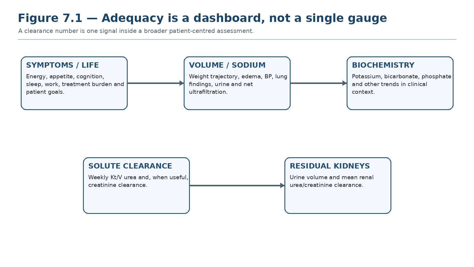
High-quality PD integrates symptoms and life goals, sodium/volume balance, biochemical control, measured small-solute clearance and residual kidney function. No single metric can substitute for the whole dashboard.
| MASTER PRINCIPLE Weekly Kt/V urea is a useful small-solute accounting tool. It is not a patient outcome, not a measure of sodium balance, and not proof that the person is clinically well. Treat the patient, preserve the kidneys, and use clearance measurements to explain or verify the clinical picture. |
|---|
0. One-page chapter map
Table 7.1 — The eight decisions that govern PD adequacy.
| Decision | Core question | Bedside output |
|---|---|---|
| 1. Patient | Are symptoms, function, nutrition and life goals acceptable? | Clinical adequacy statement |
| 2. Volume | Is sodium/water balance controlled? | Euvolemia plan |
| 3. Delivery | Was the prescription actually delivered? | Delivered-dose verification |
| 4. Kidney contribution | How much urine and renal clearance remain? | RKF measurement + trend |
| 5. Small solutes | What do weekly Kt/V and related clearance measures show? | Clearance interpretation |
| 6. Cause | If a target fails, is it measurement, delivery, RKF loss, membrane, access or prescription? | Named mechanism |
| 7. Correct | Which prescription lever addresses that mechanism? | Targeted change |
| 8. Verify | Did the patient and the relevant metric improve? | Reassessment + next trigger |
Learning outcomes
Define high-quality PD adequacy as a multidimensional clinical state rather than a single clearance target.
Explain what weekly Kt/V urea measures, how its peritoneal and renal components are obtained, and where the metric can mislead.
Apply the ISPD evidence position that higher peritoneal small-solute clearance above conventional targets has not shown survival benefit.
Measure residual kidney function using timed urine and the mean of renal urea and creatinine clearances.
Explain why residual kidney function and urine volume carry clinical value beyond their numerical contribution to total Kt/V.
Distinguish a true small-solute shortfall from collection error, nonadherence, catheter/drain dysfunction, loss of RKF and volume-driven symptoms.
Use a mechanism-based strategy to preserve residual kidney function while avoiding both chronic fluid overload and iatrogenic volume depletion.
Adjust incremental PD as residual kidney function changes without treating a single Kt/V value as the whole prescription.
| EVIDENCE POSTURE ISPD 2020 reframes adequacy as high-quality, goal-directed PD. The 2019 solute/fluid update found poor-quality evidence for small-solute targets, no survival advantage from pushing weekly Kt/V above 1.7, and only weak evidence supporting a weekly Kt/V of at least 1.7 in anuric adults. CANUSA reanalysis showed strong outcome associations with residual GFR and urine volume but not equivalent peritoneal clearance; ADEMEX and the Hong Kong randomized trial showed that higher prescribed peritoneal small-solute clearance did not improve survival. [1–7] |
|---|
1. Adequacy has changed meaning
Older PD practice often reduced adequacy to whether a calculated small-solute threshold was reached. Contemporary ISPD practice deliberately broadens the question: is the treatment achieving the person’s clinical and life goals with acceptable symptoms, volume status, nutrition, biochemical control, residual kidney function and treatment burden? Weekly Kt/V remains useful, but only as one element of that assessment. [1–3]
Table 7.2 — Five domains of adequate PD.
| Domain | What success looks like | What a Kt/V cannot tell you |
|---|---|---|
| Symptoms / function | Acceptable energy, appetite, cognition, sleep and daily function | Whether the person feels well |
| Volume / sodium | Stable weight trend, acceptable BP, no clinically important congestion | Whether sodium balance is controlled |
| Biochemistry | Acceptable potassium, bicarbonate and other relevant trends | Whether every metabolic target is controlled |
| Nutrition | No unexplained progressive nutritional deterioration | Cause of low albumin or poor intake |
| Life goals / burden | Treatment fits agreed priorities with tolerable workload | Whether the prescription is worth its burden |
| BEDSIDE TRANSLATION Document adequacy as a sentence, not a number: “Clinically well with controlled volume and biochemistry, meaningful RKF, and small-solute clearance consistent with current goals” is more useful than “Kt/V 1.82.” |
|---|
2. Kt/V urea: useful, specific and limited
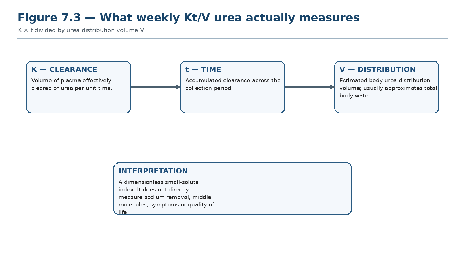
Kt/V is a dimensionless index of urea clearance scaled to urea distribution volume. It is deliberately a small-solute metric; it does not directly measure sodium balance, middle-molecule removal or quality of life.
Kt/V expresses urea clearance (K) accumulated over time (t) relative to the urea distribution volume (V), usually approximated by total body water. In PD, the weekly value usually includes a peritoneal component and, when urine is present, a renal component. The arithmetic is convenient; the biology is more complex.
Table 7.3 — What Kt/V captures and what it misses.
| Captures reasonably | Captures indirectly/poorly | Does not define |
|---|---|---|
| Urea removal | Overall nitrogenous-solute exposure | Volume status |
| Change in small-solute dose | Some consequence of missed treatments | Sodium balance |
| Renal + peritoneal urea-clearance accounting | Body-size effect through V | Middle molecules / protein-bound solutes |
| Longitudinal trend when collection is valid | Some prescription response | Symptoms, nutrition or quality of life |
| THRESHOLD DISCIPLINE The ISPD 2019 update concluded that there is no demonstrated survival advantage in targeting weekly Kt/V above 1.70. The lower limit remains uncertain; evidence supporting ≥1.7 in anuric adults is weak. Use 1.7 as a decision-support threshold—not as a claim that 1.69 is treatment failure and 1.71 proves success. [2] |
|---|
3. Why “more Kt/V” stopped being the goal
Two landmark randomized experiences changed PD adequacy thinking. In ADEMEX, increasing peritoneal urea/creatinine clearance did not improve survival. In the Hong Kong randomized study, higher Kt/V targets did not produce a clear survival advantage over lower target ranges, although very low clearance was clinically undesirable. These data weaken the assumption that every extra increment of peritoneal urea clearance improves patient outcome. [5,6]
Table 7.4 — Landmark evidence that reshaped adequacy.
| Study | Key comparison | Clinical lesson |
|---|---|---|
| CANUSA reanalysis | Residual GFR and urine output vs peritoneal clearance | Renal and peritoneal clearance are not outcome-equivalent |
| ADEMEX | Higher peritoneal clearance vs conventional CAPD | Increasing peritoneal small-solute dose did not improve survival |
| Hong Kong randomized Kt/V study | Three total Kt/V target ranges | Higher targets did not clearly improve survival; avoid interpreting “more” as automatically better |
| DO NOT OVER-DIALYZE THE NUMBER If the patient is clinically well, euvolemic and biochemically controlled, increasing exchanges solely to move Kt/V farther above an already acceptable value can add glucose exposure, connections and burden without proven survival benefit. |
|---|
4. Total clearance: additive arithmetic, unequal biology
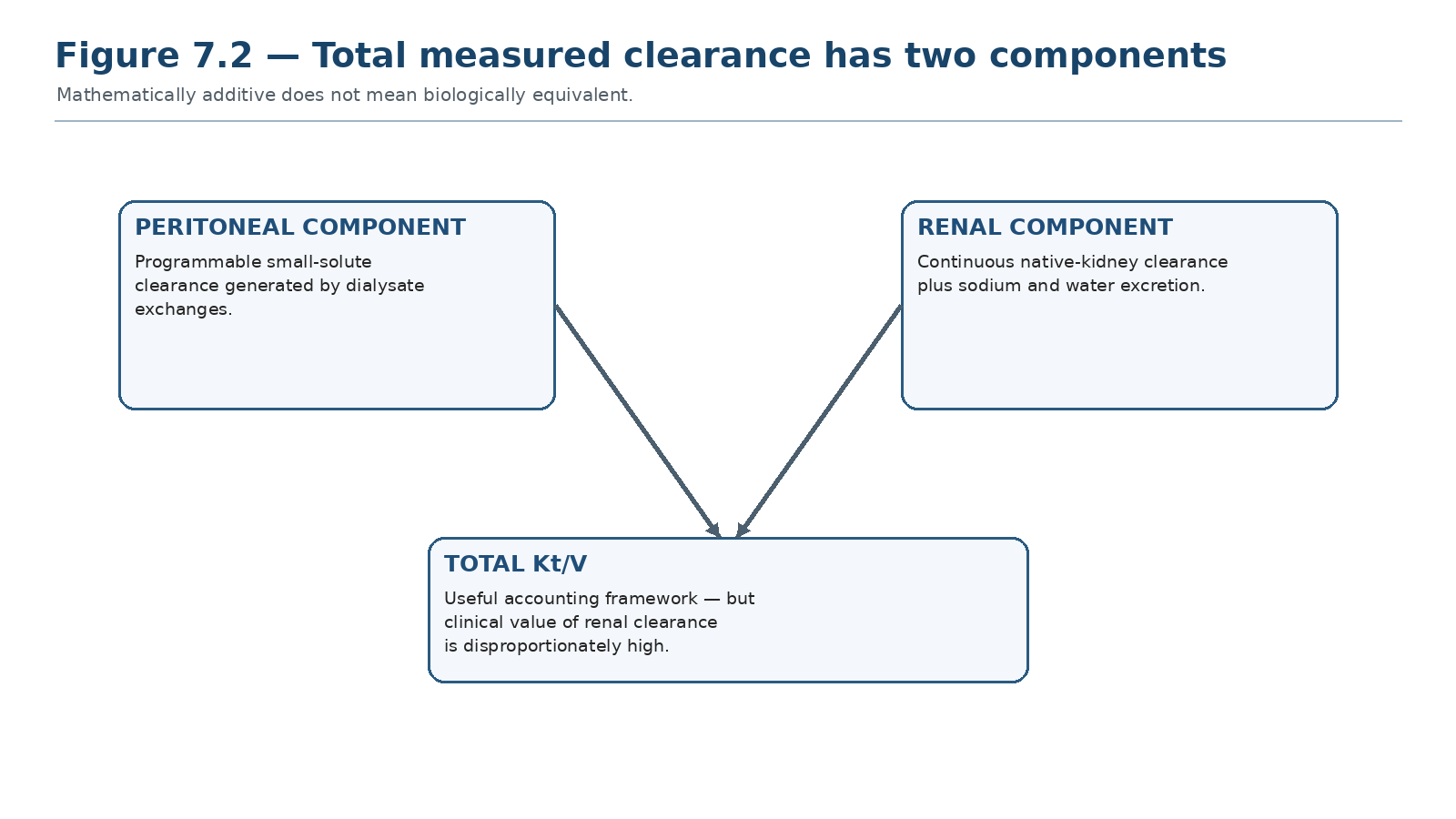
Renal and peritoneal urea clearances can be added mathematically. CANUSA reanalysis demonstrated that their clinical associations are not equivalent.
The traditional total Kt/V is calculated by adding peritoneal and renal urea-clearance components. That arithmetic is valid for urea accounting, but it must not imply biological equivalence. Residual kidneys provide continuous water, sodium and a broader spectrum of solute clearance plus endocrine and metabolic functions. [4,7]
Table 7.5 — Residual kidney clearance versus peritoneal clearance.
| Feature | Residual kidneys | Peritoneal dialysis |
|---|---|---|
| Timing | Continuous native function | Programmed exchanges/cycles |
| Water/sodium | Physiologic urine excretion | Osmotic ultrafiltration + dialysate sodium removal |
| Solute spectrum | Broad native filtration/secretion | Membrane- and dwell-dependent |
| Treatment burden | No additional exchange burden | Requires connections, dialysate and time |
| Outcome association | Consistently strong observational association | Higher small-solute dose above conventional range has not shown survival benefit |
5. How to measure peritoneal small-solute clearance
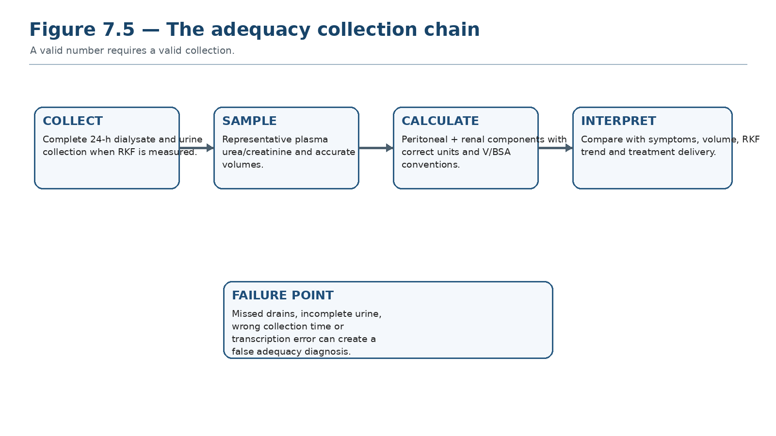
Collection integrity is part of the test. A mathematically precise calculation from an incomplete collection is still wrong.
Peritoneal clearance assessment requires complete collection of drained dialysate over the defined period, accurate total drain volume, representative dialysate sampling, and an appropriate plasma urea measurement. Automated-PD collections must account for the complete cycler and daytime prescription. A missed bag or unrecorded drain can create a false low result.
Table 7.6 — Peritoneal clearance collection checklist.
| Check | Why it matters | Failure phenotype |
|---|---|---|
| All dialysate captured | Defines total solute mass removed | Low measured clearance if drains omitted |
| Accurate total drain volume | Volume is part of clearance calculation | Dose error from guessed volumes |
| Representative mixed sample | Concentration must represent all effluent | Biased D/P concentration |
| Correct collection duration | Converts clearance to weekly value | Wrong time scaling |
| Plasma sample appropriately timed | Provides denominator concentration | Spurious result when clinical state changed markedly |
| Prescription actually delivered | Measurement should reflect real therapy | “Adequate prescription” but inadequate delivery |
| MEASUREMENT RULE When a result is surprising, verify the collection before changing the prescription. Measurement error is a treatable cause of “low adequacy.” |
|---|
6. The denominator V: the hidden source of error
Kt/V is normalized to an estimate of urea distribution volume. Anthropometric formulas are approximations, and edema, obesity, amputation, severe cachexia and unusual body composition can make V uncertain. The denominator therefore contributes error even when dialysate collection is perfect.
Table 7.7 — Situations in which V can mislead.
| Situation | Why interpretation becomes difficult | Clinical safeguard |
|---|---|---|
| Marked obesity | Adipose tissue contains less water than lean tissue | Do not equate total body weight with urea distribution volume |
| Edema/overhydration | Measured body water is temporarily expanded | Interpret clearance alongside volume assessment |
| Severe cachexia | Anthropometric equations may fit poorly | Use trends and clinical state |
| Amputation / unusual habitus | Standard equations assume usual anatomy | Document method and avoid false precision |
| NUMERICAL HUMILITY Do not manufacture precision from an uncertain V. If the number conflicts with the patient, inspect the measurement assumptions before declaring treatment failure. |
|---|
7. Creatinine clearance: complementary, not a second adequacy target to chase
Creatinine equilibrates more slowly than urea and is more sensitive to dwell time and peritoneal transport phenotype. Historically, weekly creatinine clearance normalized to 1.73 m² was used alongside Kt/V. Contemporary ISPD guidance does not restore the old concept of chasing multiple mandatory clearance targets; creatinine clearance is most useful when it helps explain discordant physiology or when renal GFR is being estimated from urinary urea and creatinine clearances. [1–3]
Table 7.8 — Kt/V versus creatinine clearance.
| Feature | Weekly Kt/V urea | Creatinine clearance |
|---|---|---|
| Solute | Urea | Creatinine |
| Normalization | Urea distribution volume V | Often body surface area |
| Transport sensitivity | Rapid equilibration | More dwell/transport dependent |
| Current role | Primary small-solute dose metric | Complementary physiologic/renal-function information |
| Major trap | Equating target achievement with total adequacy | Chasing an old numeric target without clinical reason |
8. Residual kidney function: the most valuable clearance you do not prescribe
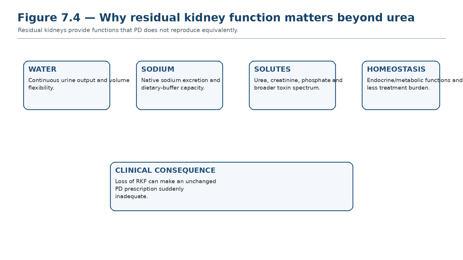
Residual kidneys add continuous urine, sodium and broad-solute clearance. Their value is not reproduced by an equal numerical increment in peritoneal Kt/V.
Residual kidney function (RKF) is consistently associated with better outcomes in PD. CANUSA reanalysis showed that increments in residual GFR and urine volume were associated with lower mortality risk, whereas equivalent changes in peritoneal creatinine clearance were not. This does not prove that preserving RKF itself causes all of the survival advantage, but it establishes RKF as a major clinical prognostic and treatment variable. [4]
Table 7.9 — Clinical benefits associated with preserved RKF.
| Domain | Why RKF helps |
|---|---|
| Volume | Urine output buffers fluid and sodium intake |
| Solute control | Adds continuous urea/creatinine and broader toxin removal |
| Phosphate/potassium | Native excretion can reduce biochemical burden |
| Nutrition | Allows lower dialysis burden and less glucose exposure in suitable patients |
| Quality of life | May permit incremental prescriptions and fewer exchanges |
| Outcomes | Observational studies consistently associate RKF with survival |
9. Measuring residual kidney function correctly
For routine PD practice, RKF should be estimated from a timed urine collection using the mean of renal urea clearance and renal creatinine clearance. Creatinine clearance alone overestimates true GFR because of tubular secretion; urea clearance alone tends to underestimate it. Their mean is a practical compromise supported by PD guidance and physiology. [7]
Table 7.10 — Timed urine collection: what is required.
| Element | Required information | Common error |
|---|---|---|
| Collection interval | Usually a complete 24-h urine collection | Missed voids |
| Urine volume | Measured, not estimated | Using diary volume without actual collection |
| Urine urea + creatinine | Laboratory concentrations | Wrong units |
| Plasma urea + creatinine | Representative blood sample | Non-contemporaneous sample during instability |
| Calculation | Renal urea and creatinine clearances, then mean | Using creatinine clearance alone as GFR |
| PRACTICE POINT Chen, Perl and Teitelbaum recommend measuring RKF from the mean 24-h urinary creatinine and urea clearances, ideally on a regular longitudinal schedule such as quarterly while clinically meaningful RKF persists. Local programs may individualize frequency. [7] |
|---|
10. Urine volume is not the same as RKF — but it matters
Urine volume and solute clearance answer different questions. A patient can produce urine with little solute clearance, and a modest urine volume may still carry meaningful sodium and water excretion. CANUSA reanalysis found a strong association between urine volume and survival, reinforcing that urine should be tracked separately from renal Kt/V. [4]
Table 7.11 — Urine volume versus renal clearance.
| Measure | Tells you | Does not tell you |
|---|---|---|
| 24-h urine volume | Water excretion and practical volume reserve | Exact solute clearance |
| Renal Kt/V | Urea-clearance contribution | Sodium removal or total kidney function |
| Mean urea/creatinine clearance | Practical RKF estimate | All tubular/endocrine functions |
11. Preserving RKF: protect a clinical asset
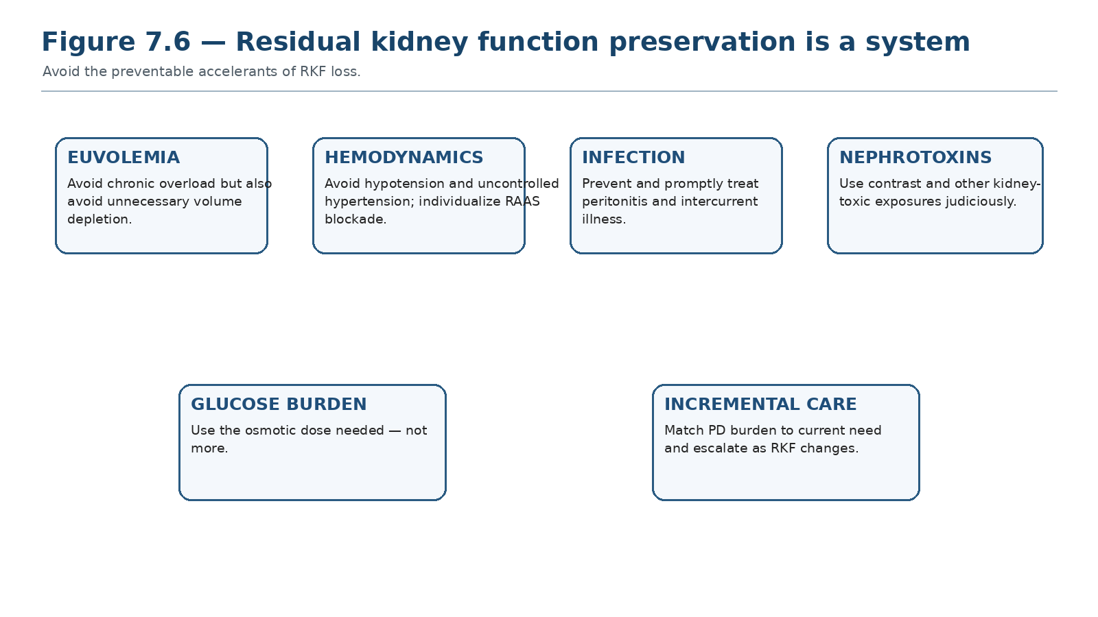
The goal is not maximal blood pressure reduction or maximal ultrafiltration; it is stable perfusion, euvolemia, infection prevention and avoidance of preventable kidney insults.
Table 7.12 — RKF-preservation strategy.
| Strategy | Physiologic logic | Clinical caution |
|---|---|---|
| Avoid volume depletion | Preserves kidney perfusion | Do not tolerate chronic overload to “protect urine” |
| Control BP appropriately | Limits vascular/glomerular injury | Avoid symptomatic hypotension |
| RAAS blockade when otherwise appropriate | May preserve RKF and has cardiovascular indications | Individualize potassium/BP tolerance |
| Loop diuretics when responsive | Maintain urine volume and sodium excretion | Do not mistake diuresis for preserved GFR |
| Prevent/treat peritonitis | Inflammation/illness can accelerate RKF loss | Peritonitis is also a technique threat |
| Judicious nephrotoxin/contrast exposure | Avoid preventable kidney injury | Do not withhold essential diagnostics reflexively |
| Incremental prescription when suitable | Avoid unnecessary glucose/burden while kidneys contribute | Requires active surveillance and escalation |
| BALANCE RULE Preserving RKF does not mean keeping the patient wet. Chronic congestion is harmful. The target is euvolemia without recurrent intravascular depletion. |
|---|
12. What accelerates RKF decline?
The evidence for individual predictors is mostly observational. A 2023 systematic review/meta-analysis identified associations between RKF loss/decline and factors including diabetes, peritonitis, proteinuria and higher systolic blood pressure; such associations do not establish that every factor is directly modifiable or causal. [10]
Table 7.13 — RKF decline: modifiable and non-modifiable signals.
| Signal | Interpretation | Action |
|---|---|---|
| Peritonitis / intercurrent illness | Inflammatory/hemodynamic insult | Prevent; treat promptly; reassess RKF afterward |
| Repeated hypotension / dehydration | Potential renal hypoperfusion | Review UF, diuretics and BP regimen |
| Uncontrolled hypertension | Vascular/kidney injury risk | Optimize BP without overcorrection |
| Nephrotoxic exposure | Potential direct injury | Risk-benefit review and monitoring |
| Diabetes / proteinuria | Higher-risk phenotype | Optimize disease-specific care |
| Time on dialysis | Expected biological decline | Plan prescription escalation rather than blame the patient |
13. Falling RKF is a prescription-changing event
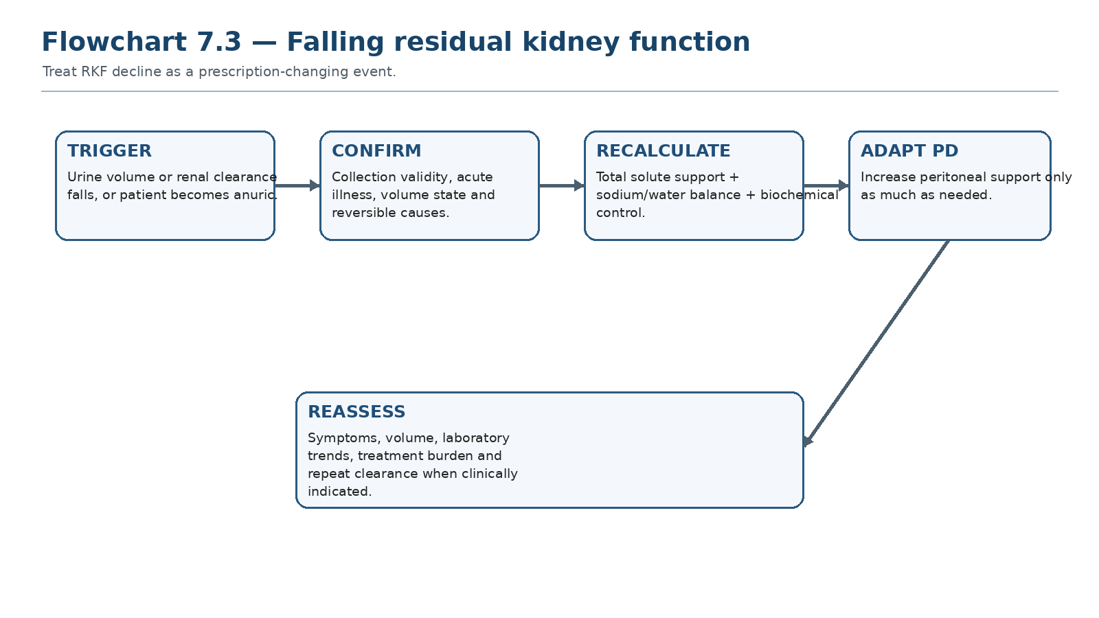
A falling urine or renal-clearance trend changes the total kidney-replacement prescription even when every PD setting is unchanged.
The most common “silent underdialysis” scenario is an unchanged PD prescription in a patient whose kidneys are providing progressively less clearance and volume control. Total measured Kt/V may fall, but the more clinically important signals may be edema, hyperphosphatemia, rising potassium, worsening appetite or increased symptom burden.
Table 7.14 — What changes when RKF falls.
| Domain | Likely direction | What to reassess |
|---|---|---|
| Total Kt/V | Falls if PD unchanged | Need for more peritoneal clearance |
| Urine volume | Often falls | Sodium/fluid prescription |
| Potassium/phosphate | May worsen | Diet, medications and dialysis contribution |
| Treatment burden needed | Often rises | CAPD/APD architecture and patient priorities |
| Glucose exposure | May rise if UF support increases | Use dwell/agent strategy rather than reflex hypertonicity |
14. Low Kt/V: diagnose before escalating
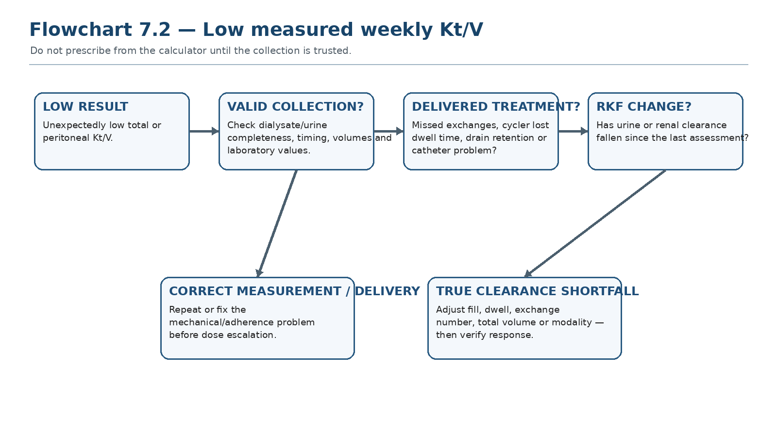
The correct response to a low number begins with collection validity, delivery and RKF trend—not automatic addition of exchanges.
Table 7.15 — Low Kt/V differential.
| Cause | Clue | Corrective direction |
|---|---|---|
| Incomplete dialysate collection | Missing bags / implausible drain total | Repeat valid collection |
| Incomplete urine collection | Urine volume inconsistent with usual output | Repeat renal component |
| Missed exchanges / cycler lost time | Treatment log discrepancy | Fix delivery problem |
| Catheter/drain retention | Poor drain, alarms, retained volume | Mechanical pathway |
| Loss of RKF | Falling urine or renal clearance | Increase peritoneal support as needed |
| Prescription insufficient for body size/transport | Valid delivery but persistent low clearance | Adjust volume, dwell, exchanges or modality |
15. “Kt/V is fine but the patient is not”
A satisfactory small-solute number does not neutralize a clinical problem. Congestion, poor sodium balance, malnutrition, recurrent peritonitis, severe treatment burden, constipation, access dysfunction or non-uremic disease can all coexist with an apparently acceptable Kt/V.
Table 7.16 — Adequate number, inadequate patient.
| Problem | Why Kt/V may remain acceptable | Next question |
|---|---|---|
| Edema / hypertension | Urea clearance can be adequate while sodium removal is poor | Volume/sodium mechanism? |
| Poor appetite / weight loss | Inflammation, depression, GI disease or dialysis burden | Is this uremia or another illness? |
| Fatigue | Anemia, sleep, heart failure, depression, infection | What competing diagnosis exists? |
| Hyperphosphatemia | Phosphate behaves differently from urea | Diet/binder/RKF/dwell strategy? |
| High burden | Clearance achieved at unacceptable workload | Can goals be met with a different architecture? |
| CLINICAL RULE Never use a Kt/V result to dismiss a symptom. Use it to narrow the differential. |
|---|
16. “The patient feels well but Kt/V is below target”
This situation requires equal caution in the opposite direction. First verify collection and delivery. Then assess RKF, volume, potassium, bicarbonate, nutrition and symptom trajectory. In an anuric adult, a persistently low weekly Kt/V deserves active prescription review because the evidence supporting ≥1.7 is weak but remains the principal guideline-based small-solute floor. The absence of symptoms is not proof that a sustained clearance shortfall is harmless. [2]
Table 7.17 — Low Kt/V with few symptoms: disciplined response.
| Step | Action |
|---|---|
| 1 | Repeat/validate the collection if unexpected |
| 2 | Confirm treatment delivery and catheter function |
| 3 | Measure RKF accurately |
| 4 | Review volume, potassium, bicarbonate, nutrition and trajectory |
| 5 | If true shortfall persists, increase peritoneal support using physiology |
| 6 | Recheck after the change; avoid endless escalation if another modality better meets goals |
17. The anuric patient: peritoneal dialysis must carry the whole load
Once RKF is absent, the peritoneal prescription becomes responsible for essentially all dialytic solute and fluid support. The margin for missed treatments, poor drains and prolonged low-UF dwells becomes smaller. This is the context in which the weekly Kt/V ≥1.7 practice point is most relevant, but clinical volume and biochemical targets remain equally important. [2]
Table 7.18 — Anuric PD checklist.
| Domain | Question |
|---|---|
| Small solutes | Is weekly peritoneal Kt/V consistently adequate by a valid collection? |
| Volume | Is net UF plus intake strategy maintaining euvolemia? |
| Sodium | Is sodium removal sufficient, especially with short APD dwells? |
| Potassium/acid-base | Are biochemical targets stable? |
| Phosphate/nutrition | Is dietary/prescription strategy adequate? |
| Delivery | Are missed cycles or drainage problems reducing dose? |
| Burden | Is achieving targets requiring an unsustainable prescription? |
18. Incremental PD: adequacy requires an escalation contract
Incremental PD deliberately uses less peritoneal dialysis while RKF contributes meaningfully. Contemporary comparative evidence is heterogeneous but generally supports feasibility in selected patients without a clear mortality penalty. The safety condition is not the starting dose; it is active monitoring with pre-defined escalation when RKF, symptoms, volume or biochemical goals change. [11]
Table 7.19 — Incremental PD: what must be monitored.
| Signal | Escalation trigger concept |
|---|---|
| Urine/RKF | Meaningful decline from the patient’s prior contribution |
| Symptoms | New uremic symptom burden |
| Volume | Worsening sodium/water control |
| Biochemistry | Persistent deterioration despite medical therapy |
| Clearance | True total small-solute shortfall |
| Life situation | Prescription no longer fits goals or support resources |
19. Adequacy assessment algorithm
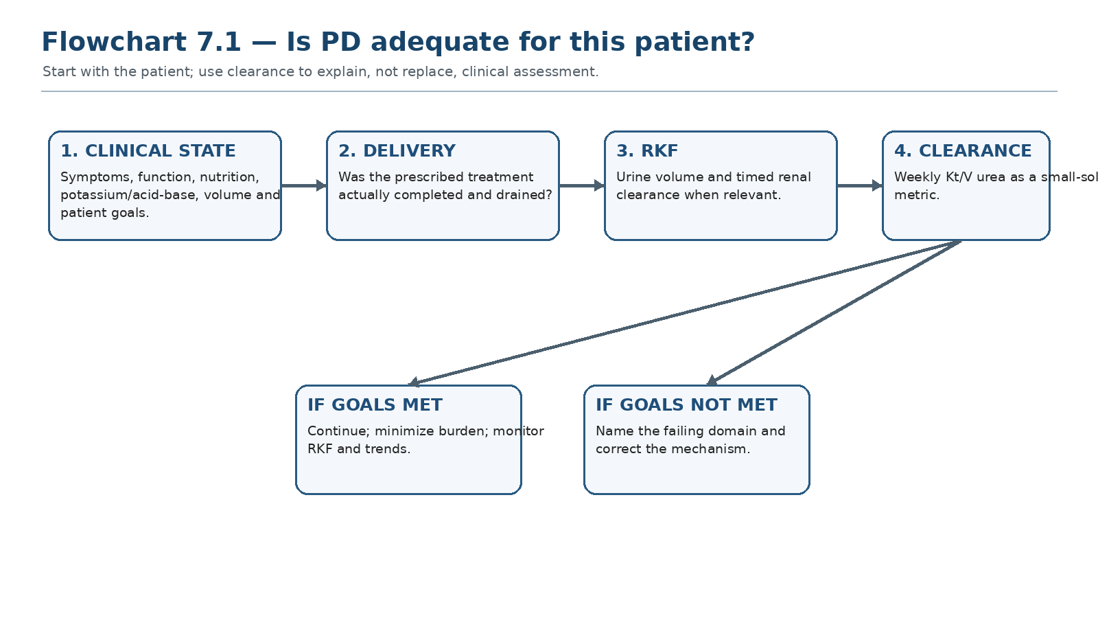
The algorithm starts with the clinical state, then verifies delivery, RKF and small-solute clearance before any prescription change.
Table 7.20 — One-minute adequacy synthesis.
| If you see… | Think… | Do now… |
|---|---|---|
| Well + euvolemic + stable labs + meaningful urine | Current total support may be appropriate | Maintain; preserve RKF; avoid unnecessary burden |
| Low Kt/V only | Measurement/delivery problem possible | Validate collection and logs |
| Falling urine + edema + same prescription | Loss of RKF | Recalculate total support and adjust PD |
| Good Kt/V + edema | Volume/sodium problem | Treat volume physiology, not urea number |
| Anuric + low Kt/V | True peritoneal dose shortfall until proven otherwise | Verify then intensify or reassess modality |
| High prescription burden + good clinical state | Possible overtreatment relative to goals | Reconsider architecture; do not sacrifice safety |
20. Clinical pearls
RKF is not “bonus dialysis”; it is a major clinical organ function that changes the prescription.
A stable PD order becomes a smaller total kidney-replacement treatment when urine and RKF fall.
Urine volume and renal clearance should both be followed; they answer different questions.
An unexpectedly low Kt/V is a collection-quality problem until the collection is proven valid.
Good urea clearance does not prove good sodium removal.
If the patient is clinically well, do not add glucose-rich exchanges solely to make an already acceptable number look larger.
In an anuric patient, missed treatments and drainage failure have greater clinical consequences because there is no kidney reserve.
The best RKF-preservation strategy is balanced: avoid congestion and avoid recurrent hypovolemia.
21. Common pitfalls — and the correction
Table 7.21 — High-frequency adequacy errors.
| Pitfall | Why it fails | Correction |
|---|---|---|
| Calling Kt/V “adequacy” | Collapses a multidimensional outcome into one solute | Use a dashboard |
| Treating 1.69 and 1.71 as different clinical worlds | False precision around low-certainty threshold | Use trend + clinical context |
| Adding renal and peritoneal clearance as biologically equal | CANUSA contradicts equivalence | Value RKF separately |
| Escalating after one low collection | Collection may be incomplete | Validate before prescribing |
| Using urine volume as GFR | Volume and solute clearance differ | Measure timed urea/creatinine clearance |
| Using creatinine clearance alone for RKF | Tubular secretion overestimates GFR | Use mean renal urea + creatinine clearance |
| Preserving urine by tolerating overload | Congestion is harmful | Target euvolemia |
| Ignoring falling RKF in incremental PD | Total support silently shrinks | Define escalation triggers |
22. Mini-cases: decisions, not trivia
Case 1 — The “adequate” congested patient
A patient has weekly Kt/V 1.84 but progressive edema, rising BP and poor daytime UF.
| BEST NEXT STEP Do not increase dialysis because the number is “too low”; it is not low. Diagnose a sodium/volume problem: intake, urine decline, dwell/transport mismatch, osmotic strategy and mechanical function. Chapter 8 develops the full volume pathway. |
|---|
Case 2 — Sudden low Kt/V
A previously stable CAPD patient has weekly Kt/V 1.42 on one collection but feels unchanged. The drain log shows one exchange was discarded before sampling.
| BEST NEXT STEP The collection is invalid. Repeat a complete collection before changing the prescription. |
|---|
Case 3 — Incremental PD and falling urine
A patient on a low-burden incremental regimen loses substantial urine over three months and develops hyperphosphatemia and fatigue.
| BEST NEXT STEP The same PD prescription now provides less total support. Confirm RKF decline, assess volume/biochemistry, and escalate the peritoneal prescription according to the failing targets. |
|---|
Case 4 — Anuric APD with low clearance
An anuric patient completes nocturnal APD but has persistently low weekly Kt/V. Cycler logs show many very short cycles and repeated drain alarms.
| BEST NEXT STEP Delivered therapy is not equivalent to the written prescription. Correct drainage/catheter and cycle-time problems before simply adding more cycles. |
|---|
Case 5 — Good numbers, severe fatigue
Kt/V and volume status are acceptable, but the patient has marked fatigue and dyspnoea.
| BEST NEXT STEP Do not label symptoms “uremia” automatically. Evaluate anemia, cardiac disease, sleep, infection, depression and other competing diagnoses. |
|---|
Case 6 — Preserving RKF badly
A patient with useful urine is repeatedly driven to very low post-dialysis weight using hypertonic glucose because edema is feared. Urine falls rapidly.
| BEST NEXT STEP Reassess true volume state and avoid recurrent intravascular depletion. RKF preservation requires euvolemia—not aggressive dehydration. |
|---|
23. Active recall
MUST MEMORIZE
Q: What does weekly Kt/V urea measure? A: Small-solute urea clearance relative to urea distribution volume over one week.
Q: What is the principal ISPD small-solute practice-point threshold for anuric adults? A: Weekly Kt/V approximately 1.7; evidence supporting the lower limit is weak.
Q: How should RKF be estimated from urine? A: Mean of timed renal urea and creatinine clearances.
Q: Why not use creatinine clearance alone for RKF? A: Tubular secretion causes creatinine clearance to overestimate true GFR.
Q: Does higher peritoneal Kt/V above conventional levels improve survival? A: Randomized evidence has not demonstrated a survival advantage.
Q: What did CANUSA reanalysis teach? A: Residual GFR/urine were strongly associated with outcome; peritoneal clearance was not equivalent.
Q: Can a patient have adequate Kt/V and poor volume control? A: Yes. Solute clearance and sodium/water balance are distinct.
Q: What should you do before changing a prescription for one low Kt/V? A: Validate collection and treatment delivery.
MUST REASON
Why can loss of 24-h urine make an unchanged PD prescription clinically inadequate?
Why might adding APD cycles worsen clearance if each dwell becomes too short?
Why can aggressive ultrafiltration threaten RKF?
Why can an obese patient have an apparently low Kt/V despite reasonable clinical dialysis?
Why is an anuric patient less tolerant of missed exchanges or catheter dysfunction?
USE AS REFERENCE
Exact local adequacy-collection protocol and sample handling.
Anthropometric equation used to estimate V.
Unit-specific interval for repeat clearance/RKF measurement.
Detailed volume-management and UF-failure pathways in Chapter 8.
Prescription optimization by CAPD/APD architecture in Chapter 9.
24. Flashcards: active recall
1. Q: Adequacy in one phrase? A: Patient-centred goals + volume + biochemistry + nutrition + RKF + appropriate clearance.
2. Q: Kt/V is a measure of? A: Urea small-solute clearance scaled to V.
3. Q: Best routine RKF clearance estimate? A: Mean urinary urea and creatinine clearance.
4. Q: Urine volume equals GFR? A: No.
5. Q: ADEMEX lesson? A: More peritoneal small-solute clearance did not improve survival.
6. Q: CANUSA reanalysis lesson? A: Kidney clearance/urine mattered more than peritoneal clearance.
7. Q: Unexpected low Kt/V: first move? A: Check collection validity and delivered treatment.
8. Q: Good Kt/V + edema? A: Volume/sodium pathway.
9. Q: Anuria changes what? A: PD must provide essentially all dialytic solute and fluid support.
10. Q: Incremental PD safety requirement? A: Active RKF/clinical monitoring with escalation triggers.
11. Q: RKF preservation volume target? A: Euvolemia — neither chronic overload nor recurrent depletion.
12. Q: Can Kt/V dismiss symptoms? A: No.
13. Q: Why track urine separately? A: Water/sodium excretion has value beyond urea clearance.
14. Q: Creatinine clearance alone for RKF? A: Avoid; use mean with urea clearance.
15. Q: Higher Kt/V always better? A: No evidence of survival benefit above conventional target range.
16. Q: True low Kt/V after verification? A: Adjust the mechanism: volume, dwell, exchanges, total time or modality.
25. Rapid troubleshooting table
Table 7.22 — Adequacy problem → likely mechanism → next step.
| Problem | Likely mechanism(s) | Next step |
|---|---|---|
| Low total Kt/V | Collection error, missed therapy, RKF loss, insufficient prescription | Validate → delivery → RKF → prescription |
| Low renal component | Incomplete urine or true RKF decline | Repeat if doubtful; assess reversible insults |
| Good Kt/V + edema | Sodium/volume problem | Chapter 8 pathway |
| Good Kt/V + symptoms | Non-uremic cause or other dialysis domain | Broaden differential |
| Anuric + poor biochemistry | Peritoneal support insufficient or delivery problem | Verify and intensify/reassess modality |
| Incremental PD + falling urine | Total support shrinking | Escalate according to failing targets |
| High burden + excellent numbers | Possible unnecessary dose intensity | Revisit patient goals and minimal effective prescription |
26. Final revision sheet
| TEN TAKE-HOME RULES 1) Adequacy is a clinical dashboard. 2) Kt/V measures urea clearance, not the whole patient. 3) Higher peritoneal clearance above conventional targets has not improved survival in randomized trials. 4) RKF has disproportionate clinical value. 5) Measure RKF from the mean urinary urea + creatinine clearance. 6) Track urine volume separately. 7) Validate a low clearance collection before escalating treatment. 8) Good Kt/V does not prove good sodium/volume control. 9) Falling RKF changes the total prescription. 10) Incremental PD is safe only with an escalation plan. |
|---|
Table 7.23 — Final revision grid.
| Core concept | Remember |
|---|---|
| Adequacy | Symptoms + function + volume + biochemistry + nutrition + life goals + RKF + clearance |
| Weekly Kt/V | Small-solute metric; ≥1.7 is a low-certainty practice point, especially relevant when anuric |
| Landmark evidence | CANUSA: RKF/urine matter; ADEMEX/Hong Kong: more peritoneal clearance not necessarily better |
| RKF measurement | Timed urine; mean renal urea + creatinine clearance |
| Low Kt/V | Collection → delivery → catheter/drain → RKF → prescription |
| Preservation | Euvolemia, hemodynamic stability, infection prevention, nephrotoxin discipline |
| Anuria | No kidney reserve: peritoneal prescription carries the full dialytic load |
| FINAL MENTAL MODEL Patient state → volume/sodium → delivered treatment → urine/RKF → small-solute measurement → identify failing domain → correct mechanism → verify response. The number supports the clinical decision; it does not replace it. |
|---|
Rapid oral viva
Explain weekly Kt/V urea to a nephrology fellow without using the word “adequacy.”
Why are renal and peritoneal clearances mathematically additive but clinically unequal?
Walk through your response to a weekly Kt/V of 1.45 in a previously stable patient.
How do you measure residual kidney function and why is urine volume also recorded separately?
Give the differential for edema in a patient whose weekly Kt/V is 1.9.
What changes in your adequacy framework when a patient becomes anuric?
| SCOPE BOUNDARY This chapter teaches adequacy, clearance measurement and RKF. Detailed sodium/volume management and ultrafiltration failure are developed in Chapter 8; CAPD/APD prescription optimization is developed in Chapter 9. |
|---|
27. Selected authoritative references
1. Brown EA, Blake PG, Boudville N, et al. International Society for Peritoneal Dialysis practice recommendations: Prescribing high-quality goal-directed peritoneal dialysis. Perit Dial Int. 2020;40(3):244–253. https://doi.org/10.1177/0896860819895364. PMID: 32063219.
2. Boudville N, de Moraes TP. 2005 Guidelines on targets for solute and fluid removal in adults being treated with chronic peritoneal dialysis: 2019 update of the literature and revision of recommendations. Perit Dial Int. 2020;40(3):254–260. https://doi.org/10.1177/0896860819898307. PMID: 32048566.
3. Teitelbaum I, Glickman J, Neu A, et al. KDOQI US Commentary on the 2020 ISPD Practice Recommendations for Prescribing High-Quality Goal-Directed Peritoneal Dialysis. Am J Kidney Dis. 2021;77(2):157–171. https://doi.org/10.1053/j.ajkd.2020.09.010. PMID: 33341315.
4. Bargman JM, Thorpe KE, Churchill DN. Relative contribution of residual renal function and peritoneal clearance to adequacy of dialysis: a reanalysis of the CANUSA study. J Am Soc Nephrol. 2001;12(10):2158–2162. https://doi.org/10.1681/ASN.V12102158. PMID: 11562415.
5. Paniagua R, Amato D, Vonesh E, et al. Effects of increased peritoneal clearances on mortality rates in peritoneal dialysis: ADEMEX, a prospective, randomized, controlled trial. J Am Soc Nephrol. 2002;13(5):1307–1320. https://doi.org/10.1681/ASN.V1351307. PMID: 11961019.
6. Lo WK, Ho YW, Li CS, et al. Effect of Kt/V on survival and clinical outcome in CAPD patients in a randomized prospective study. Kidney Int. 2003;64(2):649–656. https://doi.org/10.1046/j.1523-1755.2003.00098.x. PMID: 12846762.
7. Chen CH, Perl J, Teitelbaum I. Prescribing high-quality peritoneal dialysis: The role of preserving residual kidney function. Perit Dial Int. 2020;40(3):274–281. https://doi.org/10.1177/0896860819893821. PMID: 32063188.
8. Morelle J, Stachowska-Pietka J, Öberg C, et al. ISPD recommendations for the evaluation of peritoneal membrane dysfunction in adults: classification, measurement, interpretation and rationale for intervention. Perit Dial Int. 2021;41(4):352–372. https://doi.org/10.1177/0896860820982218.
9. Borràs Sans M, Ponz Clemente E, Rodríguez Carmona A, et al. Clinical guideline on adequacy and prescription of peritoneal dialysis. Nefrologia (Engl Ed). 2024;44 Suppl 1:1–27. https://doi.org/10.1016/j.nefroe.2024.09.001. PMID: 39341764.
10. Zhou D, Lei H, Wu S, et al. Influencing factors for residual kidney function in incident peritoneal dialysis patients: a systematic review and meta-analysis. Ren Fail. 2023;45(2):2286328. https://doi.org/10.1080/0886022X.2023.2286328. PMID: 38036948.
11. Xu S, Wu W, Cheng J. Comparison of outcomes of incremental vs. standard peritoneal dialysis: a systematic review and meta-analysis. BMC Nephrol. 2024;25:308. https://doi.org/10.1186/s12882-024-03669-w. PMID: 39285336.
12. Wang AY. The John F. Maher Award Recipient Lecture 2006. The ‘heart’ of peritoneal dialysis: residual renal function. Perit Dial Int. 2007;27(2):116–24. PMID: 17299143.
13. Li PKT, Chow KM, Cho Y, et al. ISPD peritonitis guideline recommendations: 2022 update on prevention and treatment. Perit Dial Int. 2022;42(2):110–153. https://doi.org/10.1177/08968608221080586.
14. Davies SJ. Peritoneal dialysis—current status and future challenges. Nat Rev Nephrol. 2013;9:399–408. https://doi.org/10.1038/nrneph.2013.100.
| SOURCE NOTE Guideline status and contemporary adequacy/RKF evidence were checked 2 September 2026. Exact collection procedures, V-estimation method, laboratory units, monitoring frequency and reimbursement-linked thresholds can vary by jurisdiction and program; local protocols should define operational details without replacing the patient-centred ISPD framework. |
|---|