Applied Nephrology Master Series
Chapter 8
Volume Management, Sodium Balance and Ultrafiltration Failure
Congestion | Sodium | Residual Diuresis | Ultrafiltration | Icodextrin | Membrane Dysfunction | Rescue
| CHAPTER MISSION Build a bedside volume-control system that separates sodium intake, residual kidney output, peritoneal water removal and membrane function; recognize congestion before it becomes technique failure; and diagnose true ultrafiltration failure only after mechanical, leak and prescription causes have been excluded. |
|---|
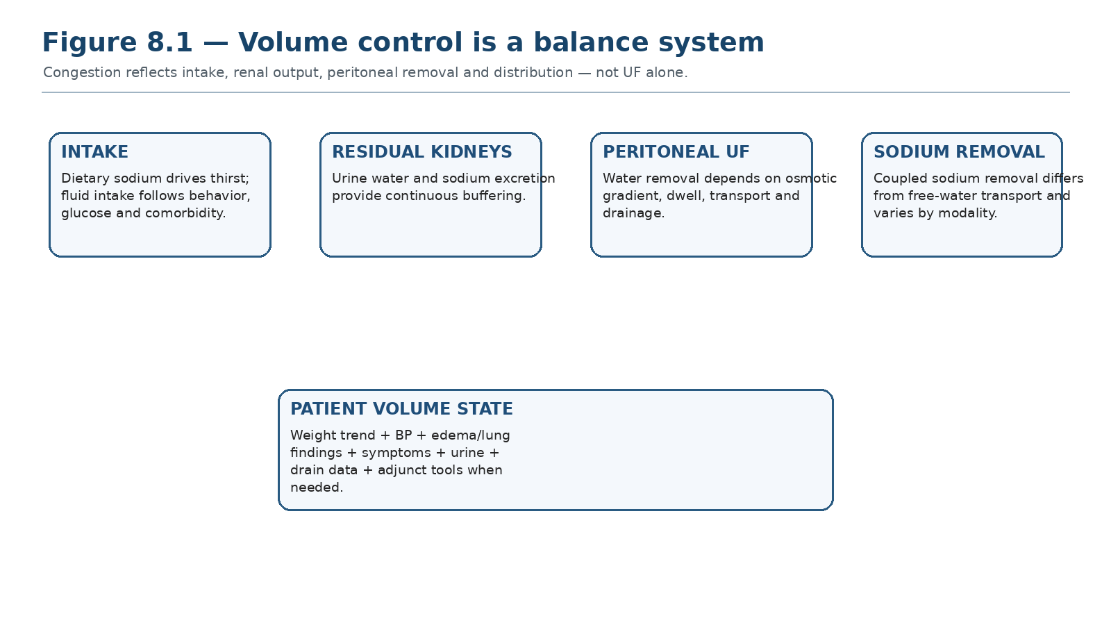
The patient becomes overhydrated when sodium and water input chronically exceed renal plus peritoneal output. Net peritoneal UF is important, but it is only one term in the balance equation.
| MASTER PRINCIPLE Treat the balance, not the bag. A patient can be overloaded with apparently “good UF,” and can have low UF without intrinsic membrane failure. Volume management requires matching intake, residual kidney function, peritoneal sodium/water removal and cardiovascular tolerance. |
|---|
0. One-page chapter map
Table 8.1 — The eight decisions that govern PD volume management.
| Decision | Core question | Bedside output |
|---|---|---|
| 1. Phenotype | Is the patient overloaded, euvolemic, depleted or uncertain? | Named volume state |
| 2. Input | How much sodium and fluid is entering the system? | Diet/thirst/hidden-sodium plan |
| 3. Kidney | How much urine and sodium excretion remain? | RKF + diuretic strategy |
| 4. Peritoneum | How much water and sodium are removed by PD? | UF + sodium-removal interpretation |
| 5. Delivery | Is the prescription actually being completed and drained? | Adherence/mechanical verification |
| 6. Membrane | Is low UF due to fast PSTR, low osmotic conductance or another membrane phenotype? | PET-based mechanism |
| 7. Correct | Which lever targets the mechanism with least glucose/burden? | Targeted prescription change |
| 8. Verify | Did congestion, weight/BP, urine and UF improve? | Reassessment + escalation trigger |
Learning outcomes
Define volume status as a clinical state created by sodium-water balance rather than by a single weight, BP or UF value.
Explain why sodium intake drives thirst and extracellular-volume expansion, and apply ISPD dietary sodium guidance without causing volume contraction.
Integrate urine volume, diuretic response and residual kidney function into the volume prescription.
Explain free-water transport, small-pore water transport, sodium sieving and why net UF does not equal sodium removal.
Compare CAPD and APD sodium-water removal and identify when short automated dwells create a “water-rich, sodium-poor” ultrafiltration pattern.
Use glucose concentration and icodextrin physiologically while minimizing unnecessary glucose exposure.
Distinguish volume overload, low daily UF and standardized ultrafiltration insufficiency.
Apply the ISPD high-glucose PET and 1-h sodium dip criteria to diagnose intrinsic membrane dysfunction without overlooking leaks or mechanical failure.
Match treatment to fast PSTR, impaired osmotic conductance, low effective surface area, high absorption and non-membrane causes.
Recognize when persistent inability to maintain safe volume control should trigger modality-transition planning.
| EVIDENCE POSTURE ISPD 2020 treats volume and sodium management as core components of high-quality goal-directed PD. ISPD cardiovascular guidance recommends regular clinical volume assessment and salt restriction below 2 g sodium/day (5 g salt/day) unless contraindicated by volume contraction or hypotension. The 2021 ISPD membrane guideline defines standardized low UF capacity and sodium-dip criteria and recommends dwell shortening, icodextrin and/or higher glucose concentration according to phenotype. Icodextrin RCT meta-analysis shows improved UF and fewer fluid-overload episodes. Observational IPOD-PD data show that overhydration is common at PD start and is associated with subsequent technique failure. [1–7] |
|---|
1. Volume management is part of dialysis adequacy
A patient with an acceptable weekly Kt/V may still have clinically important sodium and fluid overload. Conversely, a patient with low daily peritoneal UF may remain euvolemic because residual urine output is substantial and sodium intake is low. Contemporary PD practice therefore assesses volume and sodium control as a distinct adequacy domain rather than inferring it from small-solute clearance. [1,3]
Table 8.2 — Do not confuse these four concepts.
| Concept | Meaning | Why it matters |
|---|---|---|
| Volume status | The patient’s extracellular-fluid state | The clinical outcome being managed |
| Net peritoneal UF | Drain volume minus instilled volume | Water removed by PD; not total body fluid balance |
| Sodium removal | Urinary + dialytic sodium excretion | Determines sustained extracellular-volume control |
| Ultrafiltration failure | Standardized membrane dysfunction phenotype after appropriate evaluation | A diagnosis—not a synonym for edema |
| BEDSIDE TRANSLATION Start every volume review with “What is the patient’s current volume phenotype?” before asking “How much UF did the cycler report?” |
|---|
2. Sodium-water balance: the hidden driver of congestion
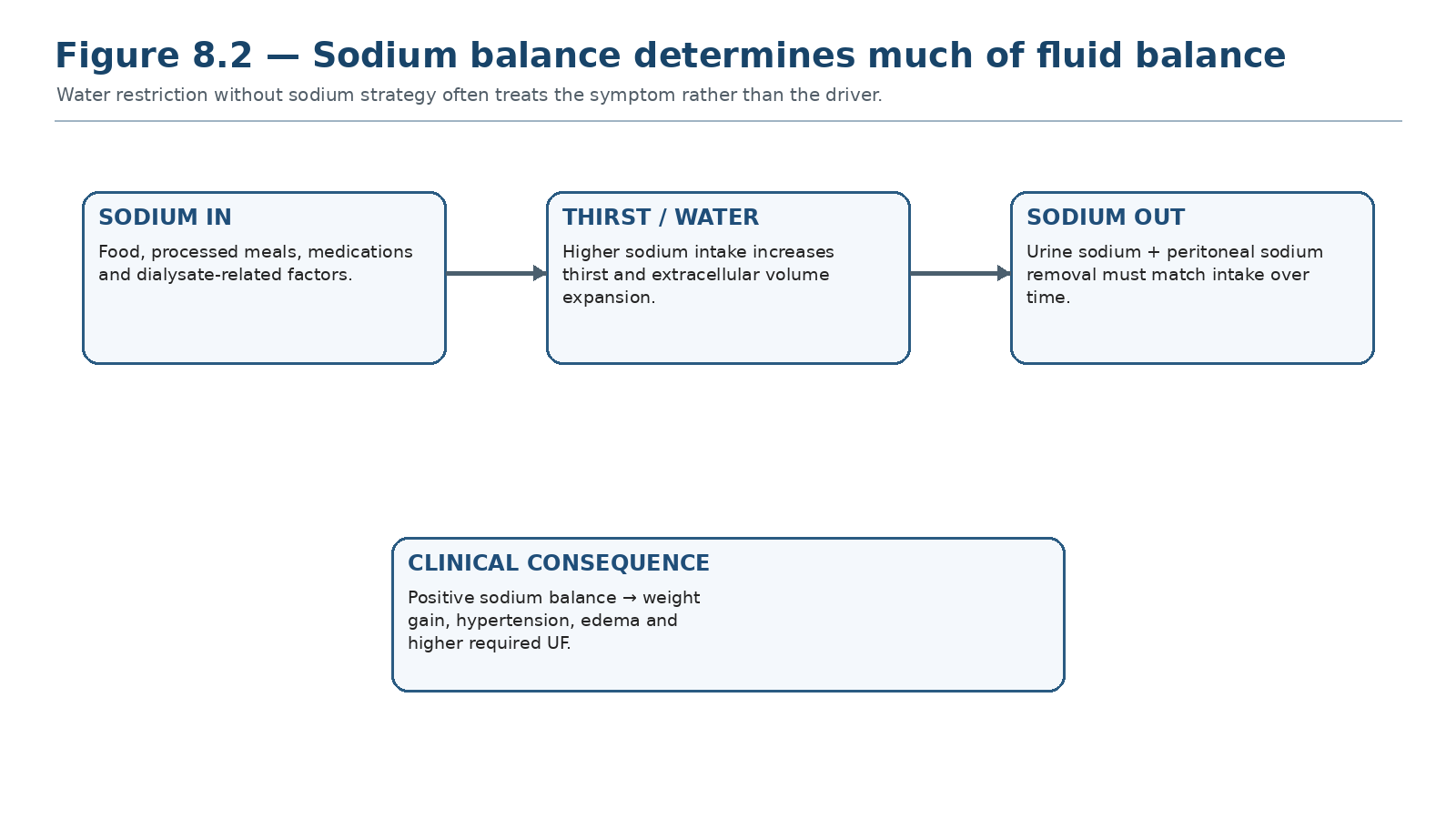
Sodium accumulation expands extracellular volume and drives thirst. Long-term control therefore depends on sodium input matching renal plus peritoneal sodium output.
In PD, water follows sodium behavior over time. High dietary sodium intake increases thirst and raises the amount of water that must be removed. A prescription that repeatedly compensates with hypertonic glucose without addressing sodium intake can control scale weight temporarily while increasing glucose exposure and treatment burden. ISPD cardiovascular guidance recommends sodium intake below 2 g/day (equivalent to approximately 5 g sodium chloride/day) unless contraindicated or there is evidence of volume contraction or hypotension. [3]
Table 8.3 — Positive sodium balance: where it comes from.
| Source | Typical clue | Corrective direction |
|---|---|---|
| Processed/high-salt food | Thirst, high intake, recurrent weight gain | Dietitian-guided sodium reduction |
| Restaurant/hidden sodium | “Fluid restriction failure” despite low stated drinks | Food-source review, label literacy |
| Medications/supplements | Effervescent products, sodium-containing drugs | Medication reconciliation |
| Low urinary sodium excretion | Falling urine/RKF | Recalculate total removal capacity |
| Insufficient dialytic sodium removal | APD short dwells, low net UF, prescription mismatch | Modify dwell pattern/long dwell/osmotic strategy |
| CLINICAL PEARL Fluid restriction without a sodium strategy is often unsustainable because thirst is physiologically driven. Treat sodium excess first unless the clinical context requires temporary fluid restriction for severe congestion. |
|---|
3. Recognizing the volume phenotype
No single bedside sign defines euvolemia. Edema is specific but insensitive; hypertension may reflect volume, arterial stiffness, medications or sympathetic activity; low BP can coexist with venous congestion in advanced heart failure. The safest approach is serial and multimodal.
Table 8.4 — Volume assessment: signal, meaning and limitation.
| Signal | What it suggests | Important limitation |
|---|---|---|
| Weight trend | Net body-mass change over days/weeks | Nutrition and bowel contents confound |
| Home BP trend | Often tracks sodium-volume state | Not specific for congestion |
| Edema | Extracellular expansion when present | May be absent despite overload |
| Orthopnea/dyspnea | Pulmonary congestion or cardiac disease | Anemia/lung disease mimic |
| JVP / venous congestion | Right-sided filling pressure | Exam skill/body habitus affect accuracy |
| Lung crackles | Pulmonary fluid | Low sensitivity; other lung disease |
| Urine volume | Residual water removal | Does not directly quantify sodium excretion |
| Daily UF | Peritoneal water removal | Does not equal sodium removal |
| SERIAL EXAM RULE A small change from a documented baseline often matters more than a one-time “dry/overloaded” label by an unfamiliar observer. |
|---|
4. Adjunctive tools: useful when the bedside picture is uncertain
Bioimpedance spectroscopy can identify subclinical overhydration and has shown prognostic associations in PD cohorts. Lung ultrasound can detect extravascular lung water and may help when symptoms and examination disagree. Neither tool should replace clinical reasoning or become a universal treatment target; intervention trials in PD remain less definitive than observational associations. [5–7,15]
Table 8.5 — Adjunctive volume tools.
| Tool | Strength | Use with caution |
|---|---|---|
| Bioimpedance | Quantifies hydration compartments; reproducible trends | Device/algorithm dependence; nutrition/body habitus |
| Lung ultrasound | Sensitive for B-lines/pulmonary congestion | B-lines are not specific to volume |
| Echocardiography | Cardiac structure/function, filling pressure clues | Not a direct “dry weight” test |
| VExUS/venous Doppler | May characterize systemic venous congestion | Emerging dialysis evidence; operator dependent |
| NT-proBNP | Can track cardiac/volume stress | High baseline in kidney failure; interpret trend/context |
| EVIDENCE CALIBRATION Bioimpedance-defined overhydration predicts adverse outcomes observationally, but a device-derived “normal” value is not a mandate for aggressive fluid removal in a frail or hypotension-prone patient. [7,15] |
|---|
5. Residual kidneys: the first fluid-removal system to protect
Residual urine output provides continuous water and sodium excretion and reduces the peritoneal osmotic burden. Volume prescriptions should therefore track urine volume and RKF as separate variables. Diuretics can increase urine volume when residual kidney function remains, but they should not be represented as preserving GFR by themselves. In the randomized CAPD trial by Medcalf et al., high-dose furosemide increased urine volume but did not demonstrate preservation of residual renal clearance. [8]
Table 8.6 — Diuretic reasoning in PD.
| Situation | Potential role | Do not assume |
|---|---|---|
| Meaningful urine output + congestion | Increase urinary sodium/water excretion | That a higher dose preserves RKF |
| Anuria | No useful diuretic fluid-removal effect | That “more diuretic” substitutes for PD UF |
| Falling urine volume | Search for reversible causes and reassess total prescription | That edema is solely membrane failure |
| Hypotension/volume depletion | Reduce iatrogenic drivers | That maximal urine output is always desirable |
| RKF PROTECTION Target euvolemia—not maximal negative balance. Recurrent hypotension and unnecessary volume depletion can accelerate loss of residual kidney function. |
|---|
6. Peritoneal ultrafiltration physiology at the bedside
Glucose generates a crystalloid osmotic gradient that moves water from the peritoneal microcirculation into dialysate. Water crosses small pores together with sodium and other small solutes, and aquaporin-1 channels as solute-free water. The balance between these pathways changes during the dwell and is modified by transport phenotype, glucose concentration, intraperitoneal pressure and membrane structure. [2,14]
Table 8.7 — Mechanism → bedside consequence.
| Mechanism | Physiologic consequence | Prescription implication |
|---|---|---|
| Higher glucose gradient | Stronger initial osmotic force | More UF but more glucose exposure |
| Fast PSTR | Rapid glucose absorption | Shorter glucose dwells may protect UF |
| Long dwell | More equilibration and reabsorption risk | Use icodextrin when long-dwell UF is poor |
| High intraperitoneal pressure | May oppose transcapillary UF / worsen leak | Review fill volume and mechanical context |
| Reduced AQP/free-water transport | Blunted early sodium dip and UF | Suggests impaired osmotic conductance |
7. Sodium sieving: why one litre of UF is not always one litre of sodium removal
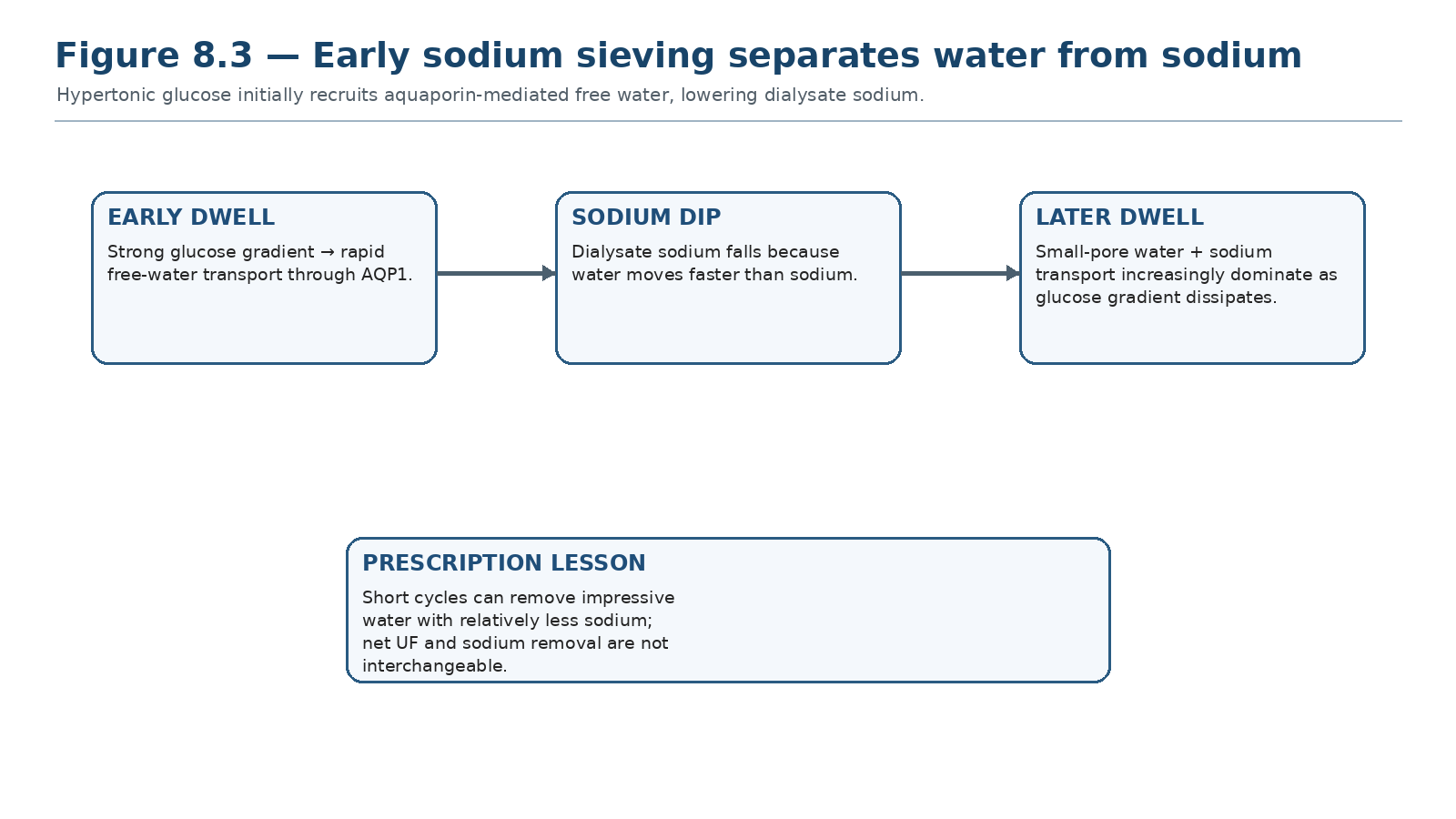
Free-water transport through AQP1 dilutes dialysate sodium early in a hypertonic glucose dwell. As the dwell continues, small-pore sodium-coupled transport becomes more prominent.
Early during a hypertonic glucose dwell, aquaporin-mediated water transport is relatively sodium-free. Dialysate sodium concentration therefore falls—the sodium dip. This is physiologically useful as a marker of free-water transport, but it also explains why short APD cycles may remove proportionally more water than sodium. [2,9,10]
Table 8.8 — Water removal versus sodium removal.
| Pattern | Interpretation | Clinical consequence |
|---|---|---|
| High early UF + prominent sodium dip | Strong free-water component | Water removal may exceed proportional sodium removal |
| Longer dwell with maintained UF | More small-pore coupled water/sodium transport | Greater sodium removal per exchange |
| Blunted sodium dip on standardized hypertonic PET | Reduced free-water transport / osmotic conductance | Investigate intrinsic membrane dysfunction |
| Good daily UF + persistent hypertension/edema | Possible sodium-removal mismatch or high intake | Do not intensify glucose before assessing sodium balance |
8. CAPD versus APD: different sodium-water phenotypes
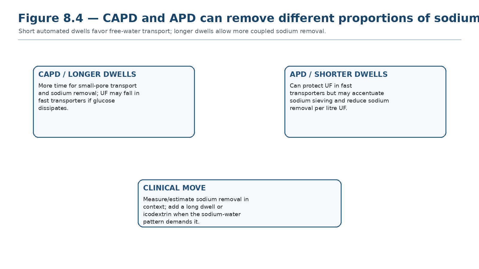
Short APD dwells can preserve osmotic UF in fast transporters but may enhance free-water–dominant removal. CAPD’s longer dwells generally permit more sodium-coupled transport.
A systematic review found lower dialytic sodium removal with APD than CAPD on average, although heterogeneity was substantial and transport status strongly modified the difference. Older physiologic cohorts similarly showed that APD sodium removal improved with longer nocturnal dwells, a daytime exchange and icodextrin. These data support measuring the sodium-water problem rather than assuming that higher net UF automatically means better sodium balance. [9,10]
Table 8.9 — Prescription patterns and sodium removal.
| Pattern | Potential advantage | Potential trade-off |
|---|---|---|
| Short APD cycles | Protect UF in fast PSTR | More sodium sieving; lower sodium removal per litre UF |
| Longer CAPD dwell | More sodium-coupled transport | Glucose gradient may dissipate in fast PSTR |
| Daytime APD long dwell | Adds sodium/solute removal | Risk of negative UF with glucose in fast PSTR |
| Icodextrin long dwell | Sustained colloid osmotic UF, useful in fast PSTR | One daily long dwell; device/glucose-meter safety considerations from Chapter 2 |
9. Dietary sodium, fluid and the “thirst loop”
The practical aim is sustained sodium balance without compromising nutrition. Sodium restriction should be specific enough to change behavior: identify processed foods, restaurant meals, sauces, preserved foods and salt substitutes rather than only advising “drink less.” Fluid restriction can be added when intake remains greater than achievable output, but overly rigid restrictions can impair quality of life and nutrition.
Table 8.10 — Counseling that changes physiology.
| Ask | Why it matters | Action |
|---|---|---|
| Where is sodium actually coming from? | Hidden food sodium drives thirst | Target the dominant sources |
| How much urine remains? | Determines allowable intake flexibility | Adjust fluid advice to real output |
| Is thirst driven by hyperglycemia? | Glucose exposure/diabetes can increase intake | Optimize glucose and diabetes strategy |
| Are there nutrition risks? | Overrestriction may reduce intake | Coordinate with renal dietitian |
| Does the patient understand salt vs sodium? | Labels may use sodium rather than salt | Teach conversion concept, not memorized lists |
10. Glucose strength: effective, immediate and biologically expensive
Increasing glucose concentration strengthens the crystalloid osmotic gradient and can rapidly increase UF. It is therefore appropriate when clinically necessary, particularly for acute congestion, but chronic reliance on hypertonic glucose increases systemic glucose absorption and peritoneal exposure. The prescription principle is to use the lowest osmotic dose that reliably maintains the patient’s volume goals. [1,2,14]
Table 8.11 — Hypertonic glucose: when it helps and what it costs.
| Benefit | Trade-off | Safeguard |
|---|---|---|
| Rapidly increases osmotic UF | Higher glucose absorption | Use as targeted intervention, not default |
| Useful when RKF falls | Greater peritoneal metabolic burden | Reassess total prescription and sodium intake |
| May rescue fast PSTR short dwells | Gradient still dissipates with time | Match dwell length to transport |
| May be necessary in intrinsic low osmotic conductance | High glucose requirement can signal membrane failure | Discuss long-term technique viability |
| GLUCOSE DISCIPLINE Escalating glucose without first checking dietary sodium, urine loss, dwell length and drain mechanics can treat the symptom while worsening the long-term prescription burden. |
|---|
11. Icodextrin: the long-dwell volume tool
Icodextrin is a glucose polymer that produces sustained colloid osmosis during a long dwell and is particularly useful when glucose-based long dwells lose the osmotic gradient, especially in fast transporters. The 2020 enriched meta-analysis of 19 randomized trials found higher UF and fewer fluid-overload episodes with icodextrin versus glucose-only long-dwell strategies. [4]
Table 8.12 — Icodextrin: mechanism-based use.
| Clinical problem | Why icodextrin fits | Reassess |
|---|---|---|
| Negative/poor long-dwell UF | Sustained colloid osmotic force | Net UF and symptoms |
| Fast PSTR | Less dependent on maintaining glucose gradient | Volume + sodium removal |
| APD with daytime congestion | Improves long-dwell fluid removal | Total 24-h balance |
| High glucose burden | Can reduce long-dwell glucose exposure | Metabolic profile and overall prescription |
| ESTABLISHED STANDARD Use icodextrin as a once-daily long-dwell alternative when maintaining euvolemia is difficult because peritoneal UF is insufficient, taking transport state and product-specific safety into account. [2–4] |
|---|
12. Volume overload: a cause-based clinical framework
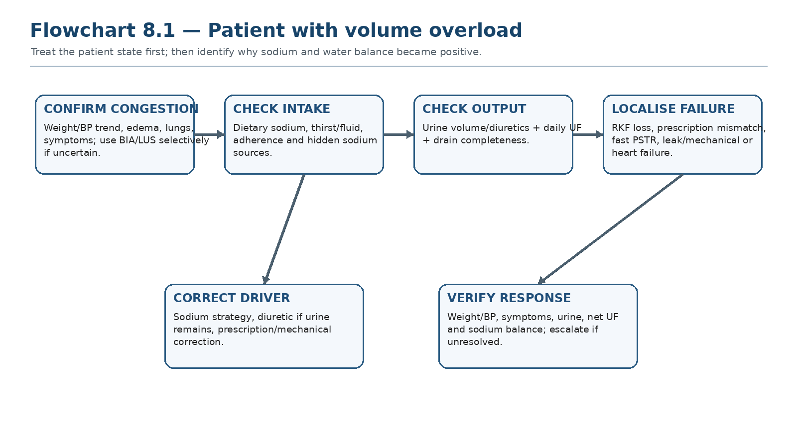
Confirm the patient state, then work through intake, residual kidneys, delivered PD and membrane/mechanical causes before escalating osmotic therapy.
Table 8.13 — Volume overload differential in bedside order.
| Domain | Clues | Next action |
|---|---|---|
| Sodium intake | Thirst, recurrent rapid weight gain | Dietary source analysis |
| Fluid intake | Intake clearly exceeds urine + UF | Behavior/goal review |
| RKF loss | Falling urine, recent illness/hypotension | Timed urine/RKF reassessment; reversible causes |
| Under-delivery | Missed exchanges, cycler lost therapy | Restore delivery |
| Mechanical/drain | Incomplete drains, alarms, constipation | Catheter/drain pathway |
| Fast transport/prescription mismatch | Good early UF, poor long dwell | Shorten glucose dwell; icodextrin long dwell |
| Intrinsic membrane dysfunction | Low standardized UF + blunted sodium dip | High-glucose PET pathway |
| Cardiac/venous disease | Congestion disproportionate to weight/UF | Heart-failure/venous assessment |
13. Low daily UF is not automatically ultrafiltration failure
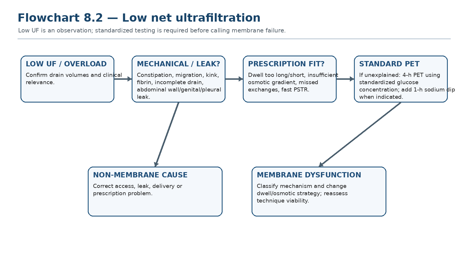
Daily drain data are screening information. True membrane dysfunction requires standardized interpretation and exclusion of non-membrane causes.
A low net UF report can be caused by incomplete drainage, catheter migration, constipation, leaks, fluid sequestration, missed exchanges, wrong solution, dwell mismatch or true membrane dysfunction. ISPD explicitly notes that low UF capacity can arise from mechanical problems, leaks or increased fluid absorption not explained by fast PSTR. [2]
Table 8.14 — Low UF: first exclusions before PET.
| Question | If yes | Why |
|---|---|---|
| Was all dialysate instilled and drained? | Correct delivery/recording | Avoid false low UF |
| Is there constipation or catheter dysfunction? | Treat mechanics | No membrane test can fix obstruction |
| Any abdominal wall/genital/pleural leak? | Leak pathway | Dialysate may leave cavity without appearing in drain |
| Was glucose strength/dwell as prescribed? | Correct prescription delivery | Standardized comparison requires known conditions |
| Recent peritonitis? | Allow recovery and reassess when appropriate | Inflammation can transiently alter transport |
14. Standardized recognition of low UF capacity
The 2021 ISPD membrane guideline provides standardized screening thresholds. Insufficient UF should be suspected when net UF after a 4-h PET is <400 mL using 3.86% glucose/4.25% dextrose, or <100 mL using 2.27% glucose/2.5% dextrose, and/or when daily UF is insufficient to maintain adequate fluid status. These are test-context thresholds, not universal daily UF targets. [2]
Table 8.15 — ISPD low-UF criteria: use exactly in context.
| Test context | Threshold | Interpretation |
|---|---|---|
| 4-h PET with 3.86% glucose / 4.25% dextrose | Net UF <400 mL | Low UF capacity suspected |
| 4-h PET with 2.27% glucose / 2.5% dextrose | Net UF <100 mL | Low UF capacity suspected |
| Routine daily therapy | UF insufficient to maintain adequate fluid status | Clinical low-UF problem; evaluate mechanism |
| THRESHOLD DISCIPLINE The 400-mL criterion belongs to a standardized 4-hour high-glucose PET. It is not a minimum daily UF target and should never be applied to a random home exchange. [2] |
|---|
15. Sodium dip: diagnosing impaired osmotic conductance
When intrinsic membrane dysfunction is suspected, ISPD recommends supplementing the high-glucose 4-h PET with a 1-h sodium measurement. A sodium dip ≤5 mmol/L and/or a sodium sieving ratio ≤0.03 at 1 hour indicates UF insufficiency due to impaired osmotic conductance/free-water transport. [2]
Table 8.16 — The diagnostic meaning of the 1-hour sodium signal.
| Finding | Physiology | Meaning |
|---|---|---|
| Preserved sodium dip | AQP-mediated free-water transport present | Low UF may be dominated by fast glucose loss or another cause |
| Dip ≤5 mmol/L | Blunted free-water transport | Supports intrinsic membrane dysfunction |
| Sodium sieving ratio ≤0.03 | Same process expressed as ratio | Supports low osmotic conductance |
| Low UF + mechanical/leak evidence | Non-membrane cause remains possible | Fix/confirm anatomy before labeling membrane failure |
16. Mechanisms of ultrafiltration insufficiency
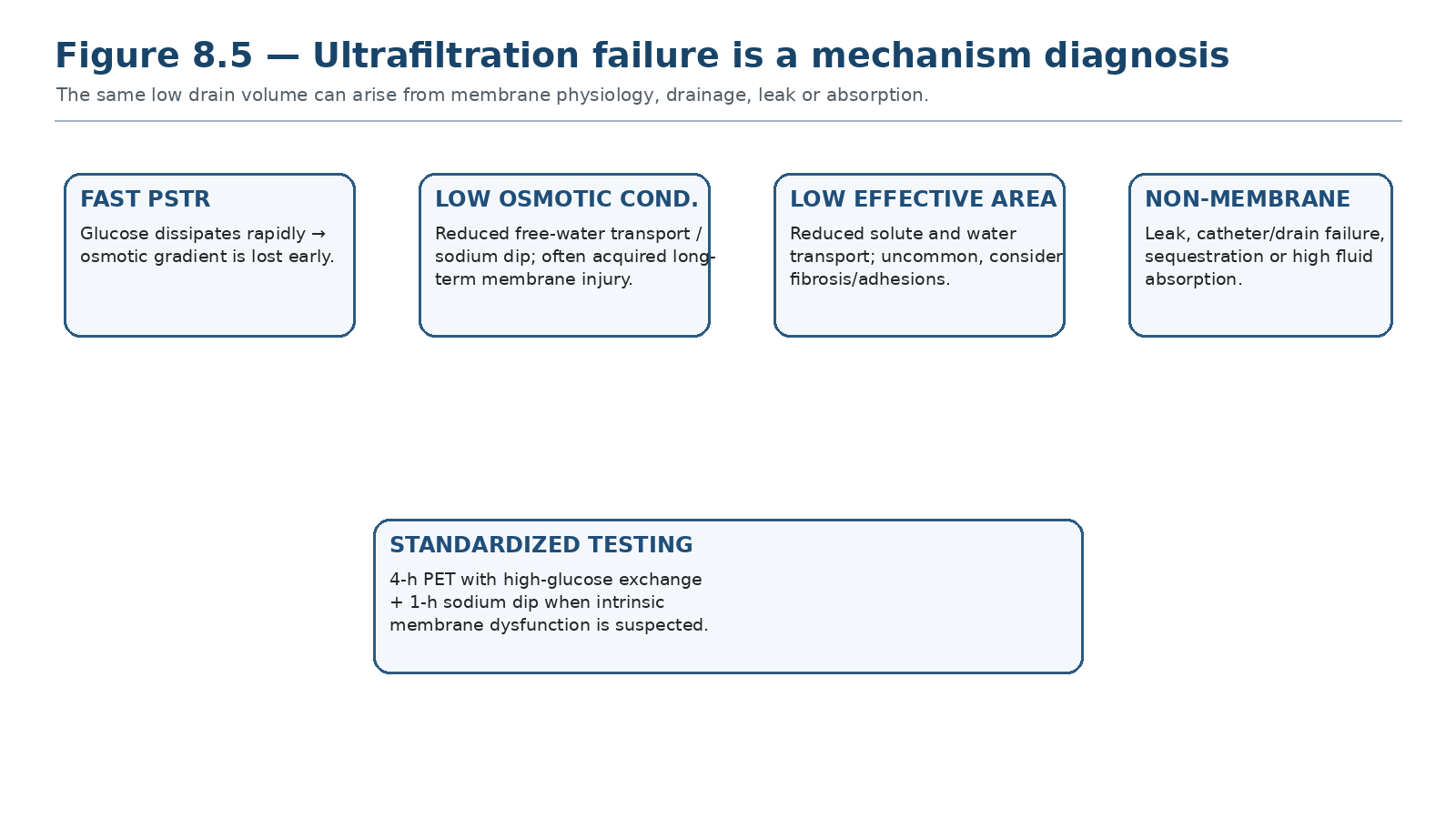
The modern ISPD framework emphasizes fast solute transport, intrinsic low osmotic conductance and other/non-membrane causes rather than relying only on older “type 1/2/3” labels.
Table 8.17 — UF phenotype → physiology → clue → response.
| Phenotype | Physiology | Key clue | Response direction |
|---|---|---|---|
| Fast PSTR | Rapid glucose absorption → osmotic gradient dissipates | High/above-average PSTR; poor long glucose dwell | Shorter glucose dwells; APD; icodextrin long dwell |
| Low osmotic conductance | Reduced free-water transport / membrane fibrosis | Low high-glucose PET UF + blunted sodium dip | Higher osmotic need; reassess long-term membrane viability |
| Low effective surface area | Reduced solute + water transport | Low transport and low UF | Consider fibrosis/adhesions; specialist evaluation |
| High fluid absorption | Peritoneal fluid loss to tissues/lymphatics | Poor UF unexplained by PSTR/sodium dip | Diagnosis of exclusion; advanced testing selectively |
| Mechanical/leak | Dialysate does not drain from cavity normally | Alarms, asymmetry, swelling, hydrothorax | Correct access/leak |
| TERMINOLOGY UPDATE Older “Type 1/2/3 UF failure” language can be useful historically, but current practice should describe the measured mechanism whenever possible. [2] |
|---|
17. Fast PSTR: shorten the glucose dwell, not the patient’s patience
Fast solute transfer rapidly equilibrates dialysate glucose with plasma, shortening the period in which glucose can pull water. The physiologic response is to avoid prolonged glucose dwells that become reabsorptive, while preserving a long dwell with icodextrin when appropriate. ISPD gives strong support to shortening glucose exchanges, using icodextrin and/or higher glucose concentrations to prevent low net UF in fast PSTR. [2]
Table 8.18 — Fast PSTR prescription levers.
| Lever | Why it works | Trade-off |
|---|---|---|
| Shorter glucose dwells | Drain before gradient fully dissipates | More cycles/connections or cycler complexity |
| APD | Automates shorter repeated dwells | Sodium sieving/low sodium removal can emerge |
| Icodextrin long dwell | Sustained long-dwell UF independent of glucose gradient | One daily dwell; product-specific safety |
| Higher glucose strength | Restores stronger gradient | Higher glucose burden |
18. Low osmotic conductance: when more glucose becomes a warning signal
Acquired intrinsic membrane dysfunction can reduce osmotic conductance to glucose, often with a blunted sodium dip and disproportionate loss of free-water transport. When RKF is absent, ISPD notes that hypertonic glucose may become necessary and transfer to hemodialysis may need to be considered. Acquired dysfunction after prolonged PD should also trigger awareness of encapsulating peritoneal sclerosis risk, developed fully in Chapter 14. [2,14]
| RED FLAG A rising requirement for hypertonic glucose to maintain the same volume state is not merely a “stronger prescription.” It can be a marker of declining membrane function and should prompt standardized reassessment. |
|---|
19. Prescription response by phenotype
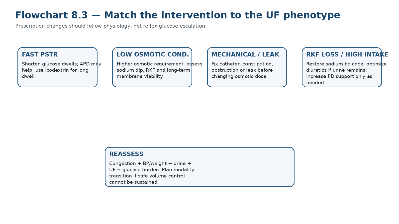
Correct intake, RKF, delivery and mechanical causes first; then use dwell and osmotic levers according to membrane physiology.
Table 8.19 — Problem → mechanism → change → reassessment.
| Problem | Likely mechanism | Targeted change | Reassess |
|---|---|---|---|
| Long-dwell negative UF | Fast PSTR / glucose absorption | Icodextrin or shorten glucose dwell | Long-dwell UF + congestion |
| APD high UF but edema persists | Low sodium removal / high intake | Longer sodium-coupled dwell + sodium strategy | Weight/BP + sodium balance |
| Falling urine + same PD | Loss of renal fluid/sodium removal | Optimize diuretic if responsive + increase PD support | Urine + total balance |
| Low high-glucose PET UF + low sodium dip | Low osmotic conductance | Higher osmotic support; technique viability review | Clinical volume + repeat membrane assessment as indicated |
| Low drains + alarms | Mechanical | Catheter/constipation pathway | Drain completeness |
| Pleural symptoms / genital edema | Leak | Stop/modify intraperitoneal pressure and investigate leak | Resolution + safe restart plan |
20. Heart failure and refractory congestion
Heart failure changes the interpretation of volume. A patient may have severe venous and pulmonary congestion with low arterial pressure, while aggressive attempts to reduce “dry weight” can compromise renal perfusion and RKF. PD can provide gentle continuous decongestion, but the prescription must be integrated with heart-failure therapy, sodium balance and hemodynamic tolerance rather than chasing a single weight target.
Table 8.20 — Congestion in heart failure: special considerations.
| Finding | Interpretation | Management implication |
|---|---|---|
| Low BP + edema/JVP | Venous congestion with low effective perfusion possible | Avoid reflex volume loading or aggressive depletion |
| Repeated pulmonary edema despite UF | Cardiac disease, sodium excess or insufficient total removal | Reassess heart, intake and PD together |
| Large diuretic response | Residual renal reserve remains valuable | Use kidney pathway while monitoring electrolytes/hemodynamics |
| Refractory congestion despite optimized PD | Technique may no longer meet fluid goals | Discuss modality support/transition early |
21. Clinical pearls and common pitfalls
Table 8.21 — High-frequency errors and the correction.
| Pitfall | Why it fails | What to do instead |
|---|---|---|
| Equating edema with UF failure | Congestion has many causes | Name the volume phenotype and balance equation |
| Calling low home drain “membrane failure” | Mechanical/leak causes common | Verify drainage and delivery first |
| Using 400 mL as a daily UF target | It is a standardized PET criterion | Use only in the 4-h high-glucose PET context |
| Treating APD UF as sodium removal | Short dwells can remove free water disproportionately | Assess sodium balance and dwell pattern |
| Escalating glucose before sodium review | Increases glucose burden without fixing input | Address sodium/thirst loop |
| Stopping diuretics because “dialysis started” | Urine output remains clinically valuable | Continue/optimize when responsive and safe |
| Using icodextrin as just “stronger glucose” | Different osmotic mechanism | Use specifically for long-dwell physiology |
| Chasing bioimpedance normality | Can cause iatrogenic depletion | Integrate device data with symptoms/hemodynamics |
| Ignoring falling RKF | Total water/sodium support falls abruptly | Recalculate full prescription |
| No transition plan for progressive membrane failure | Turns next overload into emergency HD | Discuss modality options before crisis |
22. Mini-cases: volume decisions, not trivia
Case 1 — “Good UF,” persistent edema
An APD patient reports 1.2 L nightly UF, but weight and BP rise and edema worsens. Urine is minimal and diet review reveals high sodium intake.
| BEST NEXT STEP Do not intensify glucose simply because the patient is overloaded. APD can produce water-rich UF with insufficient sodium removal; correct sodium intake and review dwell pattern/long dwell and sodium removal. |
|---|
Case 2 — Fast transporter, negative day dwell
A fast transporter on APD has acceptable overnight UF but repeatedly reabsorbs fluid during a long daytime glucose dwell.
| BEST NEXT STEP The glucose gradient is dissipating during the long dwell. Replace the long glucose dwell with icodextrin or redesign dwell duration rather than merely increasing glucose concentration. |
|---|
Case 3 — Sudden low UF after months of stability
Net UF drops abruptly with drain alarms and constipation. There is no new edema yet.
| BEST NEXT STEP Acute mechanical dysfunction is more likely than abrupt intrinsic membrane failure. Treat constipation, verify catheter position/drain completeness and reassess before ordering a membrane-failure workup. |
|---|
Case 4 — Standardized PET abnormality
A high-glucose 4-h PET produces 280 mL net UF and the 1-h sodium dip is 3 mmol/L. Mechanical causes and leaks are excluded.
| BEST NEXT STEP This meets ISPD low-UF criteria and the blunted sodium dip supports intrinsic low osmotic conductance. Reassess RKF, glucose requirement, long-term membrane status and modality viability. |
|---|
Case 5 — Falling urine volume
A patient loses most residual urine over 3 months while the PD prescription is unchanged; edema and hypertension then appear.
| BEST NEXT STEP Total sodium/water removal has fallen even if peritoneal UF is unchanged. Reassess reversible RKF loss, diuretic responsiveness, sodium intake and peritoneal prescription. |
|---|
Case 6 — Heart failure with low BP
A patient with advanced heart failure has edema, elevated JVP and dyspnea but systolic BP is low.
| BEST NEXT STEP Do not assume low BP means intravascular depletion. Integrate cardiac/venous congestion, urine response, sodium intake and PD removal; use gradual decongestion with hemodynamic reassessment and consider adjunct imaging if uncertain. |
|---|
23. Active recall
MUST MEMORIZE
What is the ISPD dietary sodium recommendation for adult PD patients, and what are the exceptions?
What standardized 4-h PET net-UF thresholds define low UF capacity for 3.86%/4.25% and 2.27%/2.5% glucose solutions?
What 1-h sodium-dip and sodium-sieving-ratio thresholds indicate intrinsic UF insufficiency?
Why can APD remove more water than sodium?
What is the preferred long-dwell osmotic agent when glucose long-dwell UF is insufficient?
MUST REASON
A patient is edematous but has 1.5 L/day UF. Give the balance-based differential.
How do you distinguish fast PSTR from low osmotic conductance clinically and with PET?
Why can shortening a glucose dwell improve UF but worsen sodium removal?
What changes when a patient becomes anuric?
When should recurrent hypertonic glucose requirement prompt modality-transition discussion?
USE AS REFERENCE
ISPD standardized UF thresholds and sodium-dip criteria.
Table 8.19 phenotype-based prescription changes.
Table 8.21 high-frequency errors.
24. Flashcards: active recall
1. Q: Volume overload equals UF failure? A: No. Volume overload is a patient state; UF failure is a membrane-function diagnosis.
2. Q: ISPD sodium limit? A: <2 g sodium/day (≈5 g sodium chloride/day) unless contraindicated or volume contracted/hypotensive.
3. Q: High-glucose 4-h PET low-UF threshold? A: Net UF <400 mL with 3.86% glucose/4.25% dextrose.
4. Q: 2.27%/2.5% 4-h PET low-UF threshold? A: Net UF <100 mL.
5. Q: 1-h sodium dip threshold? A: ≤5 mmol/L.
6. Q: Sodium sieving ratio threshold? A: ≤0.03 at 1 h.
7. Q: Why APD sodium removal may lag UF? A: Short dwells favor AQP free-water transport and sodium sieving.
8. Q: Fast PSTR long glucose dwell? A: Shorten it or use icodextrin for the long dwell.
9. Q: Best first step with abrupt low drain? A: Exclude mechanical/drain causes.
10. Q: Icodextrin role? A: Sustained long-dwell colloid osmotic UF, especially useful when glucose gradient dissipates.
11. Q: Diuretics after PD starts? A: Useful if meaningful urine output remains; they increase urine output but are not proven to preserve GFR by themselves.
12. Q: Bioimpedance role? A: Adjunct for uncertain/subclinical volume status; not an absolute fluid-removal target.
13. Q: Persistent overload despite “good” Kt/V? A: Treat sodium/volume domain independently.
14. Q: Falling RKF means? A: Total water/sodium removal has fallen; reassess the whole prescription.
15. Q: Blunted sodium dip means? A: Reduced free-water transport/low osmotic conductance.
16. Q: Low UF + hydrothorax? A: Think leak, not membrane failure.
17. Q: Rising hypertonic-glucose requirement means? A: Possible declining membrane function; reassess, do not normalize.
18. Q: When PD cannot maintain safe euvolemia? A: Plan modality transition rather than waiting for emergency congestion.
25. Rapid troubleshooting table
Table 8.22 — Volume problem → likely mechanism → next step.
| Problem | Likely mechanism(s) | Next step |
|---|---|---|
| Edema + high BP + high UF | Sodium intake / poor sodium removal | Diet + sodium-removal review |
| Edema + low urine + stable UF | RKF loss | Reassess urine/diuretic response + increase PD support as needed |
| Low UF + drain alarms | Mechanical | Constipation/catheter pathway |
| Low UF + abdominal/genital swelling | Leak | Leak assessment and intraperitoneal-pressure strategy |
| Negative long glucose dwell | Fast PSTR | Shorten dwell / icodextrin |
| Low standardized UF + low sodium dip | Low osmotic conductance | Membrane-failure pathway |
| Good Kt/V + congestion | Volume/sodium inadequacy | Do not chase small-solute clearance |
| Low BP + JVP/edema | Heart failure/venous congestion | Hemodynamic + cardiac assessment |
| Repeated hypertonic glucose dependence | Progressive membrane/RKF issue or intake mismatch | Reassess mechanism and technique viability |
26. Final revision sheet
| TEN TAKE-HOME RULES 1) Volume status is a patient state, not a UF number. 2) Sodium balance drives thirst and long-term fluid balance. 3) ISPD recommends <2 g sodium/day unless contraindicated. 4) Preserve and use residual urine whenever possible. 5) Net UF and sodium removal are not interchangeable. 6) APD short dwells can accentuate sodium sieving. 7) Icodextrin is the long-dwell tool when glucose UF is insufficient. 8) Exclude mechanical/leak causes before diagnosing membrane failure. 9) Use the ISPD high-glucose PET and 1-h sodium dip exactly in context. 10) Persistent inability to maintain euvolemia is a modality-planning problem, not an invitation to infinite glucose escalation. |
|---|
Table 8.23 — Final revision grid.
| Core concept | Remember |
|---|---|
| Clinical target | Sustained euvolemia with acceptable BP/symptoms and tolerable burden |
| Sodium | <2 g/day ISPD recommendation unless contraction/hypotension |
| Peritoneal UF | Water removal; does not equal sodium removal |
| Fast PSTR | Shorter glucose dwell + icodextrin long dwell |
| Low-UF PET | <400 mL at 4 h with 3.86%/4.25%; <100 mL with 2.27%/2.5% |
| Intrinsic dysfunction | 1-h sodium dip ≤5 mmol/L and/or sieving ratio ≤0.03 |
| First exclusions | Delivery, constipation/catheter, leak, dwell/solution mismatch |
| Transition trigger | Volume goals cannot be maintained safely despite optimized intake, RKF and PD prescription |
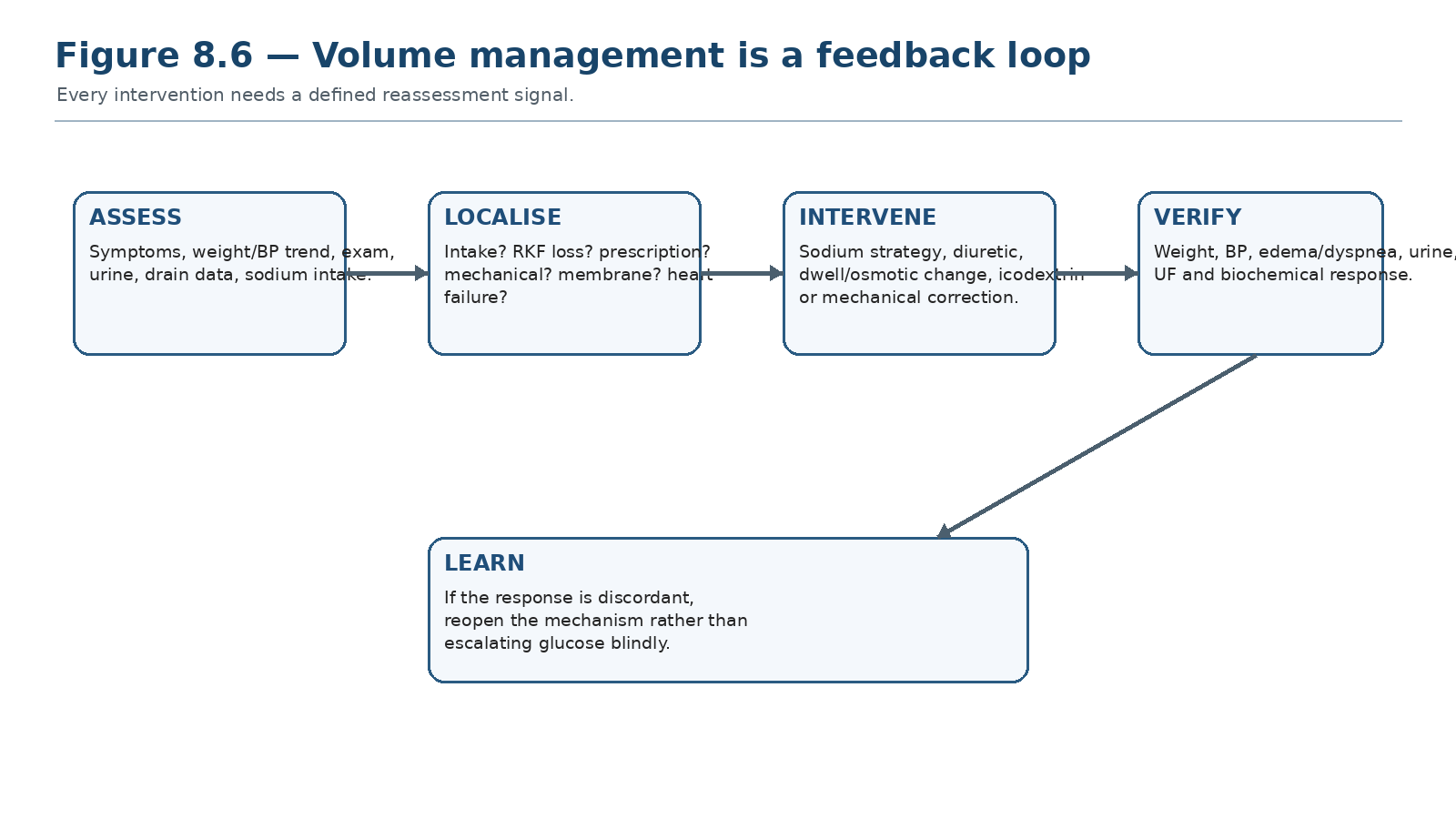
Assess → localize → intervene → verify. If the clinical response does not match the predicted physiology, reopen the diagnosis before escalating osmotic exposure.
| FINAL MENTAL MODEL Volume phenotype → sodium/water input → residual urine → peritoneal UF + sodium removal → delivery/mechanics → standardized membrane testing when needed → phenotype-specific prescription → reassessment → modality planning if goals remain unsafe. |
|---|
Rapid oral viva
Explain why a patient can have 1.5 L/day UF and still be volume overloaded.
Walk through your diagnostic sequence for an abrupt fall in net UF.
Explain sodium sieving and why it matters in APD.
Give the exact ISPD standardized criteria for low UF capacity and intrinsic low osmotic conductance.
How would you modify a prescription for a fast transporter with negative long-dwell glucose UF?
When does increasing hypertonic glucose stop being a reasonable long-term strategy?
| SCOPE BOUNDARY This chapter covers sodium/volume physiology, clinical congestion and ultrafiltration failure. Detailed CAPD/APD prescription optimization follows in Chapter 9; peritonitis in Chapter 10; mechanical complications in Chapter 12; and long-term membrane failure/EPS in Chapter 14. |
|---|
27. Selected authoritative references
1. Brown EA, Blake PG, Boudville N, et al. International Society for Peritoneal Dialysis practice recommendations: Prescribing high-quality goal-directed peritoneal dialysis. Perit Dial Int. 2020;40(3):244–253. https://doi.org/10.1177/0896860819895364. PMID: 32063219.
2. Morelle J, Stachowska-Pietka J, Öberg C, et al. ISPD recommendations for the evaluation of peritoneal membrane dysfunction in adults: classification, measurement, interpretation and rationale for intervention. Perit Dial Int. 2021;41(4):352–372. https://doi.org/10.1177/0896860820982218. PMID: 33563110.
3. Wang AYM, Brimble KS, Brunier G, et al. ISPD Cardiovascular and Metabolic Guidelines in Adult Peritoneal Dialysis Patients Part I: Assessment and Management of Various Cardiovascular Risk Factors. Perit Dial Int. 2015;35(4):379–387. https://doi.org/10.3747/pdi.2014.00279. PMID: 26228782.
4. Goossen K, Becker M, Marshall MR, et al. Icodextrin Versus Glucose Solutions for the Once-Daily Long Dwell in Peritoneal Dialysis: An Enriched Systematic Review and Meta-analysis of Randomized Controlled Trials. Am J Kidney Dis. 2020;75(6):830–846. https://doi.org/10.1053/j.ajkd.2019.10.004. PMID: 32033860.
5. Ronco C, Verger C, Crepaldi C, et al. Baseline hydration status in incident peritoneal dialysis patients: the Initiative of Patient Outcomes in Dialysis (IPOD-PD study). Nephrol Dial Transplant. 2015;30(5):849–858. https://doi.org/10.1093/ndt/gfv013. PMID: 25762355.
6. Vrtovsnik F, Verger C, Van Biesen W, et al. The impact of volume overload on technique failure in incident peritoneal dialysis patients. Clin Kidney J. 2021;14(2):570–577. https://doi.org/10.1093/ckj/sfz175. PMID: 33623681.
7. Shu Y, Liu J, Zeng X, et al. The Effect of Overhydration on Mortality and Technique Failure Among Peritoneal Dialysis Patients: A Systematic Review and Meta-Analysis. Blood Purif. 2018;46(4):350–358. PMID: 30189422.
8. Medcalf JF, Harris KP, Walls J. Role of diuretics in the preservation of residual renal function in patients on continuous ambulatory peritoneal dialysis. Kidney Int. 2001;59(3):1128–1133. https://doi.org/10.1046/j.1523-1755.2001.0590031128.x. PMID: 11231370.
9. Borrelli S, La Milia V, De Nicola L, et al. Sodium removal by peritoneal dialysis: a systematic review and meta-analysis. J Nephrol. 2019;32(2):231–239. https://doi.org/10.1007/s40620-018-0507-1. PMID: 29978446.
10. Rodríguez-Carmona A, Fontán MP. Sodium removal in patients undergoing CAPD and automated peritoneal dialysis. Perit Dial Int. 2002;22(6):705–713. PMID: 12556073.
11. Fischbach M, Zaloszyc A, Schaefer B, Schmitt CP. Optimizing peritoneal dialysis prescription for volume control: increasing sodium removal by applying dialysis mechanics. Perit Dial Int. 2016;36(5):512–517. PMID: 26924063.
12. Fischbach M, Issad B, Dubois V, Taamma R. The beneficial influence on the effectiveness of automated peritoneal dialysis of varying the dwell time and fill volume: a randomized controlled trial. Kidney Int. 2011;80(10):1087–1094. PMID: 21454393.
13. Teitelbaum I. Ultrafiltration failure in peritoneal dialysis: a pathophysiologic approach. Blood Purif. 2015;39(1-3):70–73. https://doi.org/10.1159/000368972. PMID: 25661912.
14. Krediet RT. Physiology of peritoneal dialysis; pathophysiology in long-term patients. Front Physiol. 2024;15:1322493. https://doi.org/10.3389/fphys.2024.1322493. PMID: 39193440.
15. Davies SJ, Davenport A. The role of bioimpedance and biomarkers in helping to aid clinical decision-making of volume assessments in dialysis patients. Kidney Int. 2014;86(3):489–496.
16. Davies SJ, Mushahar L, Yu Z, Lambie M. Determinants of peritoneal membrane function over time. Semin Nephrol. 2011;31(2):172–182. https://doi.org/10.1016/j.semnephrol.2011.01.006. PMID: 21439431.
17. Krediet RT. Acquired decline in ultrafiltration in peritoneal dialysis: the role of glucose. J Clin Med. 2021;10(17):3929. PMID: 34321252.
18. Khursigara MR, Liu P, Kaur R, Mavrakanas TA. Role of SGLT-2 inhibitors in ultrafiltration failure in peritoneal dialysis: a narrative review. Can J Kidney Health Dis. 2024;11:20543581241293500. https://doi.org/10.1177/20543581241293500. PMID: 39502166.
| SOURCE NOTE Guidelines and contemporary evidence were checked 2 September 2026. The ISPD 2021 high-glucose PET and sodium-dip thresholds are reproduced only in their standardized test context. Emerging approaches such as intraperitoneal/systemic SGLT2 inhibition remain investigational and are not presented as established treatment. Local product labeling, glucose-meter compatibility, diuretic dosing and heart-failure protocols remain program specific. |
|---|