Applied Nephrology Master Series
Chapter 10
Peritoneal Dialysis-Associated Peritonitis
Prevention | Recognition | Effluent Analysis | Empiric Therapy | Organism-Specific Treatment | Catheter Decisions | Recovery
| CHAPTER MISSION Recognise PD-associated peritonitis at the first clinically meaningful signal, obtain high-yield microbiology without delaying treatment, start appropriate empiric therapy quickly, use organism and response trajectory to guide treatment duration and catheter decisions, identify surgical and catheter-related sources early, and turn every episode into a prevention intervention. |
|---|
| MASTER PRINCIPLE Cloudy effluent is an emergency signal, not a final diagnosis. Treat suspected infectious peritonitis promptly after obtaining proper specimens, but keep asking a second question in parallel: “What source or organism would make antibiotics alone insufficient?” |
|---|
0. One-page chapter map
Table 10.1 — The eight decisions that govern PD-peritonitis care.
| Decision | Core question | Bedside output |
|---|---|---|
| 1. Threat | Is the patient septic, haemodynamically unstable, or presenting with a surgical abdomen? | Hospital / sepsis / surgical pathway when needed. |
| 2. Confirm | Does the presentation meet the ISPD diagnostic framework? | Named syndrome: peritonitis, mimic, or uncertain. |
| 3. Sample | Was the effluent collected and cultured correctly before antibiotics? | High-yield microbiology + cell-count baseline. |
| 4. Cover | What empiric regimen gives prompt local Gram-positive and Gram-negative coverage? | Documented first-dose time and delivery plan. |
| 5. Identify | What organism and source explain the episode? | Organism-specific therapy + source hypothesis. |
| 6. Track | Is pain, cloudiness and effluent WBC trajectory improving? | Continue, re-investigate, or declare treatment failure. |
| 7. Catheter | Is the catheter a salvageable conduit or an infected biofilm reservoir? | Retain, remove, exchange, or reinsert deliberately. |
| 8. Prevent | Why did this episode occur and what changes before the next exchange? | Root-cause action, retraining, prophylaxis and CQI. |
Learning outcomes
Diagnose PD-associated peritonitis using the current ISPD two-of-three diagnostic framework and apply the APD-specific PMN caveat correctly.
Distinguish infectious peritonitis from hemoperitoneum, eosinophilic/chemical inflammation, chylous effluent and intra-abdominal surgical disease.
Obtain cell count, differential, Gram stain and culture using culture techniques that minimize culture-negative episodes.
Start centre-specific empiric therapy promptly after sampling, covering both Gram-positive and Gram-negative organisms without waiting for laboratory confirmation.
Use local resistance patterns, APD/CAPD schedule, residual kidney function and dwell exposure to judge whether antibiotic delivery is actually adequate.
Interpret the 48-hour, day-3 and day-5 response trajectory and apply the corrected 2024 refractory-peritonitis definition.
Match treatment duration and catheter strategy to common organisms, culture-negative disease, polymicrobial/enteric disease, fungi and mycobacteria.
Distinguish relapsing, recurrent and repeat peritonitis and know when simultaneous catheter removal/reinsertion may be considered.
Apply primary and secondary prevention including wet-contamination management, antifungal prophylaxis during antibiotic courses, procedure prophylaxis, training review and apparent-cause analysis.
Document medical cure and unit-level peritonitis outcomes consistently for quality improvement.
| EVIDENCE POSTURE The current adult ISPD peritonitis guideline remains the 2022 update; the ISPD guideline repository still lists it as the active adult prevention/treatment standard in 2026. Two corrigenda (2023 and 2024) must be read with it, especially the corrected refractory-peritonitis leukocyte threshold. The 2023 catheter-related infection guideline and 2025 PD-training position paper refine source control and prevention. Local microbiology and antimicrobial stewardship govern the final regimen. [1–5] |
|---|
1. Core concept: peritonitis is time-sensitive infection plus source control
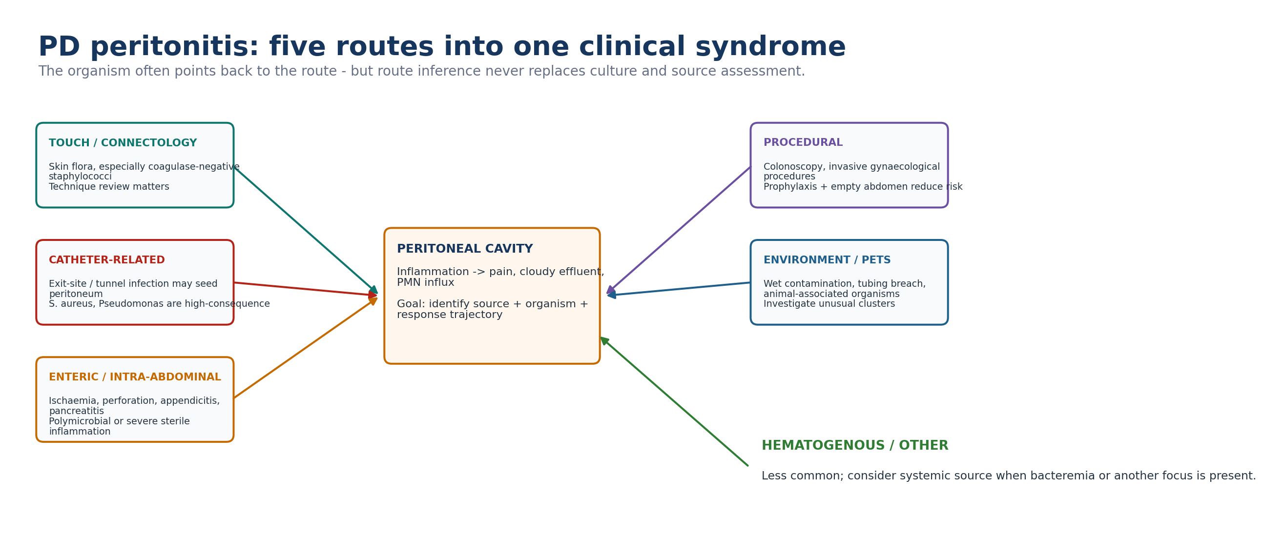
PD-associated peritonitis is inflammation of the peritoneal cavity in a patient receiving peritoneal dialysis, most often caused by infection introduced through touch contamination, a catheter-related route, an enteric/intra-abdominal source or less commonly an environmental or procedural exposure. It matters because an episode can produce severe pain, hospitalisation, catheter removal, transition to haemodialysis, peritoneal membrane injury and death. The acute clinical task is therefore dual: suppress microbial growth quickly and identify whether there is a source that antibiotics cannot eradicate. [1,6]
The first therapeutic error is delay. In the prospective PROMPT study, longer contact-to-treatment time after presentation to a healthcare facility was independently associated with catheter removal or death; the signal is clinically important even though the observational design does not prove a precise causal dose-response. The practical rule is simpler: sample correctly, then treat promptly. [8]
Table 10.2 — Peritonitis is three problems at once.
| Problem | What is happening | Clinical question |
|---|---|---|
| Inflammation | Mesothelial and leukocyte response produces pain, cloudy effluent and transport changes. | How sick is the patient and is the trajectory improving? |
| Microbial burden | Bacteria/fungi may be free in dialysate or embedded in biofilm. | What organism is present and is the antibiotic active at the site? |
| Source | Touch, catheter, bowel, procedure or environmental exposure may continue to seed the cavity. | Will antibiotics alone eradicate the cause? |
| BEDSIDE TRANSLATION The antibiotic name is only one part of treatment. Timing, route, dwell exposure, susceptibility, source control and follow-up determine whether the prescription becomes a cure. |
|---|
2. Diagnosis: use the two-of-three rule
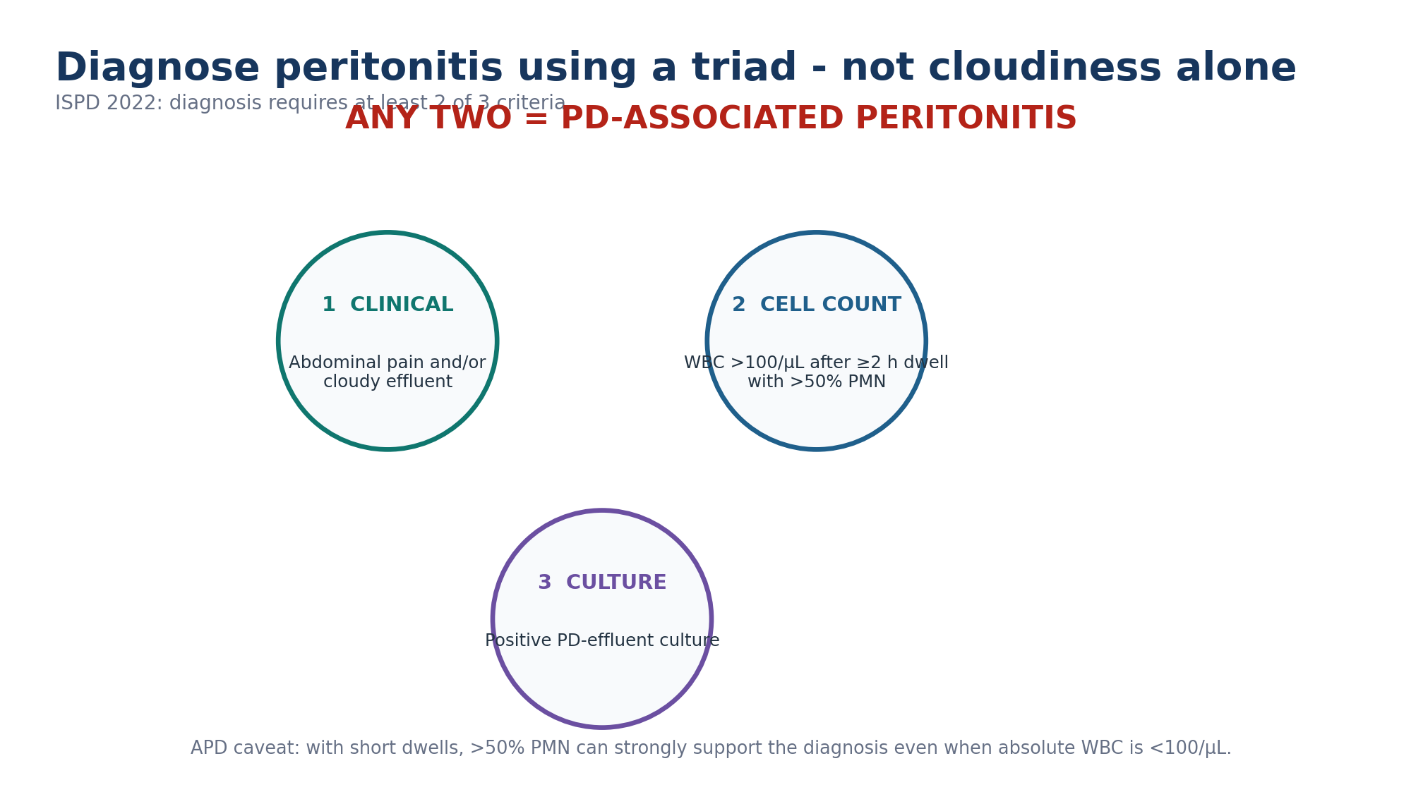
Table 10.3 — ISPD diagnostic criteria.
| Criterion | Operational definition | Interpretation |
|---|---|---|
| Clinical | Abdominal pain and/or cloudy dialysis effluent. | Either may be absent early; cloudiness should trigger urgent evaluation. |
| Effluent inflammation | WBC >100/µL (>0.1 × 10⁹/L) after dwell ≥2 h, with >50% PMN. | Strongly supports infectious peritonitis in the correct context. |
| Microbiology | Positive dialysis-effluent culture. | Identifies cause and guides narrowing/source inference. |
The absolute effluent WBC count depends on dwell duration. In APD patients with rapid cycles, a PMN proportion above 50% is strong evidence even when the absolute WBC is below 100/µL. If an APD patient has no daytime effluent available, the guideline describes instilling 1 L of dialysis solution, dwelling for 2 hours and then draining for inspection and testing. [1]
| CLOUDY-BAG RULE A PD patient presenting with cloudy effluent should be presumed to have peritonitis and treated as such until the diagnosis is confirmed or excluded. Do not wait for fever, severe tenderness or a positive culture. [1] |
|---|
3. Cloudy effluent: infectious until proven otherwise, but not always infection
Table 10.4 — Rapid differential for cloudy or abnormal effluent.
| Pattern | Clues | What separates it from routine bacterial peritonitis |
|---|---|---|
| Bacterial peritonitis | Pain, cloudiness, PMN-predominant leukocytosis. | Usually meets ≥2 diagnostic criteria; culture may be positive or negative. |
| Hemoperitoneum | Pink/red effluent; RBC predominance. | Blood rather than neutrophilic inflammation; investigate gynaecologic, traumatic or intra-abdominal cause. |
| Eosinophilic reaction | Often early after catheter exposure/air/drug; eosinophil predominance. | May be noninfectious; infection still needs exclusion. |
| Chemical / icodextrin-associated inflammation | Temporal relation to solution/exposure; variable cell differential. | Review solution lot/exposure and culture; do not assume sterility. |
| Chylous effluent | Milky appearance; triglyceride-rich. | Not equivalent to neutrophilic cloudy dialysate. |
| Enteric inflammatory process | Severe/localised pain, ileus, systemic toxicity; may be polymicrobial or culture-negative. | Imaging/surgical assessment may be more important than effluent appearance. |
| Pancreatitis / intra-abdominal inflammation | Pain pattern and systemic biochemical clues. | Contiguous sterile inflammation may mimic peritonitis. |
| DO NOT MISCLASSIFY Culture-negative effluent does not automatically mean “culture-negative infectious peritonitis.” Enteric inflammation, chemical/eosinophilic reactions and other mimics must stay in the differential when the clinical course is atypical. |
|---|
4. Pathogenesis: use the organism to reconstruct the source
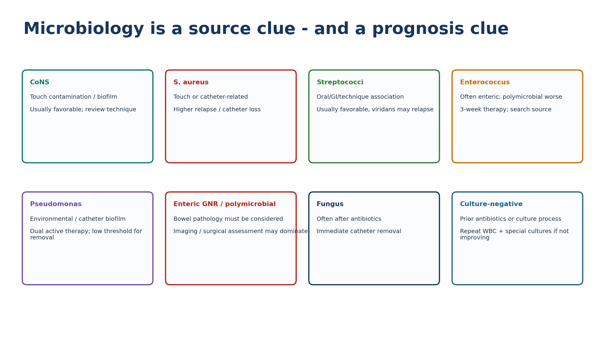
Table 10.5 — Source phenotypes worth recognising.
| Source phenotype | Typical clues | Immediate implication |
|---|---|---|
| Touch contamination | Technique lapse; CoNS; no tunnel abnormality. | Review exchange technique; retraining may prevent recurrence. |
| Catheter-related | Exit-site/tunnel inflammation with same organism or temporal association. | Source-control threshold is lower; cross-reference Chapter 11. |
| Enteric / surgical | Severe or focal abdominal findings, ileus, polymicrobial growth, Gram-positive + Gram-negative mix. | Imaging and surgical assessment; broaden antimicrobial coverage appropriately. |
| Wet contamination | Open-system breach, infused fluid after contamination, proximal tubing leak. | Contact PD team urgently; prophylactic antibiotic pathway if no peritonitis yet. |
| Procedural | Recent colonoscopy or invasive gynaecological procedure. | Assess prophylaxis history and possible enteric flora. |
| Pet/environmental | Animal contact with equipment or tubing; unusual organism. | Environmental history and prevention redesign. |
| Post-antibiotic fungal | Recent antibiotic course. | Immediate catheter removal if fungus identified; prevention should include antifungal prophylaxis during antibiotic courses. |
5. The first 30 minutes: threat assessment before routine treatment
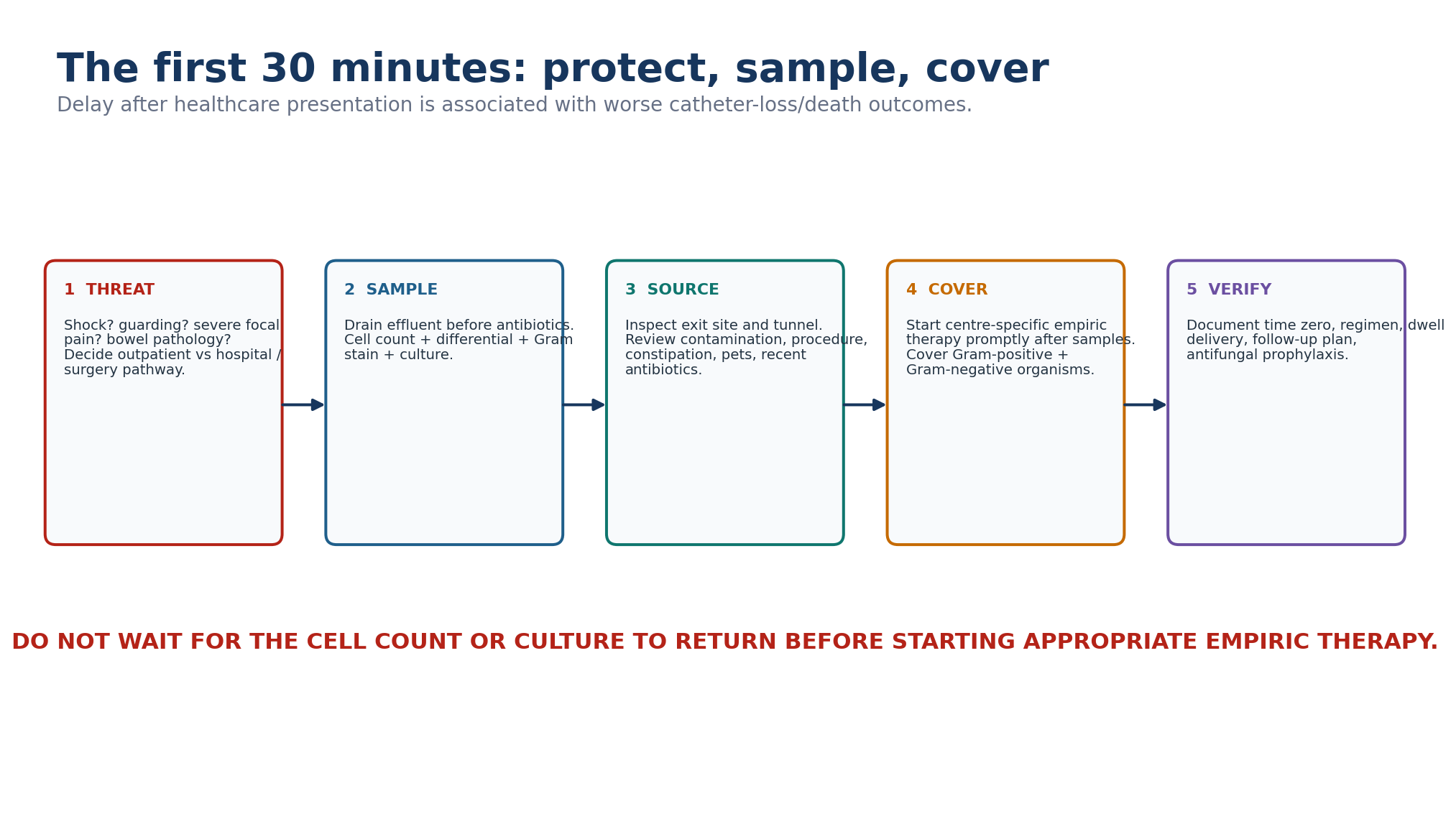
Table 10.6 — Features that should lower the threshold for hospitalisation or urgent escalation.
| Finding | Why it matters |
|---|---|
| Haemodynamic instability / sepsis | Requires resuscitation and systemic sepsis management in parallel with PD-specific treatment. |
| Guarding, rebound, severe focal tenderness or rigid abdomen | Raises concern for bowel pathology or another surgical source. |
| Persistent vomiting, ileus or inability to perform PD safely at home | Outpatient IP therapy may be unreliable. |
| Fungal, polymicrobial enteric, difficult non-fermenter or NTM suspicion | Source control and complex antimicrobial management are more likely. |
| Uncontrolled pain or rapidly worsening clinical status | Clinical deterioration overrides routine outpatient algorithms. |
| Limited social support / inability to administer IP antibiotics | Logistics can make an otherwise stable episode unsafe at home. |
Peripheral blood cultures are usually unnecessary in uncomplicated PD peritonitis but should be obtained when the patient is clinically septic, immunosuppressed or bacteraemia/another systemic focus is suspected. Bacteraemia during PD peritonitis should prompt consideration of another intra-abdominal or systemic source. Routine abdominal radiography is not required merely because peritonitis is suspected; imaging should answer a specific clinical question. [1]
6. Effluent sampling: culture quality is part of treatment
Table 10.7 — What to send whenever peritonitis is suspected.
| Specimen/test | Purpose | Common failure |
|---|---|---|
| Effluent WBC count | Quantifies inflammatory burden and establishes response baseline. | Sampling after a very short dwell can understate WBC. |
| Differential | PMN predominance supports acute infectious inflammation. | Ignoring differential in APD rapid-cycle patients. |
| Gram stain | Low sensitivity but can guide urgency and fungal/Gram pattern clues. | Using a negative result to exclude infection. |
| Effluent culture | Identifies organism and susceptibility. | Wrong container, delayed transport, low inoculum. |
| Special cultures when indicated | Mycobacteria, fungi, Nocardia, fastidious bacteria. | Waiting too long in persistent culture-negative disease. |
ISPD recommends that effluent be sent for cell count with differential, Gram stain and culture whenever peritonitis is suspected. Direct inoculation into blood-culture bottles at the bedside is standard practice in many units. Where resources permit, concentration/centrifugation of effluent and prompt laboratory transport improve organism recovery and help centres meet the culture-negative target. [1]
Table 10.8 — When culture-negative rates are telling you about the system.
| Signal | Interpretation | Unit action |
|---|---|---|
| Culture-negative >15% of peritonitis episodes | Above ISPD target. | Audit collection, inoculation, transport, laboratory methods and recent pre-culture antibiotic exposure. |
| Sudden cluster of culture-negative episodes | Potential process/environmental issue. | Epidemiologic investigation; inspect solution, equipment and sampling chain. |
| Culture negative after 3 days + poor response | Possible unusual organism or wrong diagnosis. | Repeat WBC/differential and request targeted special cultures; reassess source. |
| QUALITY RULE A culture-negative rate is partly a microbiology outcome and partly a unit-process outcome. Treat persistent culture negativity as a diagnostic signal and a quality signal. |
|---|
7. Empiric therapy: cover first, then narrow
Table 10.9 — ISPD empiric-therapy architecture.
| Requirement | Guideline principle | Bedside implementation |
|---|---|---|
| Timing | Start as soon as possible after appropriate specimens are obtained. | Document presentation time, sample time and first-antibiotic time. |
| Spectrum | Cover both Gram-positive and Gram-negative organisms. | Use centre antibiogram and previous patient isolates. |
| Gram-positive | First-generation cephalosporin or vancomycin. | Vancomycin use depends on local methicillin resistance and patient factors. |
| Gram-negative | Third-generation cephalosporin or aminoglycoside. | Consider ESBL/AmpC epidemiology and prior resistant organisms. |
| Alternative | Cefepime monotherapy may be acceptable. | Use only where local susceptibility and programme protocol support it. |
| Route | IP or systemic may be used initially; IP delivers high local exposure when feasible. | Do not delay urgent treatment simply because IP logistics are unavailable. |
Empiric choice should be centre-specific. The largest practical error is a regimen that fails to cover one side of the microbiologic spectrum or is administered so late that a theoretically “correct” prescription becomes ineffective. PDOPPS data support the principle that complete empiric coverage is associated with better cure. [1,6]
Table 10.10 — Route and delivery questions before you say “the patient is on antibiotics.”
| Question | Why it changes treatment |
|---|---|
| Was the antibiotic actually instilled into a dwell long enough for intermittent IP delivery? | ISPD intermittent IP regimens generally assume one exchange daily with a dwell of at least 6 h. |
| Is the patient on APD? | Rapid cycling can shorten antibiotic dwell exposure; regimen may need adaptation. |
| Does substantial residual kidney function increase drug clearance? | Underdosing can occur for renally eliminated antibiotics; follow centre/pharmacy protocol. |
| Is the patient septic or unable to perform PD? | Systemic therapy may be necessary initially; IP can be incorporated when feasible. |
| Are concentrations/levels needed? | Vancomycin/aminoglycoside monitoring is drug- and protocol-specific; do not extrapolate HD targets blindly. |
| THRESHOLD DISCIPLINE This chapter teaches the regimen architecture and guideline durations. Exact drug doses must follow the current ISPD dosing table, local pharmacy protocol, PD modality, dwell schedule, residual kidney function, susceptibility and formulation stability. Do not copy an isolated dose into a different APD/CAPD context. |
|---|
8. Adjunctive care during the acute episode
Table 10.11 — Useful adjuncts and what they do not replace.
| Adjunct | Use | Boundary |
|---|---|---|
| Analgesia | Controls pain and enables exchanges. | Does not substitute for source assessment. |
| IP heparin for fibrin | ISPD notes 500 units/L may help prevent catheter occlusion while effluent is cloudy. | Treats fibrin, not infection. |
| Reduced fill volume when pain is severe | May improve tolerance temporarily. | Antibiotic mass/concentration must remain appropriate. |
| One or two rapid initial exchanges | Sometimes used for pain relief before IP antibiotics. | Extensive rapid lavage has not improved cure/relapse in randomized trials. |
| Antifungal prophylaxis | Co-prescribe during any antibiotic course in PD patients. | Choose nystatin/fluconazole according to availability, interactions and local policy. |
| N-acetylcysteine with aminoglycoside exposure | 2022 guideline introduced an ototoxicity-mitigation recommendation in selected contexts. | Does not eliminate need to minimize prolonged/repeated aminoglycoside exposure. |
9. Follow the response: 48 hours, day 3, day 5
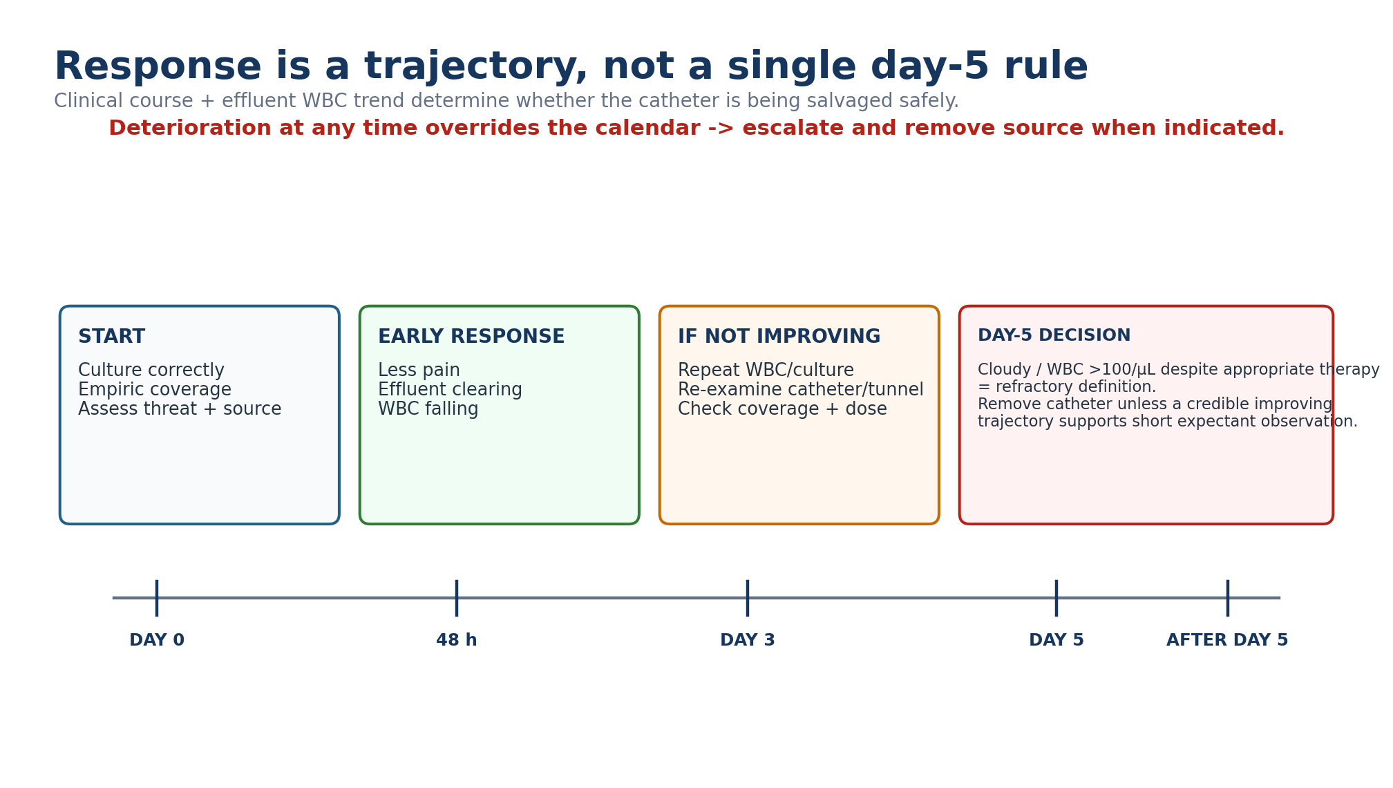
Table 10.12 — Response checkpoints.
| Time | What should be happening | If not |
|---|---|---|
| First 24–48 h | Pain and systemic features begin improving; effluent starts clearing. | Confirm antibiotic delivery, susceptibility, source and need for admission/imaging. |
| ~48 h with poor response | Clinical improvement absent. | Repeat effluent WBC and culture; re-examine exit site/tunnel; reconsider organism/resistance/surgical source. |
| Day 3 | WBC trajectory should be clearly downward in most successful episodes. | High persistent WBC is prognostic, not an automatic catheter-removal threshold; intensify reassessment. |
| Day 5 | If effluent remains cloudy or WBC >0.1 ×10⁹/L after appropriate therapy, episode meets refractory definition. | Catheter removal recommended, unless a credible decreasing trajectory toward normal supports short expectant observation in a stable patient. |
| Any time | Clinical deterioration, sepsis, new surgical signs. | Escalate immediately; do not wait for day 5. |
| CORRIGENDUM ALERT The 2024 corrigendum corrected the refractory-peritonitis leukocyte definition to persistent effluent leukocytes >0.1 × 10⁹/L after 5 days of appropriate antibiotics, not >100 × 10⁹/L. Use the corrected definition. [2] |
|---|
10. Organism-specific treatment: duration reflects biology, not convenience
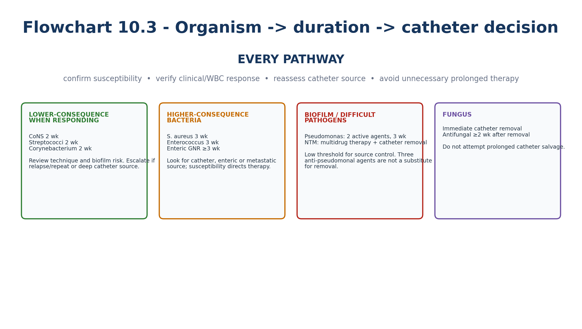
Table 10.13 — High-yield ISPD organism-specific treatment durations.
| Organism / category | Guideline direction | Catheter/source emphasis |
|---|---|---|
| Coagulase-negative Staphylococcus | IP cephalosporin or vancomycin by susceptibility for 2 weeks. | Review technique; consider biofilm/catheter removal in repeat or refractory disease. |
| Staphylococcus aureus | Effective antibiotics for 3 weeks. | Inspect exit site/tunnel carefully; catheter-related disease has lower salvage probability. |
| Streptococci | Appropriate antibiotics for 2 weeks. | Usually favorable; viridans group may relapse and can suggest oral source. |
| Corynebacterium | Effective antibiotics for 2 weeks; vancomycin for beta-lactam-resistant strains. | Early removal if poor response or catheter infection; repeat episodes may reflect biofilm. |
| Enterococcus | 3 weeks: amoxicillin if ampicillin-susceptible or IP vancomycin; VRE requires active alternative. | Polymicrobial enterococcal disease should trigger enteric-source concern. |
| Pseudomonas | Two active antibiotics with different mechanisms for 3 weeks. | Remove catheter with concomitant exit-site/tunnel infection or poor response by day 5. |
| Enteric Gram-negative bacilli | Effective antibiotics for at least 3 weeks. | ESBL/AmpC/resistance and bowel source may drive regimen and removal threshold. |
| Culture-negative, improving | Continue effective initial therapy; 2 weeks is sufficient. | No growth by day 3 + poor response -> special cultures / alternative diagnosis. |
| Fungal | Immediate catheter removal + appropriate antifungal for at least 2 weeks after removal. | Catheter salvage is not recommended. |
| Mycobacterium tuberculosis | Antituberculous therapy is primary treatment. | Routine removal is not automatically required if diagnosis established and patient responding. |
| Non-tuberculous mycobacteria | Effective multidrug therapy + catheter removal. | Request acid-fast studies in persistent culture-negative disease when clinically plausible. |
10.1 Coagulase-negative staphylococci: technique plus biofilm
Coagulase-negative staphylococci are commonly linked to touch contamination. Two weeks of susceptibility-guided therapy is generally sufficient when antibiotic exposure is adequate. A repeat episode—especially after an apparently successful course—should shift the mental model from “another touch event” toward catheter biofilm. Retraining is a treatment intervention, not a punishment. [1]
10.2 Staphylococcus aureus: search for the catheter source
S. aureus peritonitis carries a higher risk of catheter loss and relapse than low-virulence skin flora. The guideline recommends 3 weeks of effective therapy. Simultaneous exit-site or tunnel infection changes the source-control threshold and should be managed with the 2023 catheter-infection guidance. [1,3]
10.3 Pseudomonas: dual therapy and early source control
Pseudomonas behaves as a biofilm-forming difficult pathogen. ISPD suggests two active agents with different mechanisms for 3 weeks. Concomitant exit-site/tunnel infection warrants catheter removal, and lack of clinical response after 5 days should trigger early removal rather than escalation to three anti-pseudomonal drugs. [1]
10.4 Polymicrobial and enteric disease: antibiotics may not be the definitive treatment
Multiple enteric organisms, severe abdominal findings or a mixed Gram-positive/Gram-negative pattern should trigger urgent consideration of bowel pathology. Broad antimicrobial coverage, abdominal imaging and surgical consultation may dominate the case. By contrast, polymicrobial episodes containing only Gram-positive organisms can behave more like touch-contamination peritonitis and may respond to medical therapy. [1]
10.5 Fungal peritonitis: the catheter is the reservoir
Fungal peritonitis is the clearest example of source-control biology. ISPD recommends immediate catheter removal when fungi are identified and continuation of appropriate antifungal therapy for at least 2 weeks after removal. Delayed salvage attempts expose the patient to persistent biofilm infection and high mortality. [1]
11. Culture-negative peritonitis: a diagnosis that should trigger two audits
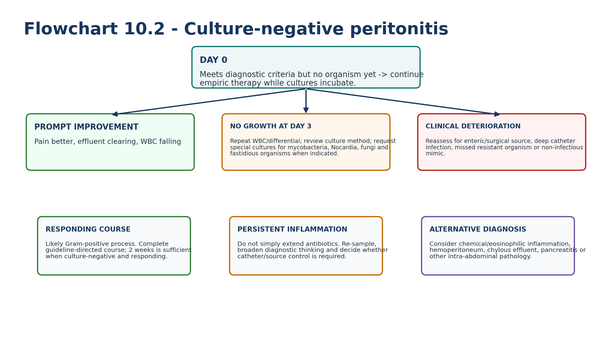
Table 10.14 — Culture-negative peritonitis: two parallel questions.
| Question | Why it matters | Action |
|---|---|---|
| Is this truly infectious peritonitis? | Chemical, eosinophilic, enteric and other inflammatory mimics can meet part of the phenotype. | Reassess differential if course is atypical. |
| Did the laboratory have a fair chance to identify the organism? | Recent antibiotics, poor inoculation, delayed transport and inadequate culture methods increase false-negative results. | Audit collection and repeat/special cultures when needed. |
For episodes with no growth after 3 days, ISPD advises repeat effluent WBC/differential and special culture requests when unusual organisms are plausible. If the patient improves promptly, the initial regimen can be continued, with a 2-week treatment duration. [1,11]
12. Catheter decisions: source control is part of antimicrobial therapy
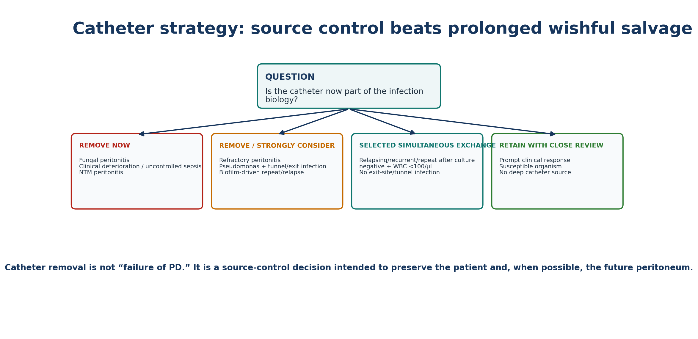
Table 10.15 — Catheter removal / exchange decision matrix.
| Situation | Guideline direction | Reason |
|---|---|---|
| Fungal peritonitis | Immediate removal. | High biofilm burden and mortality with retention. |
| Refractory peritonitis | Remove catheter; short expectant observation only in stable patient with clearly improving WBC trajectory. | Persistent inflammatory burden harms peritoneum and patient. |
| Clinical deterioration before day 5 | Remove/escalate earlier as indicated. | Calendar threshold must not delay source control. |
| Pseudomonas + exit-site/tunnel infection | Remove catheter. | Concordant catheter source is difficult to eradicate. |
| Pseudomonas not responding after 5 days effective therapy | Early removal favored. | A third antibiotic is not a substitute for source control. |
| NTM peritonitis | Antibiotics + catheter removal. | Difficult biofilm-forming infection. |
| Relapsing/recurrent/repeat episodes | Timely removal should be considered. | Biofilm or unresolved source may be driving recurrence. |
| Selected relapse/repeat after control | Simultaneous removal/reinsertion may be considered when culture negative, WBC <100/µL and no exit-site/tunnel infection. | Allows continued PD while removing colonized catheter. |
13. Refractory, relapsing, recurrent and repeat are not synonyms
Table 10.16 — ISPD outcome-specific definitions.
| Term | Definition | Clinical meaning |
|---|---|---|
| Medical cure | Complete resolution with none of: relapse/recurrent peritonitis, catheter removal, HD transfer ≥30 d or death. | Patient-important success, not merely temporary clearing. |
| Refractory | Persistently cloudy bags or effluent leukocytes >0.1 ×10⁹/L after 5 days of appropriate therapy. | Treatment/source-control failure unless trajectory strongly supports delayed response. |
| Relapsing | Within 4 weeks after therapy, same organism or specific organism/culture-negative pair. | Think persistence/biofilm. |
| Recurrent | Within 4 weeks after therapy, different organism. | New episode but often marks vulnerable technique/host/unit. |
| Repeat | >4 weeks after therapy, same organism. | Think recurrent exposure or persistent catheter biofilm. |
| Peritonitis-associated catheter removal | Catheter removed as part of treatment of active episode. | Outcome to report. |
| Peritonitis-associated HD transfer | Any transfer to HD as part of episode. | Capture even if temporary. |
| Peritonitis-associated death | Death attributable/associated according to guideline definition. | Unit quality outcome. |
| LANGUAGE MATTERS “Relapse” means same-organism persistence within 4 weeks; “recurrent” means a different organism within 4 weeks; “repeat” means the same organism after more than 4 weeks. These labels drive different causal thinking. |
|---|
14. Prevention begins before the first episode
Table 10.17 — Primary prevention bundle.
| Intervention | Current evidence/guidance | Practice translation |
|---|---|---|
| Antibiotic prophylaxis before catheter insertion | ISPD 1A recommendation. | Choose agent according to local resistance and access protocol. |
| High-quality training and competency assessment | ISPD guidance; PDOPPS links training practices with peritonitis risk. | Individualized teaching, return-demonstration, reassessment after events. |
| Exit-site/tunnel prevention and prompt treatment | 2023 ISPD catheter guideline. | Cross-reference Chapter 11; catheter infection is a peritonitis risk. |
| Connection-system integrity | Wet contamination carries peritonitis risk. | Immediate PD-team contact after breach; do not improvise at home. |
| Procedure prophylaxis | ISPD suggests prophylaxis for colonoscopy and invasive gynaecological procedures; drain abdomen before procedure. | Create a pre-procedure PD checklist. |
| Pet/environmental precautions | Guideline recognizes pet-associated risk. | Keep animals away from equipment/exchanges; investigate unusual organisms. |
| Medication/host review | Hypokalaemia and H2-receptor antagonist use are modifiable associations highlighted by ISPD. | Correct clinically relevant hypokalaemia; review unnecessary H2RA exposure rather than automatic PPI substitution. |
| Unit surveillance | Track overall/organism-specific rates and susceptibilities. | Local antibiogram should change empiric therapy when microbiology changes. |
15. Wet contamination: prevention before peritonitis develops
Table 10.18 — Dry versus wet contamination.
| Contamination type | Operational idea | Action direction |
|---|---|---|
| Dry contamination | Contamination outside a closed system; clamp remained closed and no contaminated fluid entered. | Contact PD team; prophylactic antibiotics not routinely indicated solely for dry contamination. |
| Wet contamination | Open system with possibility that contaminated fluid entered; proximal leak/breach, open catheter set or touch during active connection. | Immediate PD-team advice; ISPD suggests prophylactic antibiotics. |
| Uncertain clamp status | Cannot confirm whether system was open. | Treat as wet contamination until proven otherwise in local protocol. |
| PREVENTION RULE A patient should never be left to decide alone whether a contamination event “counts.” Every breach gets immediate unit contact and a named dry-vs-wet decision. |
|---|
16. Antifungal prophylaxis: make it automatic when antibiotics are prescribed
ISPD recommends antifungal prophylaxis whenever a PD patient receives an antibiotic course, regardless of the indication. Randomized trials of nystatin or fluconazole and a Cochrane synthesis support reduced subsequent fungal peritonitis. The guideline cites oral nystatin 500,000 units four times daily or fluconazole 200 mg every 48 hours as studied prophylaxis regimens, but local availability, interactions, QT/hepatic considerations, resistance ecology and pharmacy policy must guide the chosen regimen. [1,14,15]
Table 10.19 — Antifungal prophylaxis safety check.
| Question | Why |
|---|---|
| Is nystatin available? | Avoids systemic azole interactions where available. |
| If fluconazole is used, what interacts? | Warfarin, calcineurin inhibitors, some statins and other drugs may require review. |
| Is prophylaxis prescribed for non-peritonitis antibiotic courses too? | ISPD recommendation applies regardless of antibiotic indication. |
| Is the stop date linked to the antibacterial course? | Prevents unplanned prolonged antifungal exposure. |
17. Every episode gets an apparent-cause analysis
Table 10.20 — Root-cause review after peritonitis.
| Domain | Questions | Corrective action examples |
|---|---|---|
| Technique | Any touch contamination, rushing, visual/hand limitation, skipped steps? | Observed exchange + targeted retraining. |
| Connection/equipment | Bag, tubing, transfer set or cycler breach? | Replace component, clarify wet-contamination plan, manufacturer review if cluster. |
| Exit site/tunnel | Recent drainage, pain, cuff/tunnel abnormality? | Treat catheter infection; cross-reference Chapter 11. |
| Bowel | Constipation, diarrhoea, GI procedure, diverticular disease symptoms? | Bowel regimen, procedure prophylaxis, surgical/GI assessment where indicated. |
| Medication | Recent antibiotic course without antifungal prophylaxis? Hypokalaemia? H2RA? | Correct medication/process gap. |
| Environment/pets | Animals near exchange area? water/air/environment cluster? | Environmental redesign; unit investigation for unusual clusters. |
| Microbiology process | High culture-negative rate or delayed transport? | Laboratory/process intervention. |
| Patient capability | Vision, dexterity, cognition, caregiver support changed? | Retraining, assisted PD or modality redesign rather than blame. |
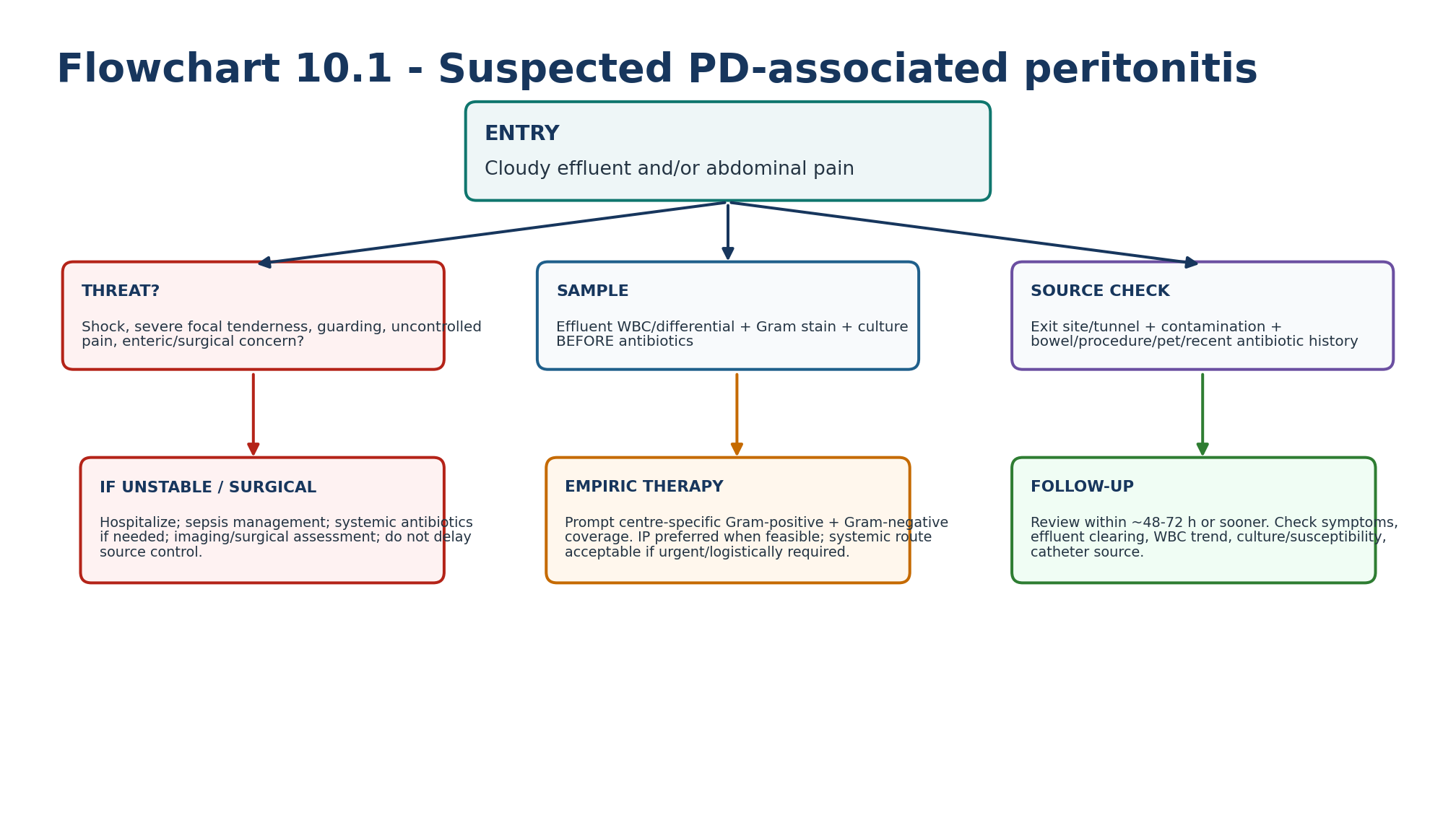
18. Major clinical algorithms
19. Retention tables: pattern recognition
Table 10.21 — If you see this, think this first.
| Finding | First hypothesis | Immediate action |
|---|---|---|
| Cloudy bag + mild diffuse pain | PD peritonitis. | Send effluent and start empiric therapy promptly. |
| Cloudy bag + rigid/focal abdomen | Enteric/surgical source until excluded. | Hospital assessment, imaging/surgery as indicated. |
| Pain on APD with WBC 80/µL but 85% PMN | Peritonitis still likely despite low absolute WBC. | Use PMN proportion and dwell context; treat after sampling. |
| No growth day 3 + improving WBC | Culture-negative bacterial episode likely. | Continue effective initial therapy; complete 2 weeks. |
| No growth day 3 + WBC rising | Unusual organism, wrong regimen, wrong source or noninfectious mimic. | Repeat studies; special cultures; source re-evaluation. |
| Same CoNS 6 weeks later | Repeat peritonitis. | Think biofilm/re-exposure; consider catheter strategy. |
| Same S. aureus 2 weeks after therapy | Relapsing peritonitis. | Think persistent catheter/source; timely removal consideration. |
| Pseudomonas + tunnel drainage | Catheter-related difficult infection. | Dual active therapy + catheter removal. |
| Yeast on culture | Fungal peritonitis. | Immediate catheter removal + antifungal. |
| Recent systemic antibiotics without prophylaxis + fungal peritonitis | Preventable secondary risk. | Treat episode and fix antifungal-prophylaxis process. |
Table 10.22 — What not to confuse.
| Do not confuse | With | Correction |
|---|---|---|
| Cloudy effluent | Automatic culture-positive bacterial infection | Use diagnostic triad and differential. |
| Culture negative | No infection | Could reflect prior antibiotics or poor culture method. |
| Day-5 WBC >100/µL | Automatic removal regardless of trajectory | Refractory definition matters, but stable delayed responders with falling WBC may be observed briefly. |
| Relapse | Recurrent | Relapse = same organism/sterile pairing within 4 weeks; recurrent = different organism within 4 weeks. |
| Antibiotics completed | Medical cure | Medical cure excludes relapse/recurrent, catheter removal, HD transfer ≥30 d and death. |
| More antibiotics | Better source control | Fungal/NTM/refractory/biofilm disease may require removal. |
| Exit site looks normal | No catheter source | Tunnel infection or intraluminal biofilm can still exist. |
| IP route | Guaranteed adequate exposure | Dwell time, APD cycle, drug clearance and preparation matter. |
20. Clinical pearls
1. Peritonitis diagnosis is clinical + cellular + microbiologic; culture is not required if the other two criteria are met.
2. In APD, PMN percentage can be more informative than absolute WBC when dwell times are short.
3. Obtaining the specimen first is important; waiting for the result before starting therapy is not.
4. If the patient is unstable, systemic sepsis care and surgical evaluation proceed in parallel with PD-specific treatment.
5. Culture-negative peritonitis that is not improving by day 3 is a diagnostic problem, not a reason to blindly continue the same plan.
6. Fungal peritonitis is a source-control emergency: remove the catheter immediately.
7. Pseudomonas plus tunnel/exit infection is a catheter-removal phenotype.
8. Repeated prolonged antibiotic courses can select fungus and resistance without curing catheter biofilm.
9. Every antibiotic course in a PD patient should automatically trigger an antifungal-prophylaxis decision.
10. Every peritonitis episode should end with a prevention action, even when the immediate cure is uncomplicated.
21. Common pitfalls — and the correction
Table 10.23 — High-frequency errors in PD-peritonitis care.
| Pitfall | Why it fails | Correction |
|---|---|---|
| Waiting for lab confirmation before antibiotics | Delays treatment in a time-sensitive infection. | Sample promptly, then cover empirically. |
| No differential count | Misses APD/atypical diagnostic signal. | Always request WBC + differential. |
| Culture in a sterile pot only | Lower organism recovery. | Use validated blood-culture bottle / concentration method. |
| Empiric regimen covers only Gram-positive organisms | Misses severe Gram-negative disease. | Cover both sides according to local antibiogram. |
| Copying CAPD dosing into rapid APD | May underexpose the organism. | Check dwell and modality-specific protocol. |
| Calling day-5 cloudiness “refractory” without checking trend | May remove a catheter in a delayed responder. | Use symptoms + WBC trajectory, while respecting safety threshold. |
| Trying a third drug for Pseudomonas instead of removing source | Biofilm persists. | Use two active agents + timely source control. |
| Treating fungus with catheter retention | High failure/mortality risk. | Immediate removal. |
| No antifungal prophylaxis with antibiotics | Increases secondary fungal risk. | Hardwire prophylaxis into antibiotic order set. |
| No post-episode technique review | Misses preventable recurrence. | Apparent-cause analysis + retraining when indicated. |
22. Mini-cases: decisions, not trivia
Case 1 — Short-dwell APD
A patient on nocturnal APD presents at noon with diffuse abdominal pain. A freshly instilled 2-hour dwell returns slightly cloudy; WBC is 82/µL with 88% PMN.
| BEST NEXT STEP The low absolute WBC does not exclude peritonitis. The dwell is adequate and PMN predominance is strong evidence. Obtain culture and start prompt empiric Gram-positive/Gram-negative coverage. |
|---|
Case 2 — Culture negative but improving
Day 3: cultures remain negative. Pain has nearly resolved, effluent is clearer and WBC has fallen markedly.
| BEST NEXT STEP This is a responding culture-negative episode, commonly due to Gram-positive organisms or culture-process limitations. Continue effective initial therapy and complete the guideline 2-week course; review culture technique if the unit rate is high. |
|---|
Case 3 — Day-5 delayed responder
On day 5 the patient is clinically well. Effluent is still faintly cloudy and WBC is 180/µL, down from 4,500/µL on day 1.
| BEST NEXT STEP The episode technically meets the refractory threshold, but the patient has a clear downward trajectory. ISPD allows short expectant observation beyond day 5 in a stable patient when WBC is decreasing toward normal. Continue close review; deterioration changes the decision immediately. |
|---|
Case 4 — Pseudomonas plus tunnel pain
Pseudomonas grows from effluent. The exit site is erythematous and there is tenderness along the tunnel.
| BEST NEXT STEP This is a high-probability catheter-related difficult infection. Use two active anti-pseudomonal agents and remove the catheter rather than attempting prolonged salvage. |
|---|
Case 5 — Same organism after 2 weeks
Two weeks after completing treatment for S. aureus peritonitis, the patient develops another episode with the same organism.
| BEST NEXT STEP This is relapsing peritonitis. Search for catheter/tunnel source and biofilm; timely catheter removal should be considered rather than repeating the same course indefinitely. |
|---|
Case 6 — Yeast after antibiotics
A patient completed antibiotics for pneumonia without antifungal prophylaxis. Two weeks later, peritonitis culture grows Candida.
| BEST NEXT STEP Remove the PD catheter immediately and treat with an appropriate antifungal for at least 2 weeks after removal. The event also exposes a prevention-system failure: antifungal prophylaxis should accompany future antibiotic courses. |
|---|
23. Active recall
MUST MEMORIZE
Table 10.24 — Core facts.
| Prompt | Answer |
|---|---|
| ISPD diagnosis? | At least 2 of 3: compatible symptoms/cloudy effluent; WBC >100/µL after ≥2 h with >50% PMN; positive culture. |
| APD caveat? | >50% PMN strongly supports diagnosis even if absolute WBC <100/µL with short dwells. |
| Overall unit peritonitis target? | ≤0.40 episodes per patient-year at risk. |
| Culture-negative target? | <15% of all peritonitis episodes. |
| Patients free of peritonitis target? | >80% per year. |
| Refractory definition? | Persistently cloudy bags or effluent leukocytes >0.1 ×10⁹/L after 5 days of appropriate antibiotics. |
| CoNS duration? | 2 weeks. |
| S. aureus duration? | 3 weeks. |
| Streptococcal duration? | 2 weeks. |
| Enterococcus duration? | 3 weeks. |
| Pseudomonas? | Two active agents, 3 weeks; catheter source-control logic. |
| Fungal? | Immediate catheter removal + antifungal ≥2 weeks after removal. |
| Culture-negative responding? | 2 weeks. |
| Antibiotic course in any PD patient? | Co-prescribe antifungal prophylaxis. |
MUST REASON
Why a rapid-cycle APD effluent WBC of 80/µL with 90% PMN can still represent peritonitis.
Why a culture-negative episode can be either an infection-management problem or a laboratory-process problem.
Why clinical deterioration should trigger catheter/source-control escalation before day 5.
Why a day-5 WBC of 180/µL can justify different decisions depending on its trajectory and the patient’s clinical state.
Why Pseudomonas tunnel infection and fungal peritonitis are biologically different from uncomplicated CoNS peritonitis.
Why a “successful” antibiotic course is not medical cure if the episode relapses or forces prolonged HD transfer.
How the same organism occurring at 2 weeks versus 8 weeks changes the label and the source hypothesis.
USE AS REFERENCE
Exact IP and systemic antibiotic doses by agent, CAPD/APD schedule and residual kidney function.
Local antibiogram thresholds for empirical vancomycin or resistant Gram-negative coverage.
Drug compatibility/stability in specific PD solutions and bags.
Antifungal selection in Candida versus mould peritonitis.
NTM/TB multidrug regimens and duration.
Procedure-specific prophylaxis protocols and local surgical pathways.
24. Flashcards: spaced repetition
1. Q: Cloudy effluent: first assumption? A: Peritonitis until confirmed or excluded.
2. Q: Three diagnostic criteria? A: Symptoms/cloudy effluent; inflammatory effluent WBC/PMN; positive effluent culture.
3. Q: What makes APD diagnosis tricky? A: Short dwells can keep absolute WBC low; PMN proportion is key.
4. Q: First microbiology bundle? A: Effluent WBC/differential, Gram stain and culture.
5. Q: Empiric spectrum? A: Gram-positive + Gram-negative.
6. Q: Empiric Gram-positive choices? A: First-generation cephalosporin or vancomycin.
7. Q: Empiric Gram-negative choices? A: Third-generation cephalosporin or aminoglycoside; cefepime monotherapy may be acceptable.
8. Q: Why prompt antibiotics? A: Delay after healthcare presentation is associated with catheter loss/death.
9. Q: Day-5 threshold? A: Cloudy effluent or leukocytes >0.1×10⁹/L despite appropriate therapy = refractory definition.
10. Q: Same organism within 4 weeks? A: Relapsing.
11. Q: Different organism within 4 weeks? A: Recurrent.
12. Q: Same organism after >4 weeks? A: Repeat.
13. Q: Fungal catheter management? A: Immediate removal.
14. Q: Pseudomonas with tunnel infection? A: Remove catheter.
15. Q: Culture-negative + improving? A: Continue effective initial therapy for 2 weeks.
16. Q: No growth after 3 days + poor response? A: Repeat WBC, special cultures and rethink diagnosis/source.
17. Q: Antifungal prophylaxis timing? A: Whenever a PD patient receives any antibiotic course.
18. Q: Best prevention response after an episode? A: Apparent-cause analysis + targeted intervention/retraining.
25. Rapid differential / troubleshooting
Table 10.25 — Peritonitis troubleshooting from problem to action.
| Problem | Differential | First actions |
|---|---|---|
| Cloudy effluent, minimal pain | Early bacterial peritonitis; chemical/eosinophilic; chylous effluent. | Cell count/differential + culture; inspect effluent appearance; treat as peritonitis until excluded. |
| Severe pain but low WBC | Very early infection; short APD dwell; bowel pathology; pancreatitis. | Repeat adequate dwell sample; assess PMN; examine abdomen; image if surgical concern. |
| No clinical response by 48 h | Wrong drug, resistant organism, catheter source, enteric disease, fungal/NTM, wrong diagnosis. | Repeat WBC/culture; review susceptibility/delivery; inspect catheter/tunnel; image/escalate as indicated. |
| Effluent remains cloudy day 5 | Refractory episode or delayed responder. | Assess trend; remove catheter unless stable with clearly decreasing WBC toward normal. |
| Recurrent CoNS episodes | Repeated touch contamination or catheter biofilm. | Observed technique + retraining; consider catheter exchange/removal. |
| Polymicrobial Gram+ and Gram− | Enteric source. | Broad coverage, imaging, surgical assessment. |
| Culture-negative cluster in unit | Sampling/laboratory/environmental problem. | CQI investigation, culture-process audit, environmental review. |
| Fungal after recent antibiotics | Secondary fungal peritonitis. | Immediate catheter removal + antifungal; fix prophylaxis process. |
26. Final revision sheet
| CORE CONCEPT PD-associated peritonitis is a time-sensitive inflammatory syndrome in which success depends on five linked tasks: diagnose, culture, cover, identify source, and verify response. Catheter removal is a source-control tool, not a defeat. |
|---|
Table 10.26 — One-minute revision.
| Domain | Must remember |
|---|---|
| Diagnosis | ≥2 of 3 criteria; PMN >50% is crucial in rapid-cycle APD. |
| Sampling | WBC/differential + Gram stain + culture before antibiotics. |
| Empiric therapy | Prompt, centre-specific, Gram+ and Gram− coverage. |
| Response | Clinical improvement should begin by 48–72 h; follow WBC trajectory. |
| Refractory | Day-5 cloudy/WBC >0.1×10⁹/L despite appropriate therapy; remove unless stable delayed responder with falling WBC. |
| Durations | CoNS 2 wk; S. aureus 3 wk; strep 2 wk; Coryne 2 wk; Enterococcus 3 wk; Pseudomonas dual therapy 3 wk; culture-negative responding 2 wk. |
| Catheter | Fungus remove now; NTM remove; Pseudomonas + catheter infection remove; relapse/repeat/recurrent consider timely removal. |
| Prevention | Insertion prophylaxis, training, exit-site care, wet-contamination plan, procedure prophylaxis, antifungal with every antibiotic course. |
| Quality | Unit target ≤0.40 episodes/patient-year; culture-negative <15%; >80% patients peritonitis-free/year. |
| After cure | Apparent-cause analysis and prevention intervention. |
TEN TAKE-HOME RULES 1) Cloudy effluent is peritonitis until proven otherwise. 2) Diagnose using two of three ISPD criteria. 3) In APD, trust the PMN proportion when the dwell is short. 4) Sample first, then treat promptly. 5) Cover Gram-positive and Gram-negative organisms empirically. 6) Reassess at 48 h, day 3 and day 5 using the WBC trajectory. 7) Fungus and NTM require catheter source control; Pseudomonas often does. 8) Relapse/recurrent/repeat are different entities. 9) Give antifungal prophylaxis with every antibiotic course. 10) Every episode ends with apparent-cause analysis and prevention redesign.
Table 10.27 — One-minute bedside synthesis.
| If you see… | Think… | Do now… |
|---|---|---|
| Cloudy effluent + pain | PD peritonitis | Effluent studies + prompt empiric therapy. |
| WBC <100/µL, PMN >50% on rapid APD | Peritonitis still likely | Use dwell context; culture and treat. |
| Rigid/focal abdomen | Enteric/surgical disease | Hospital + imaging/surgical review. |
| No growth day 3, improving | Culture-negative bacterial episode | Continue effective course; 2 weeks. |
| No growth day 3, worsening | Unusual organism / wrong source / mimic | Special cultures + diagnostic reset. |
| Same organism 2 weeks later | Relapse | Biofilm/source; catheter strategy. |
| Pseudomonas + tunnel infection | Catheter-related difficult infection | Dual therapy + remove catheter. |
| Fungus | Biofilm source-control emergency | Remove catheter immediately. |
| Any antibiotic course | Secondary fungal risk | Add antifungal prophylaxis. |
| Episode resolved | Prevention opportunity | Apparent-cause analysis + retraining/process fix. |
| FINAL MENTAL MODEL Threat -> diagnostic triad -> correct effluent sample -> prompt Gram+/Gram− coverage -> organism/source -> response trajectory -> catheter decision -> medical cure -> apparent-cause analysis. |
|---|
Rapid oral viva
Give the ISPD diagnostic criteria and explain the APD short-dwell exception.
Walk through the first 30 minutes of a cloudy-bag presentation.
Differentiate refractory, relapsing, recurrent and repeat peritonitis.
Explain why culture-negative peritonitis can be a laboratory-quality problem.
Compare catheter strategy for CoNS, Pseudomonas, fungal and NTM peritonitis.
Explain why extensive lavage and indiscriminate prolonged antibiotics are not routine solutions.
Build a prevention plan after a touch-contamination CoNS episode and after fungal peritonitis following systemic antibiotics.
| SAFETY BOUNDARY This chapter teaches clinical reasoning and guideline architecture. Exact antimicrobial dosing, compatibility, stability, therapeutic drug monitoring, allergy substitution, resistant-organism therapy, fungal species treatment and timing of catheter reinsertion require the current ISPD tables, local microbiology, renal pharmacy, infectious-disease and PD programme protocols. Surgical abdomen and sepsis pathways supersede routine outpatient PD logic. |
|---|
27. Selected authoritative references
1. Li PKT, Chow KM, Cho Y, et al. ISPD peritonitis guideline recommendations: 2022 update on prevention and treatment. Perit Dial Int. 2022;42(2):110–153. https://doi.org/10.1177/08968608221080586. PMID: 35264029.
2. Corrigendum to 2022 ISPD Peritonitis Guidelines: 2022 update on prevention and treatment. Perit Dial Int. 2024;44(3):223. https://doi.org/10.1177/08968608241251453. PMID: 38643390.
3. Chow KM, Li PKT, Cho Y, et al. ISPD Catheter-related Infection Recommendations: 2023 Update. Perit Dial Int. 2023;43(3):201–219. https://doi.org/10.1177/08968608231172740. PMID: 37232412.
4. Chow JSF, Brunier G, Figueiredo AE, et al. Teaching peritoneal dialysis: A position paper for the International Society for Peritoneal Dialysis. Perit Dial Int. 2025;45(6):327–343. https://doi.org/10.1177/08968608251375512. PMID: 40966019.
5. Hurst H, Figueiredo AE, Perez Moran D, et al. Peritoneal dialysis training and interventions: A narrative review. Perit Dial Int. 2026;46(1):6–15. https://doi.org/10.1177/08968608251328517. PMID: 40221963.
6. Perl J, Fuller DS, Bieber BA, et al. Peritoneal Dialysis-Related Infection Rates and Outcomes: Results From the Peritoneal Dialysis Outcomes and Practice Patterns Study (PDOPPS). Am J Kidney Dis. 2020;76(1):42–53. https://doi.org/10.1053/j.ajkd.2019.09.016. PMID: 31932094.
7. Al Sahlawi M, Zhao J, McCullough K, et al. Variation in Peritoneal Dialysis-Related Peritonitis Outcomes in the Peritoneal Dialysis Outcomes and Practice Patterns Study (PDOPPS). Am J Kidney Dis. 2022;79(1):45–55.e1. https://doi.org/10.1053/j.ajkd.2021.03.022. PMID: 34052357.
8. Muthucumarana K, Howson P, Crawford D, Burrows S, Swaminathan R, Irish A. The Relationship Between Presentation and the Time of Initial Administration of Antibiotics With Outcomes of Peritonitis in Peritoneal Dialysis Patients: The PROMPT Study. Kidney Int Rep. 2016;1(2):65–72. https://doi.org/10.1016/j.ekir.2016.05.003. PMID: 29142915.
9. Kitrungphaiboon T, Puapatanakul P, Chuengsaman P, et al. Intraperitoneal Cefepime Monotherapy Versus Combination Therapy of Cefazolin Plus Ceftazidime for Empirical Treatment of CAPD-Associated Peritonitis: A Multicenter, Open-Label, Noninferiority, Randomized, Controlled Trial. Am J Kidney Dis. 2019;74(5):601–609. https://doi.org/10.1053/j.ajkd.2019.05.011. PMID: 31331757.
10. Htay H, Cho Y, Pascoe EM, et al. Multicentre registry data analysis comparing outcomes of culture-negative peritonitis and different subtypes of culture-positive peritonitis in peritoneal dialysis patients. Perit Dial Int. 2020;40(1):47–56. PMID: 32063153.
11. Szeto CC, Wong TY, Chow KM, et al. The clinical course of culture-negative peritonitis complicating peritoneal dialysis. Am J Kidney Dis. 2003;42(3):567–574.
12. Htay H, Cho Y, Pascoe EM, et al. Outcomes of Corynebacterium Peritonitis: A Multicenter Registry Analysis. Perit Dial Int. 2017;37(6):619–626. https://doi.org/10.3747/pdi.2017.00028. PMID: 28698253.
13. Szeto CC, Chow KM, Kwan BCH, et al. Staphylococcus aureus peritonitis complicates peritoneal dialysis: review of 245 consecutive cases. Clin J Am Soc Nephrol. 2007;2(2):245–251.
14. Chang TI, Kim HW, Park JT, et al. Early catheter removal improves patient survival in peritoneal dialysis patients with fungal peritonitis: results of ninety-four episodes. Perit Dial Int. 2011;31(1):60–66.
15. Lo WK, Chan CY, Cheng SW, et al. A prospective randomized control study of oral nystatin prophylaxis for Candida peritonitis complicating continuous ambulatory peritoneal dialysis. Am J Kidney Dis. 1996;28(4):549–552.
16. Restrepo C, Chacon J, Manjarres G. Fungal peritonitis in peritoneal dialysis patients: successful prophylaxis with fluconazole, as demonstrated by prospective randomized control trial. Perit Dial Int. 2010;30(6):619–625.
17. Kraft L, Oppold J, Kitterer D, et al. Changing microbiology and outcomes of PD-associated peritonitis over four decades. Clin Kidney J. 2026;19(1):sfaf387. https://doi.org/10.1093/ckj/sfaf387. PMID: 41536568.