Applied Nephrology Master Series
Chapter 11
Exit-Site and Tunnel Infections
Recognition | Depth | Culture | Ultrasound | Antibiotics | Pseudomonas | Source Control | Prevention
| CHAPTER MISSION Recognise catheter-related infection before superficial inflammation becomes tunnel disease or peritonitis; distinguish true exit-site infection from trauma, dermatitis, colonisation and granuloma; define the anatomical depth of infection; obtain useful microbiology; select and reassess antimicrobial treatment according to organism and response; and recognise the point at which an infected foreign body requires surgical source control rather than another antibiotic course. |
|---|
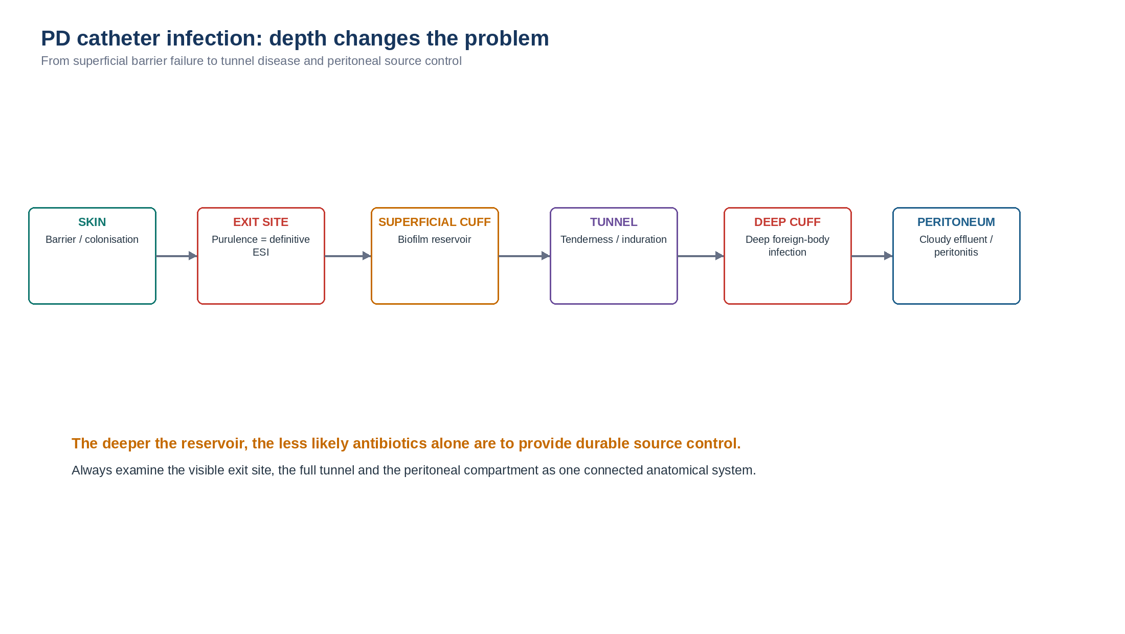
| MASTER PRINCIPLE Do not treat the redness; treat the anatomical infection phenotype. Every suspected catheter infection requires four linked questions: Is this truly infection? How deep does it extend? What organism is present? Is the catheter still salvageable? |
|---|
0. One-page chapter map
Table 11.1 — The eight decisions that govern PD catheter-infection care.
| Decision | Core question | Bedside output |
|---|---|---|
| 1. Confirm | Is this definitive exit-site infection or noninfectious inflammation/colonisation? | Named exit-site phenotype |
| 2. Depth | Is disease confined to the exit site or extending along the tunnel/cuff? | ESI versus TI |
| 3. Peritoneum | Is there cloudy effluent, abdominal pain or same-organism peritonitis? | Isolated catheter infection versus catheter-related peritonitis |
| 4. Culture | What organism is present and is the isolate clinically meaningful? | Culture + susceptibility interpretation |
| 5. Cover | What empiric treatment is appropriate while culture is pending? | Organism-risk-adjusted antimicrobial plan |
| 6. Track | Is purulence, inflammation and tunnel disease resolving? | Response at approximately 1 week |
| 7. Source control | Is the catheter now an infected foreign-body reservoir? | Preserve, salvage, replace or remove |
| 8. Prevent | Why did infection occur and how is recurrence prevented? | Exit-site care + immobilisation + CQI |
Learning outcomes
Define definitive exit-site infection using the current ISPD clinical definition.
Explain why erythema, tenderness, crusting or granuloma without purulent discharge do not alone establish definitive ESI.
Define tunnel infection and recognise when tunnel ultrasonography adds clinically useful information.
Distinguish true infection from colonisation, mechanical trauma, contact dermatitis and granulation tissue.
Perform a systematic exit-site-tunnel-peritoneum assessment.
Obtain and interpret catheter-related microbiology without treating colonisation as infection.
Select empiric treatment that covers Staphylococcus aureus while modifying treatment for prior MRSA or Pseudomonas risk and local susceptibility patterns.
Apply the 2023 ISPD response-adapted duration strategy for uncomplicated ESI.
Recognise why Pseudomonas, tunnel infection and nontuberculous mycobacteria require different management logic.
Identify refractory catheter-related infection and escalate from antimicrobial therapy to catheter source control appropriately.
Use cuff procedures, exit-site relocation, simultaneous catheter replacement and catheter removal selectively rather than interchangeably.
Build an exit-site prevention and catheter-infection surveillance programme.
| EVIDENCE POSTURE The 2023 ISPD Catheter-related Infection Recommendations remain the principal adult guideline framework for ESI and TI. They clarified diagnostic definitions, introduced an overall ESI target of <=0.40 episodes/year at risk, replaced the routine fixed 2-week treatment concept with response-adapted treatment for uncomplicated ESI, retained >=3 weeks for Pseudomonas ESI and any TI, and expanded catheter-salvage options. The 2022 ISPD peritonitis guideline governs concomitant peritonitis and antifungal prophylaxis. Contemporary salvage and NTM evidence remains predominantly observational, so source-control decisions are anatomy- and patient-specific. [1–6] |
|---|
1. Core concept: catheter infection is infection plus foreign-body biology
A PD catheter is not merely an opening in the skin. It is a continuous foreign body traversing the exit site, superficial cuff, subcutaneous tunnel and deep cuff before entering the peritoneal cavity. Organisms can therefore remain superficial, track inward along tissue planes, adhere to catheter material or cuff fibres, and persist within biofilm. [1,3]
This explains the clinical continuum from skin-barrier failure to exit-site infection, tunnel infection and catheter-related peritonitis. The continuum is not inevitable, but observational data show that ESI is followed by an increased risk of subsequent peritonitis, particularly in the period soon after the catheter infection. [7]
Table 11.2 — Catheter infection is three problems at once.
| Problem | What is happening | Clinical question |
|---|---|---|
| Skin barrier failure | Trauma, colonisation or local inflammation permits microbial entry. | Is this truly infection? |
| Anatomical extension | Infection may spread from the exit site toward one or both cuffs. | How deep is it? |
| Foreign-body persistence | Catheter/cuff biofilm may prevent eradication despite in-vitro susceptibility. | Can antibiotics cure this without source control? |
| BEDSIDE TRANSLATION A susceptible organism plus a correct antibiotic does not guarantee cure. If infection persists, ask whether the problem is drug failure - or an infected catheter that the drug cannot sterilise. |
|---|
2. Definitions: use the ISPD language precisely
Definitive exit-site infection
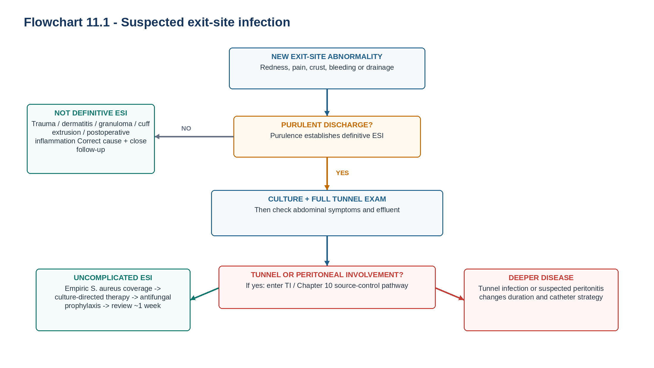
ISPD suggests defining definitive ESI as purulent discharge at the catheter-epidermal interface, with or without surrounding erythema. In the absence of purulent discharge, erythema, tenderness, swelling, granuloma or crust formation are insufficient by themselves to establish definitive ESI. [1]
Table 11.3 — Exit-site findings and what they mean.
| Finding | Interpretation | Clinical consequence |
|---|---|---|
| Purulent discharge +/- erythema | Definitive ESI | Culture and treat; examine tunnel |
| Erythema alone | Not sufficient for definitive ESI | Consider irritation/trauma; monitor for evolution |
| Tenderness alone | Inflammation; cause uncertain | Assess traction, tunnel and local lesion |
| Granuloma without pus | Granulation/inflammation | Treat local tissue problem; do not assume infection |
| Crust alone | Does not define infection | Inspect after appropriate site care |
| Positive culture + normal site | Colonisation | Do not diagnose ESI from microbiology alone |
| DEFINITION DISCIPLINE Do not convert every red exit site into ESI. Conversely, erythema without pus can represent evolving early infection and deserves surveillance rather than dismissal. |
|---|
Tunnel infection
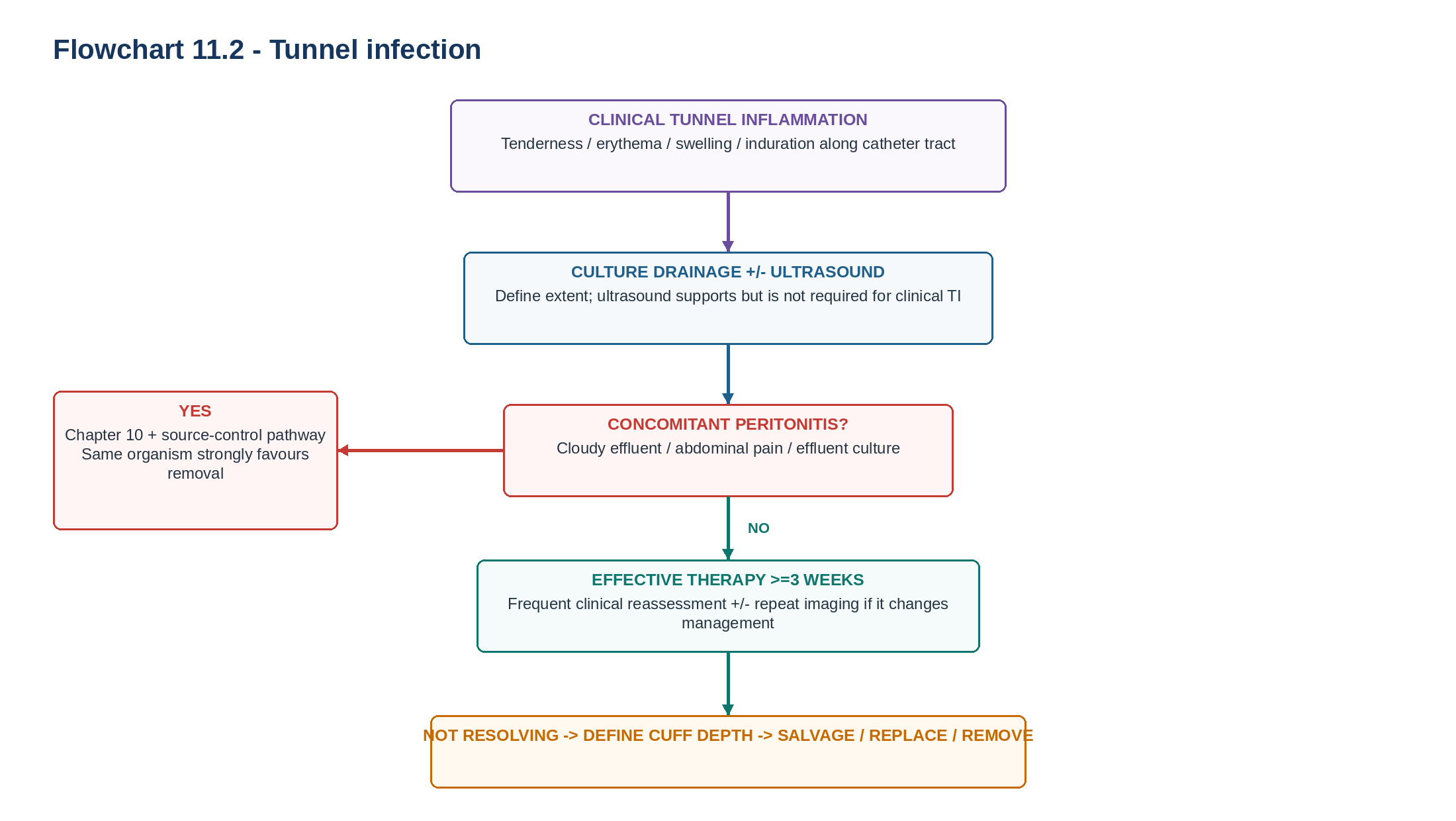
Tunnel infection is defined clinically by erythema, swelling, tenderness and/or induration along the subcutaneous catheter tunnel, with or without ultrasonographic evidence of fluid surrounding the catheter. Ultrasound can support the diagnosis and define extent but is not required for the clinical definition. [1]
Catheter insertion-related infection
ESI or TI occurring within 30 days of catheter insertion is classified as catheter insertion-related infection. This quality marker helps identify problems in insertion, perioperative prophylaxis, postoperative wound care and early catheter handling. [1]
Refractory catheter-related infection
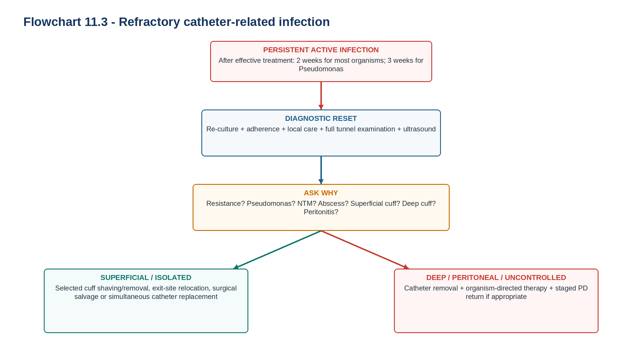
ISPD defines refractory catheter-related infection as failure to respond after 2 weeks of effective antibiotic therapy plus appropriately intensified exit-site care, or after 3 weeks when the infection is caused by Pseudomonas species. [1]
| WORDING MATTERS "Two weeks of antibiotics" is not the definition. The antimicrobial must have been effective against the organism and accompanied by appropriate catheter-site care. |
|---|
3. A red exit site is a differential diagnosis - not a prescription
Exit-site appearance should be interpreted relative to the patient's usual baseline. Several noninfectious processes can resemble ESI, and unnecessary antibiotic treatment creates resistance, adverse effects and fungal risk without correcting the underlying mechanical or dermatologic problem.
Table 11.4 — Red or abnormal exit site: rapid differential.
| Phenotype | Typical clues | Corrective direction |
|---|---|---|
| True ESI | Purulent discharge +/- erythema | Culture + antimicrobial pathway |
| Mechanical trauma | Recent pull/twist, bleeding, tenderness, no pus | Immobilise catheter; correct traction |
| Contact dermatitis | Pruritus, geometric erythema, new adhesive/cleanser | Remove offending product |
| Granulation tissue | Friable tissue, minor bleeding, moisture | Local granuloma care; assess separately for infection |
| Early postoperative inflammation | Recent insertion; expected wound evolution possible | Assess timing, trajectory and surgical complications |
| Cuff extrusion | Visible superficial cuff, chronic local irritation | Determine whether infection coexists |
| Colonisation | Positive swab but clinically healthy site | Do not treat as ESI automatically |
Catheter traction deserves particular attention. Repetitive movement can produce microtrauma at the epithelial interface and compromise the local barrier. ISPD therefore recommends immobilising the catheter as an infection-prevention strategy. [1]
| CLINICAL PEARL If the redness exactly follows a new adhesive, cleansing product or repeated catheter pull, the diagnostic question changes from "Which antibiotic?" to "What damaged the barrier?" |
|---|
4. Examine the catheter as a tract - not as a hole in the skin
The common examination error is inspecting the visible exit site and stopping. A complete catheter-infection examination moves from outside inward: catheter-epidermal interface -> superficial cuff -> full palpable tunnel -> deeper cuff region -> peritoneal symptoms and effluent.
Table 11.5 — Bedside catheter examination.
| Step | What to assess | Why it matters |
|---|---|---|
| Look | Purulence, erythema, crust, bleeding, granuloma, visible cuff | Defines the exit-site phenotype |
| Palpate | Tenderness, swelling, induration | Detects occult tunnel disease |
| Trace | Entire catheter course | Prevents missing proximal infection |
| Express gently when appropriate | Drainage appearing from the tract | Supports tunnel involvement |
| Inspect fixation | Tension, twisting, mobile external segment | Identifies a recurring mechanical driver |
| Check effluent | Cloudiness +/- abdominal pain | Detects possible peritonitis |
| TUNNEL RULE Every definitive ESI deserves a tunnel examination. A clean tunnel cannot be assumed merely because the visible exit site appears mild. |
|---|
5. Tunnel ultrasound: image a clinical question
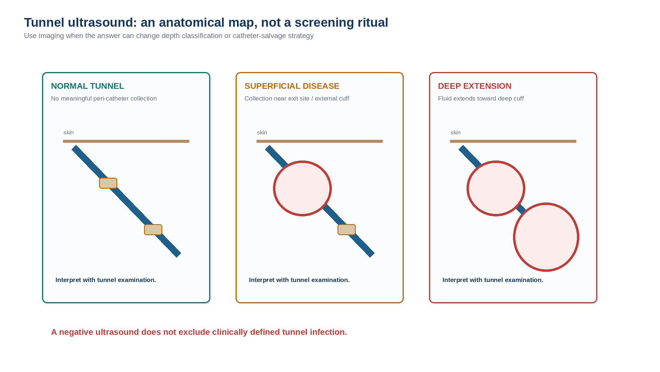
Ultrasonography is most useful when the result can change the diagnosis, duration of treatment or source-control strategy. It can demonstrate peri-catheter fluid, localise disease around the superficial cuff and identify extension toward the deep cuff. Routine screening of an asymptomatic tunnel is not required. [1,13,14]
Table 11.6 — When tunnel ultrasound is most useful.
| Clinical question | Why ultrasound helps | Management consequence |
|---|---|---|
| ESI + tunnel tenderness | Defines deeper extension | Reclassify as TI when appropriate |
| Persistent infection despite active therapy | Searches for a hidden reservoir | Moves toward source-control assessment |
| Pseudomonas or S. aureus with concerning examination | Detects cuff/tunnel disease | Lowers threshold for escalation |
| External cuff infection | Tests whether disease is truly superficial | Selects potential salvage candidate |
| Planning cuff/exit-site salvage | Maps remaining uninfected catheter | Avoids leaving deep infection behind |
| Residual focal tenderness after surface improvement | Assesses incomplete anatomical resolution | Continue/escalate rather than declaring cure |
| DO NOT AUTOMATE Tunnel ultrasound is not a ritual test for every catheter. Use it when the answer can change depth classification, response assessment or catheter-salvage strategy. |
|---|
6. Microbiology: culture infection, not colonisation
When purulent drainage is present, obtain an exit-site swab for culture and susceptibility testing before or at antimicrobial initiation when feasible. Gram stain may be useful when there is a drainable purulent component. A positive swab from a clinically normal exit site represents colonisation rather than definitive ESI. [1]
Table 11.7 — Microbiology result -> interpretation.
| Result | Clinical interpretation | Next question |
|---|---|---|
| S. aureus + purulent ESI | Clinically important pathogen | MRSA? tunnel involvement? recurrent source? |
| MRSA + ESI | Requires MRSA-active treatment | What is local susceptibility and allergy profile? |
| Pseudomonas + ESI | Difficult catheter pathogen | Is there occult tunnel/cuff involvement? |
| Corynebacterium/diphtheroid + healthy site | Colonisation may be more likely | Is there true purulence/inflammation? |
| Repeated diphtheroid + refractory drainage | NTM misidentification possible | Request AFB/mycobacterial studies |
| No growth + improving ESI | Prior antibiotic or limited yield possible | Is clinical resolution complete? |
| No growth + worsening disease | Atypical organism or wrong diagnosis | Repeat/special cultures and diagnostic reset |
Culture-negative ESI can follow recent antibiotics, suboptimal specimen collection, slowly growing organisms, atypical mycobacteria, fungal infection or misclassification of a noninfectious lesion. [1]
| MICROBIOLOGY RULE The swab identifies the organism after the clinical syndrome is established. It does not independently create the diagnosis. |
|---|
7. The first visit: six tasks before the patient leaves
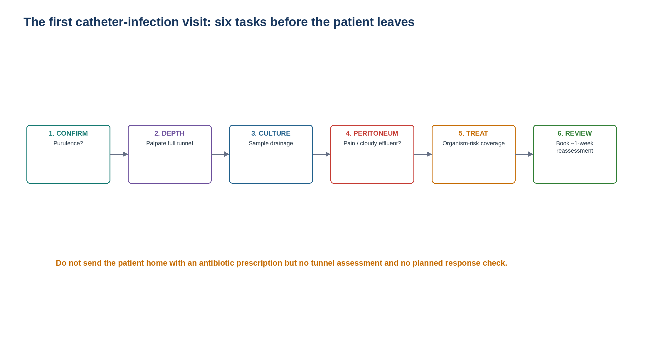
Table 11.8 — First-visit checklist.
| Task | Bedside action | Failure if omitted |
|---|---|---|
| 1. Confirm phenotype | Is purulent discharge present? | Overdiagnosis or missed true ESI |
| 2. Examine tunnel | Palpate the entire subcutaneous tract | Occult TI is missed |
| 3. Culture | Swab purulent drainage | Empiric treatment cannot be narrowed |
| 4. Check peritoneum | Ask about pain and inspect effluent | Catheter-related peritonitis is missed |
| 5. Treat | Use organism-risk and local microbiology | Initial coverage may be inappropriate |
| 6. Follow | Review at approximately 1 week | Duration/failure decisions are delayed |
During active ESI, ISPD suggests at least daily exit-site inspection and cleansing. The purpose is not simply hygiene; it allows recognition of worsening inflammation, tunnel extension and early treatment failure. [1]
8. Empiric therapy: cover the likely organism without losing stewardship
For a typical ESI, ISPD recommends empiric oral treatment providing appropriate S. aureus coverage, commonly with a first-generation cephalosporin or an antistaphylococcal penicillin. The empirical strategy changes when there is previous MRSA or Pseudomonas infection/colonisation, relevant allergy or resistant local microbiology. [1]
Table 11.9 — Empiric treatment reasoning.
| Clinical context | Empiric priority | Do not assume |
|---|---|---|
| Typical uncomplicated ESI | S. aureus coverage | That every patient needs broad Gram-negative therapy |
| Previous MRSA | MRSA-active regimen | That a first-generation cephalosporin is adequate |
| Previous Pseudomonas | Antipseudomonal coverage | That surface appearance predicts depth |
| Severe systemic illness | Escalate beyond routine outpatient ESI pathway | That oral therapy alone is sufficient |
| Concomitant peritonitis | Chapter 10 regimen + source-control assessment | That ESI treatment alone treats the peritoneum |
| Refractory/atypical infection | Diagnostic reset and special microbiology | That another routine ESI antibiotic is the answer |
Antifungal prophylaxis
The 2022 ISPD peritonitis guideline recommends antifungal prophylaxis whenever a PD patient receives an antibiotic course, irrespective of the indication. Catheter infection therefore triggers the same secondary-fungal-prevention reflex as any other antibiotic exposure. [2]
| SAFETY HABIT ESI antibiotic started -> antifungal-prophylaxis decision automatically triggered. Do not cure one infection while creating avoidable fungal-peritonitis risk. |
|---|
9. Duration: response determines uncomplicated ESI treatment
The 2023 ISPD update changed the old fixed-duration habit. A mandatory 2-week course is no longer required for every uncomplicated ESI. Duration should be adjusted to clinical response, culture and susceptibility. If clinical reassessment at approximately 1 week confirms complete resolution of an uncomplicated ESI, a total course of 7-10 days may be sufficient. [1]
Table 11.10 — Duration framework.
| Phenotype | Current ISPD direction | Clinical safeguard |
|---|---|---|
| Uncomplicated ESI with confirmed resolution | 7-10 days may suffice | Resolution must be verified clinically at approximately 1 week |
| ESI not resolved at ~1 week | Continue/reassess according to organism and mechanism | Search for resistance, adherence or tunnel disease |
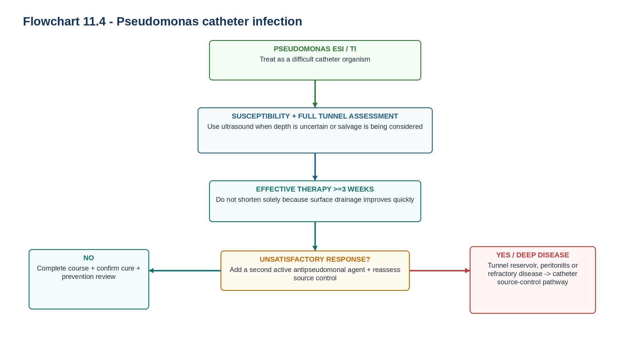
| Pseudomonas ESI | >=3 weeks effective therapy | Do not shorten because surface drainage improves early |
| Any tunnel infection | >=3 weeks effective therapy | Follow anatomy and response |
| Refractory infection | Source-control assessment | Avoid indefinite antibiotic extension |
| THRESHOLD DISCIPLINE Seven to ten days applies only when uncomplicated ESI has actually resolved on clinical reassessment. It is not permission to give every catheter infection a short course. |
|---|
10. Response trajectory: the 1-week review is part of treatment
The antimicrobial prescription is incomplete until response is checked. At approximately 1 week, reassess purulent drainage, erythema, tenderness, the entire tunnel, culture/susceptibility, adherence, local care, adverse effects and any new peritoneal symptoms. [1]
Table 11.11 — One-week response phenotypes.
| Finding | Interpretation | Action |
|---|---|---|
| Purulence gone; inflammation resolved | Good uncomplicated response | Complete appropriate 7-10-day total course |
| Improving but residual inflammation | Partial response | Confirm active drug; continue and reassess |
| Persistent purulence | Ongoing infection | Re-culture; assess tunnel and source |
| New tunnel tenderness | Disease progression | TI pathway + ultrasound when useful |
| New cloudy effluent | Possible peritonitis | Enter Chapter 10 pathway immediately |
| Worsening despite susceptible isolate | Biofilm/depth/adherence/wrong diagnosis | Diagnostic reset |
| Repeated culture-negative failure | Atypical organism possible | Special microbiology including NTM |
| TRAJECTORY RULE The important question is not "Has the patient finished the antibiotics?" but "Has the infected phenotype disappeared?" |
|---|
11. Pseudomonas: a high-risk catheter phenotype
Pseudomonas infection deserves deliberate escalation because eradication from a foreign body can be difficult and tunnel extension, recurrence and catheter loss are clinically important. ISPD recommends at least 3 weeks of effective antibiotic therapy for Pseudomonas ESI. If response is unsatisfactory, a second active antipseudomonal agent should be added. [1]
Table 11.12 — Pseudomonas checklist.
| Step | Required question | Reason |
|---|---|---|
| Culture | Is species identification and susceptibility available? | Ensures true active therapy |
| Tunnel | Is there tenderness or induration? | Finds deeper infection |
| Ultrasound | Is cuff/tunnel involvement present? | Defines anatomy when clinically useful |
| Therapy | Is treatment genuinely antipseudomonal? | Avoids ineffective exposure |
| Duration | Has >=3 weeks been planned? | Prevents premature cessation |
| Response | Is the site and tunnel clearly improving? | Identifies failure early |
| Escalation | Is a second active agent required? | ISPD recommendation for unsatisfactory response |
| Source control | Is the catheter sustaining infection? | Prevents endless antibiotic cycling |
| PSEUDOMONAS RULE Do not repeatedly extend single-agent treatment while a tunnel reservoir persists. Once adequate antimicrobial treatment is failing, the problem has shifted toward source control. |
|---|
12. Staphylococcus aureus and MRSA: think skin source, tunnel and biofilm
S. aureus remains a major catheter pathogen because of skin/nasal carriage, adhesion to foreign material, tunnel extension and association with subsequent peritonitis. Treatment should be narrowed according to susceptibility while the catheter anatomy is assessed in parallel. [1,7]
Table 11.13 — S. aureus questions that matter.
| Question | Why it matters | Direction |
|---|---|---|
| Previous MRSA? | Changes empirical therapy | Use MRSA-active local protocol |
| Tunnel tenderness? | Detects deeper source | Classify/treat as TI when present |
| Recurrent same organism? | Suggests persistence or biofilm | Reassess catheter source |
| Concomitant peritonitis? | Raises source-control urgency | Chapter 10 + catheter decision |
| Slow response despite susceptibility? | Drug activity may not be the limiting problem | Recheck adherence, depth and source |
For slowly responding S. aureus catheter infection, ISPD notes that rifampicin may be considered as an adjunct in selected circumstances. Evidence is limited, rifampicin should never be used as monotherapy, and clinically important drug interactions must be reviewed. [1]
13. Refractory or unusual infection: think beyond ordinary bacteria
Persistent catheter infection should broaden the diagnostic differential. Rapidly growing nontuberculous mycobacteria may initially be culture negative, reported as diphtheroids or mistaken for Corynebacterium, and they frequently respond poorly to standard antibacterial regimens. [1,11,12]
Table 11.14 — When to suspect NTM.
| Clue | Why it matters | Next step |
|---|---|---|
| Refractory ESI/TI | Standard organism or strategy may be wrong | Request special microbiology |
| Repeated culture-negative drainage | Routine media may miss NTM | AFB stain + mycobacterial culture |
| "Diphtheroids" repeatedly reported | Possible misidentification | Species-level review |
| Granulation + persistent drainage | Chronic NTM phenotype possible | Do not keep recycling routine antibiotics |
| Failure despite apparently active standard antibiotic | Foreign body/atypical organism likely | Define anatomy + organism |
| Repeated recent antibiotic exposure | Can suppress routine organisms and delay diagnosis | Broaden diagnostic strategy |
Important PD pathogens include Mycobacterium fortuitum, M. abscessus and M. chelonae. A 2024 systematic review of 335 published PD-related NTM infections identified M. abscessus most frequently and found that catheter removal was common, particularly when peritonitis was present. Treatment is species- and susceptibility-dependent and often requires multidrug therapy plus source control. [11]
| ATYPICAL-ORGANISM RULE Persistent "diphtheroid" catheter infection is not automatically benign Corynebacterium. Ask whether the laboratory needs to look specifically for NTM. |
|---|
14. When antibiotics are not enough: recognise a source-control problem
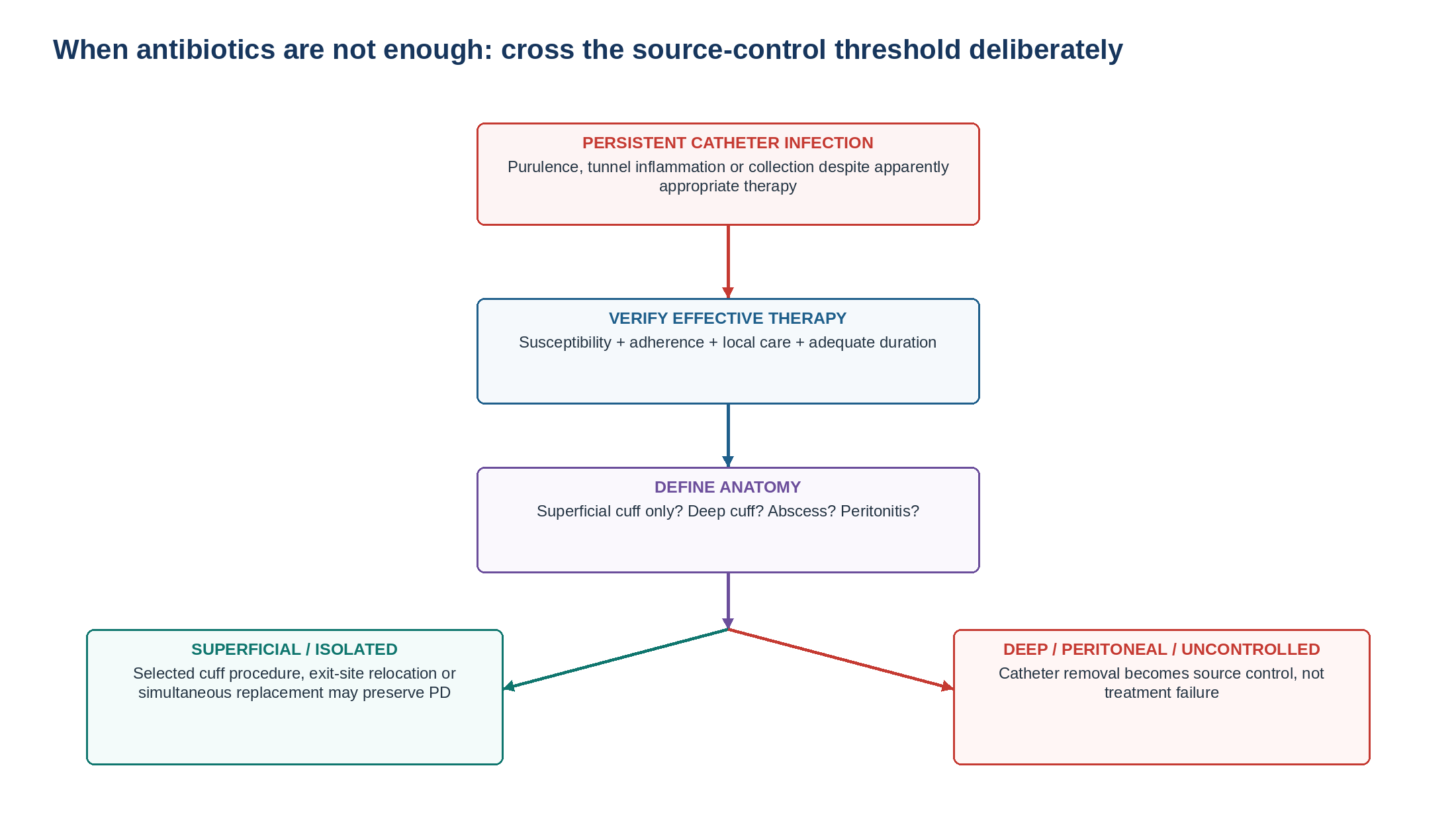
Antibiotic failure should not automatically generate another antibiotic prescription. Confirm that the organism is susceptible, treatment was delivered and taken, local care was appropriate and sufficient time has elapsed. Then search systematically for tunnel infection, cuff involvement, abscess, biofilm, Pseudomonas, NTM, concomitant peritonitis or an incorrect original diagnosis. [1]
Table 11.15 — Failure mechanism -> corrective direction.
| Failure mechanism | Corrective direction | Why |
|---|---|---|
| Resistant isolate | Correct antimicrobial therapy | The prescribed drug was not truly effective |
| Poor adherence/delivery | Correct practical barrier | Apparent resistance may be underexposure |
| Tunnel infection | >=3-week treatment + anatomical assessment | Disease is deeper than ESI |
| External cuff-limited disease | Consider selected salvage | Infected compartment may be removable |
| Deep cuff infection | Move toward catheter removal | Limited superficial salvage leaves source behind |
| Same-organism peritonitis | Catheter removal/source control | Catheter likely connects infectious compartments |
| NTM / difficult organism | Specialist multidrug + source control | Routine ESI therapy is inadequate |
15. Catheter interventions: preserve PD when safe, not infection at any cost
Current ISPD guidance recognises several catheter strategies rather than a binary choice between antibiotics and abandoning PD. The appropriate strategy depends on whether infection is isolated or accompanied by peritonitis, whether disease is limited to the superficial catheter and whether effective antimicrobial treatment has failed. [1,10]
Table 11.16 — Source-control options.
| Situation | Direction | Key safeguard |
|---|---|---|
| Responsive uncomplicated ESI | Preserve catheter | Complete treatment and prevention review |
| Responsive TI | Preserve while completing >=3 weeks | Confirm clinical resolution |
| Refractory superficial ESI | Consider cuff procedure / exit-site relocation | Define deeper catheter as uninfected |
| Refractory isolated catheter infection | Consider simultaneous removal/reinsertion with new exit site | Use selected cases without uncontrolled peritonitis |
| Deep cuff involvement | Removal increasingly favoured | Avoid leaving infected foreign material |
| Catheter infection + same-organism peritonitis | Catheter removal | Treat peritonitis in parallel |
| Unresolved significant peritonitis | Staged source control | Do not place new catheter into uncontrolled infection |
| M. abscessus catheter infection | Strong removal bias | Difficult eradication with foreign material retained |
Selected techniques include external cuff shaving/removal, exit-site relocation, deroofing, en-bloc resection and catheter diversion. A systematic review of 409 patients undergoing 445 salvage procedures reported an overall catheter-salvage rate of approximately 73% and a procedure-related complication rate of approximately 2.7%, but the evidence was predominantly observational and no single technique can be considered universally superior. [10]
| SALVAGE PRINCIPLE Catheter salvage is justified when it removes the infected compartment. Preserving an infected deep cuff is not successful salvage - it is delayed source control. |
|---|
16. Catheter infection plus peritonitis: two syndromes become one source
Always ask a patient with ESI or TI about abdominal pain, cloudy effluent and systemic illness. If peritonitis is suspected, enter the Chapter 10 PD-Associated Peritonitis pathway immediately. Catheter-related infection and peritonitis caused by the same organism strongly support a shared source and lower the threshold for catheter removal. [1,2]
Table 11.17 — Catheter infection + peritoneal findings.
| Pattern | Interpretation | Immediate direction |
|---|---|---|
| ESI, clear effluent, no abdominal symptoms | Isolated catheter infection possible | ESI pathway + tunnel assessment |
| TI + clear effluent | Deep catheter infection without proven peritonitis | >=3-week treatment + source-control surveillance |
| ESI/TI + cloudy effluent | Possible catheter-related peritonitis | Chapter 10 diagnostic pathway immediately |
| Same organism in catheter + effluent | Strong catheter-source phenotype | Catheter removal/source control |
| Severe focal abdomen / polymicrobial effluent | Enteric/surgical source possible | Hospital imaging/surgical pathway |
| CROSS-REFERENCE Chapter 10 governs effluent diagnosis, empiric intraperitoneal therapy, organism-specific peritonitis treatment, refractory-peritonitis definitions and peritonitis-associated catheter-removal decisions. |
|---|
17. Prevention and programme quality: protect the barrier every day
Catheter infection prevention is not a single antiseptic product. It is a system combining topical antimicrobial prophylaxis, appropriate cleansing, catheter immobilisation, patient/caregiver competency, early recognition, continued care while the catheter remains in situ and unit-level surveillance. [1,4–6,8,9,17,18]
Table 11.18 — Primary prevention bundle.
| Intervention | Current guidance/evidence | Practice translation |
|---|---|---|
| Topical exit-site prophylaxis | ISPD recommends daily mupirocin or gentamicin | Choose according to local protocol/resistance and product compatibility |
| Exit-site cleansing | No single cleansing agent has consistent superiority | Clean at least twice weekly and after showering/vigorous exercise or relevant water exposure |
| Catheter immobilisation | ISPD prevention recommendation | Prevent traction and microtrauma |
| Long-term dressing | Not mandatory for every patient | Individualise to environment, skin and fixation needs |
| Training/competency | ISPD teaching framework | Return-demonstration and event-triggered reassessment |
| Catheter temporarily unused | Exit-site care still required | Continue while catheter remains in situ |
| Surveillance/CQI | Track overall and organism-specific infection rates | Use data to change practice |
A landmark randomised trial found daily gentamicin exit-site cream highly effective against Pseudomonas catheter infection and comparable with mupirocin for S. aureus prevention, although subsequent antimicrobial-resistance and ecological concerns mean that local policy remains important. [8,16]
Antibacterial honey did not reduce PD-related infection in the HONEYPOT randomised trial and is not recommended as routine exit-site prophylaxis. [15]
Quality targets
ISPD recommends an overall ESI rate of <=0.40 episodes per year at risk and suggests that catheter insertion-related ESI/TI within 30 days should occur in <5% of inserted catheters. Programmes should also track tunnel infection, organism-specific infection, catheter removal and peritonitis following catheter infection. [1]
Table 11.19 — PD-unit catheter-infection dashboard.
| Metric | Why track it | What a rise should trigger |
|---|---|---|
| Overall ESI rate | Core benchmark | Exit-site-care/prophylaxis review |
| TI rate | Measures deeper catheter morbidity | Examination/ultrasound/source-control audit |
| Organism-specific rate | Detects S. aureus, Pseudomonas and atypical clusters | Microbiology/environmental review |
| Early insertion-related infections | Tests insertion/postoperative process | Surgical/wound-care audit |
| Catheter removals for infection | Captures technique consequence | Treatment/source-control review |
| Peritonitis after ESI/TI | Measures inward progression | Link Chapter 10 and 11 CQI |
| Refractory infections | Identifies treatment/process failure | Adherence, microbiology and salvage audit |
| CQI RULE Infection surveillance is useful only if a change in the number triggers a change in practice: measure -> identify pattern -> intervene -> remeasure. |
|---|
18. Major clinical algorithms
19. Retention tables: pattern recognition
Table 11.20 — If you see this, think this first.
| Finding | First hypothesis | Immediate action |
|---|---|---|
| Purulent exit-site discharge | Definitive ESI | Culture + tunnel assessment + treatment |
| Redness without pus after new adhesive | Contact dermatitis possible | Remove trigger; reassess |
| ESI + tenderness 4 cm along tract | Tunnel infection | TI pathway +/- ultrasound |
| Positive swab + normal exit site | Colonisation | Do not automatically treat |
| Pseudomonas ESI | Difficult catheter organism | >=3 weeks + tunnel assessment |
| Persistent "diphtheroid" drainage | NTM possible | AFB/mycobacterial culture |
| ESI + cloudy effluent | Possible catheter-related peritonitis | Chapter 10 immediately |
| Same organism exit site + peritoneum | Catheter source | Removal/source-control pathway |
| External cuff extrusion + refractory ESI | Potential superficial source | Define depth; access review |
| Persistent infection despite active antibiotic | Biofilm/deep source | Anatomical reassessment |
Table 11.21 — What not to confuse.
| Do not confuse | With | Correction |
|---|---|---|
| Erythema | Definitive ESI | Purulence defines definitive ESI |
| Positive culture | Infection | Clinical phenotype comes first |
| Negative ultrasound | No TI | Clinical TI can exist despite negative scan |
| Susceptible organism | Guaranteed cure | Foreign-body biofilm may persist |
| Better-looking exit site | Resolved tunnel disease | Re-examine the full tunnel |
| 7-10-day duration | Universal ESI duration | Only uncomplicated resolved disease |
| Two weeks of any antibiotic | Effective therapy | Refractory definition requires active treatment |
| More antibiotics | Better source control | Persistent foreign-body infection may require intervention |
| Cuff shaving | Treatment for deep TI | Use only in selected superficial disease |
| Stopping PD exchanges | Stopping catheter care | Continue exit-site care while catheter remains |
20. Clinical pearls
Purulence carries more diagnostic weight than erythema.
Every ESI examination includes the entire tunnel.
A positive culture from a normal site is colonisation, not definitive ESI.
Tunnel ultrasound answers an anatomical question; it does not replace clinical examination.
Review uncomplicated ESI at approximately 1 week because duration depends on that review.
Properly resolved uncomplicated ESI can often finish treatment at 7-10 days.
Pseudomonas ESI requires at least 3 weeks of effective therapy.
Any tunnel infection requires at least 3 weeks of effective therapy.
Every antibiotic course in PD triggers an antifungal-prophylaxis decision.
Persistent infection despite susceptibility should trigger biofilm/source-control thinking.
Refractory diphtheroid or culture-negative infection should trigger NTM thinking.
Same-organism catheter infection and peritonitis is a catheter-removal phenotype.
External cuff disease can sometimes be salvaged; deep cuff infection changes the equation.
Catheter immobilisation is an infection-prevention intervention, not merely a comfort measure.
A PD catheter still requires exit-site care when temporarily unused.
21. Common pitfalls - and the correction
Table 11.22 — High-frequency errors in catheter-infection care.
| Pitfall | Why it fails | Correction |
|---|---|---|
| Treating every red site with antibiotics | Overdiagnosis and resistance | Look for purulent discharge and alternative causes |
| Swabbing healthy sites and treating the result | Treats colonisation | Culture clinical infection |
| Inspecting only the external opening | Misses TI | Palpate the full tunnel |
| Assuming negative ultrasound excludes TI | False reassurance | Integrate scan with clinical findings |
| Giving every ESI exactly 14 days | Ignores response-adapted guidance | Review at ~1 week and individualise |
| Giving Pseudomonas a short course | Higher failure risk | Treat >=3 weeks |
| Treating TI like superficial cellulitis | Ignores depth and foreign body | >=3 weeks + anatomical reassessment |
| Repeated antibiotics despite persistent pus | Delays source control | Search for cuff/tunnel/biofilm |
| Forgetting antifungal prophylaxis | Increases secondary fungal risk | Pair prophylaxis with antibiotic course |
| Ignoring repeated diphtheroid cultures | Delays NTM diagnosis | Request AFB/mycobacterial studies |
| Cuff shaving without defining depth | Leaves deeper infection behind | Use anatomy/ultrasound to select salvage |
| Immediate replacement during uncontrolled peritonitis | Risks contaminating a new foreign body | Achieve appropriate source control first |
22. Mini-cases: decisions, not trivia
Case 1 - Red but not infected
A patient on APD develops erythema around the exit site 24 hours after changing to a new adhesive. There is no purulent discharge, swelling, tunnel tenderness or systemic symptom.
| BEST NEXT STEP This does not satisfy the definition of definitive ESI. Remove the likely irritant, ensure catheter immobilisation and arrange reassessment rather than automatically prescribing antibiotics. |
|---|
Case 2 - Uncomplicated MSSA ESI
Purulent drainage is present at the catheter-skin interface. The tunnel is nontender and effluent is clear. Culture grows susceptible MSSA. At the 1-week review, drainage and inflammation have completely resolved.
| BEST NEXT STEP Complete an appropriate response-adapted course. A total duration of 7-10 days is consistent with current ISPD guidance when uncomplicated ESI resolution is confirmed. |
|---|
Case 3 - The hidden tunnel
A patient has modest purulent drainage but describes pain several centimetres proximal to the exit site. Palpation reproduces tenderness. Ultrasound shows peri-catheter fluid around the superficial cuff.
| BEST NEXT STEP Diagnose TI. This is no longer short-course uncomplicated ESI: use effective antimicrobial treatment for at least 3 weeks, follow response closely and reassess source control if the tunnel abnormality persists. |
|---|
Case 4 - Pseudomonas looks better quickly
Culture from ESI grows susceptible P. aeruginosa. Purulent drainage almost disappears after 8 days.
| BEST NEXT STEP Do not stop treatment because the surface looks healed. Pseudomonas ESI requires at least 3 weeks of effective therapy. Examine the tunnel and consider ultrasound if deeper disease is possible. |
|---|
Case 5 - Persistent susceptible organism
A patient with MSSA ESI has received an active antibiotic with good adherence for 2 weeks but still has purulent drainage and focal tenderness near the external cuff.
| BEST NEXT STEP This is refractory catheter-related infection. Reassess tunnel/cuff anatomy, obtain ultrasound when useful and begin source-control planning rather than simply extending routine therapy. |
|---|
Case 6 - The "Corynebacterium" that never disappears
A chronic draining exit site has failed several antibacterial courses. Two earlier cultures were reported as diphtheroids/Corynebacterium.
| BEST NEXT STEP Request acid-fast staining, mycobacterial culture and species identification. Rapidly growing NTM must be excluded. |
|---|
Case 7 - Same organism in exit site and peritoneum
A patient being treated for S. aureus ESI develops abdominal pain and cloudy effluent. Effluent culture subsequently grows the same organism.
| BEST NEXT STEP Treat peritonitis according to Chapter 10 and manage this as a catheter source-control problem. Same-organism ESI/TI and peritonitis strongly favours catheter removal. |
|---|
Case 8 - A potentially salvageable catheter
A patient has refractory infection around an extruded external cuff. Ultrasound suggests disease remains confined to the superficial cuff with no deep extension and no peritonitis.
| BEST NEXT STEP This is a potential surgical-salvage phenotype. An experienced PD-access team may consider external cuff removal/shaving or exit-site relocation rather than automatically abandoning PD. |
|---|
23. Active recall
| MUST MEMORIZE Use the table below as the minimum bedside knowledge set. Exact antimicrobial doses, resistant-organism regimens and surgical details remain reference-level information. |
|---|
Table 11.23 — Core facts.
| Prompt | Answer |
|---|---|
| Definitive ESI? | Purulent discharge +/- erythema at catheter-epidermal interface |
| Is erythema alone enough? | No |
| Positive culture + normal site? | Colonisation |
| TI definition? | Tunnel erythema/swelling/tenderness/induration +/- ultrasound fluid |
| Does negative ultrasound exclude TI? | No |
| What should be cultured? | Purulent exit-site drainage |
| Main routine empirical target? | S. aureus |
| First major treatment review? | Approximately 1 week |
| Responsive uncomplicated ESI duration? | 7-10 days may suffice |
| Pseudomonas ESI duration? | >=3 weeks |
| Any TI duration? | >=3 weeks |
| Refractory non-Pseudomonas infection? | Failure after 2 weeks effective therapy + intensified care |
| Refractory Pseudomonas? | Failure after 3 weeks effective therapy |
| Antibiotic-course companion? | Antifungal prophylaxis |
| Persistent diphtheroid infection? | Consider NTM |
| Same-organism catheter infection + peritonitis? | Catheter source-control/removal phenotype |
| Overall ESI quality target? | <=0.40 episodes/year at risk |
| Early insertion-related infection target? | <5% within 30 days |
| Key mechanical prevention? | Immobilise catheter |
| PD stopped but catheter remains? | Continue exit-site care |
| USE AS REFERENCE Exact antibiotic doses, MRSA regimen selection, antipseudomonal combinations, allergy substitutions, local resistance thresholds, NTM species-specific multidrug therapy, drug interactions, catheter-compatible topical preparations and operative timing should be checked against the current ISPD tables and local PD, microbiology, renal-pharmacy and surgical protocols. |
|---|
24. Flashcards: spaced repetition
1. Q: Definitive ESI finding? A: Purulent discharge +/- erythema.
2. Q: Redness without pus? A: Not definitive ESI.
3. Q: Healthy exit site + positive swab? A: Colonisation.
4. Q: What must be examined after the exit site? A: The entire tunnel.
5. Q: Tunnel infection clinical signs? A: Erythema, swelling, tenderness or induration along the tunnel.
6. Q: Ultrasound required to diagnose TI? A: No.
7. Q: Negative ultrasound excludes TI? A: No.
8. Q: Routine empirical organism target? A: S. aureus.
9. Q: Review after starting ESI therapy? A: Approximately 1 week.
10. Q: Uncomplicated resolved ESI duration? A: 7-10 days may suffice.
11. Q: Pseudomonas ESI? A: At least 3 weeks effective antibiotics.
12. Q: Tunnel infection? A: At least 3 weeks effective antibiotics.
13. Q: Poor Pseudomonas response? A: Add a second active antipseudomonal agent and reassess source.
14. Q: Refractory ordinary catheter infection? A: Failure after 2 weeks effective treatment.
15. Q: Refractory Pseudomonas? A: Failure after 3 weeks effective treatment.
16. Q: Antibiotics in PD - secondary prevention? A: Antifungal prophylaxis.
17. Q: Repeated diphtheroid drainage? A: Consider NTM.
18. Q: Same-organism ESI/TI + peritonitis? A: Catheter-removal/source-control pathway.
19. Q: Refractory superficial cuff infection? A: Consider selected cuff/exit-site salvage.
20. Q: Deep cuff involvement? A: Strong argument against limited superficial salvage.
21. Q: ESI rate target? A: <=0.40 episodes/year at risk.
22. Q: Catheter insertion-related infection window? A: Within 30 days.
23. Q: Best protection against traction injury? A: Immobilise the catheter.
24. Q: Exit-site care after temporary PD cessation? A: Continue while the catheter remains.
25. Rapid differential / troubleshooting
Table 11.24 — Catheter-infection troubleshooting from problem to action.
| Problem | Differential | First actions |
|---|---|---|
| Red site, no pus | Trauma, dermatitis, granuloma, early infection | Review products/traction; close follow-up |
| Purulent ESI, no tunnel signs | Uncomplicated bacterial ESI | Culture + empiric therapy + 1-week review |
| Purulent ESI + proximal pain | Tunnel infection | Ultrasound when useful + >=3-week pathway |
| Culture negative + improving | Recent antibiotics/limited culture yield | Continue clinically effective plan and verify cure |
| Culture negative + worsening | NTM, fungus, wrong specimen, wrong diagnosis | Repeat/special cultures; diagnostic reset |
| No response at ~1 week | Resistance, nonadherence, tunnel disease, biofilm | Check susceptibility/delivery; tunnel examination |
| Persistent infection at 2 weeks | Refractory disease | Source-control assessment |
| Pseudomonas not improving | Inadequate activity/deep infection | Second active drug + anatomical/source-control review |
| Diphtheroids repeatedly isolated | Corynebacterium vs NTM | AFB/mycobacterial studies |
| External cuff extrusion + ESI | Superficial cuff reservoir | Ultrasound when useful; consider cuff salvage |
| Tunnel collection persists | Infected cuff/biofilm | PD-access/surgical review |
| ESI/TI + cloudy effluent | Catheter-associated peritonitis | Chapter 10 urgently |
| Same organism in effluent/site | Catheter source | Removal pathway |
| Recurrent catheter infections in unit | Technique/prophylaxis/environmental process | CQI audit + organism surveillance |
26. Final revision sheet
| CORE CONCEPT PD catheter infection is a depth + organism + response + source-control problem. Confirm true infection, examine the entire tunnel, culture purulence, exclude peritonitis, treat according to organism, reassess early and stop expecting antibiotics to sterilise an infected foreign body when the clinical phenotype remains active. |
|---|
Table 11.25 — One-minute revision.
| Domain | Must remember |
|---|---|
| ESI definition | Purulent discharge +/- erythema |
| Not ESI by itself | Erythema, tenderness, granuloma or crust without purulence |
| TI | Clinical tunnel inflammation +/- ultrasound collection |
| Culture | Purulent drainage; normal site + positive culture = colonisation |
| Empiric therapy | Cover S. aureus; modify for MRSA/Pseudomonas/local resistance |
| Review | Approximately 1 week |
| Uncomplicated ESI | 7-10 days if resolution confirmed |
| Pseudomonas | >=3 weeks |
| Tunnel infection | >=3 weeks |
| Refractory | 2 weeks effective therapy; 3 weeks for Pseudomonas |
| Atypical failure | Think NTM |
| Peritonitis | Same-organism catheter infection -> source control/removal |
| Salvage | Selected superficial disease only |
| Prevention | Topical antimicrobial + cleansing + immobilisation + competency |
| Antibiotics | Add antifungal prophylaxis |
| Quality | ESI <=0.40/year at risk; early catheter infection <5% |
| TEN TAKE-HOME RULES 1) Purulence - not redness alone - defines definitive ESI. 2) Every ESI gets a complete tunnel examination. 3) A positive swab from a normal site is colonisation. 4) Ultrasound defines depth but a negative scan does not rule out TI. 5) Review treatment at approximately 1 week. 6) Resolved uncomplicated ESI may require only 7-10 days; Pseudomonas ESI and TI require >=3 weeks. 7) Every antibiotic course in PD triggers antifungal prophylaxis. 8) Persistent diphtheroid/culture-negative infection should trigger NTM investigation. 9) Persistent infection despite effective antibiotics is a source-control problem until proved otherwise. 10) Same-organism catheter infection and peritonitis strongly favours catheter removal. |
|---|
Table 11.26 — One-minute bedside synthesis.
| If you see... | Think... | Do now... |
|---|---|---|
| Pus at exit site | Definitive ESI | Culture + tunnel exam |
| Redness only | Infection not yet established | Differential + close observation |
| Tender tunnel | TI | >=3-week pathway +/- ultrasound |
| Pseudomonas | Difficult catheter infection | >=3 weeks + early source assessment |
| No improvement after adequate treatment | Refractory/biofilm | Re-image/re-culture/source control |
| Persistent diphtheroids | NTM | AFB/mycobacterial culture |
| External cuff + superficial infection | Potential salvage phenotype | Define depth |
| Same organism + peritonitis | Catheter reservoir | Remove/source control |
| Any antibiotic | Secondary fungal risk | Antifungal prophylaxis |
| Episode resolved | Prevention opportunity | Apparent-cause analysis + CQI |
| FINAL MENTAL MODEL Purulence -> tunnel -> culture -> peritoneum -> organism -> 1-week trajectory -> source control -> prevention. |
|---|
Rapid oral viva
Define definitive ESI and explain why erythema alone is insufficient.
Define TI and explain the role and limitation of tunnel ultrasound.
Walk through the first visit for a patient with purulent catheter drainage.
Explain why a positive exit-site culture does not necessarily mean infection.
Compare treatment duration for uncomplicated ESI, Pseudomonas ESI and TI.
Define refractory catheter-related infection precisely.
Explain why persistent susceptible S. aureus infection may still require source control.
Describe the clues that should prompt investigation for NTM.
Compare cuff shaving, exit-site relocation, simultaneous replacement and catheter removal.
Explain how management changes when the same organism causes catheter infection and peritonitis.
Build a prevention programme including topical prophylaxis, cleansing, immobilisation, competency and surveillance.
State the ISPD catheter-infection quality targets.
| SAFETY BOUNDARY This chapter teaches diagnostic architecture and guideline-based management reasoning. Exact antimicrobial doses, allergy substitutions, MRSA and resistant Gram-negative treatment, antipseudomonal combinations, NTM multidrug regimens, drug interactions, catheter-compatible topical products and operative timing require the current ISPD treatment tables, local antibiogram, renal pharmacy, infectious-disease expertise and PD-access protocol. Sepsis, abscess and concomitant peritonitis supersede routine outpatient ESI logic. |
|---|
27. Selected authoritative references
1. Chow KM, Li PKT, Cho Y, et al. ISPD Catheter-related Infection Recommendations: 2023 Update. Perit Dial Int. 2023;43(3):201–219. https://doi.org/10.1177/08968608231172740. PMID: 37232412.
2. Li PKT, Chow KM, Cho Y, et al. ISPD peritonitis guideline recommendations: 2022 update on prevention and treatment. Perit Dial Int. 2022;42(2):110–153. https://doi.org/10.1177/08968608221080586. PMID: 35264029.
3. Crabtree JH, Shrestha BM, Chow KM, et al. Creating and maintaining optimal peritoneal dialysis access in the adult patient: 2019 update. Perit Dial Int. 2019;39(5):414–436. https://doi.org/10.3747/pdi.2018.00232. PMID: 31028108.
4. Chow JSF, Brunier G, Figueiredo AE, et al. Teaching peritoneal dialysis: A position paper for the International Society for Peritoneal Dialysis. Perit Dial Int. 2025;45(6):327–343. https://doi.org/10.1177/08968608251375512. PMID: 40966019.
5. Perl J, Teitelbaum I, Warady BA, et al. Core Interventions for the Prevention of Peritoneal Dialysis-Related Infections. Clin J Am Soc Nephrol. 2026;21(8):1439–1444. https://doi.org/10.2215/CJN.0000000976. PMID: 41348491.
6. Perl J, Fuller DS, Bieber BA, et al. Peritoneal Dialysis-Related Infection Rates and Outcomes: Results From the Peritoneal Dialysis Outcomes and Practice Patterns Study (PDOPPS). Am J Kidney Dis. 2020;76(1):42–53. https://doi.org/10.1053/j.ajkd.2019.09.016. PMID: 31932094.
7. van Diepen ATN, Tomlinson GA, Jassal SV. The association between exit-site infection and subsequent peritonitis among peritoneal dialysis patients. Clin J Am Soc Nephrol. 2012;7(8):1266–1271. https://doi.org/10.2215/CJN.00980112. PMID: 22745277.
8. Bernardini J, Bender F, Florio T, et al. Randomized, double-blind trial of antibiotic exit-site cream for prevention of exit-site infection in peritoneal dialysis patients. J Am Soc Nephrol. 2005;16(2):539–545. https://doi.org/10.1681/ASN.2004090773. PMID: 15625071.
9. Htay H, Johnson DW, Wu SY, et al. Regional variation in the treatment and prevention of peritoneal dialysis-related infections in the Peritoneal Dialysis Outcomes and Practice Patterns Study. Perit Dial Int. 2019;39(3):212–220.
10. Soon JJY, Ng NZP, Lee SQW, Tan SG. Are salvage techniques safe and effective in the treatment of peritoneal dialysis catheter-related exit-site and tunnel infections? A systematic review. Perit Dial Int. 2022;42(6):591–601. https://doi.org/10.1177/08968608221116689. PMID: 35945909.
11. Kadota N, Ishikawa K, Kubono Y, et al. Systematic literature review of the diagnosis, prognosis, and treatment of peritoneal dialysis-related infection caused by nontuberculous mycobacteria. BMC Nephrol. 2024;25(1):432. https://doi.org/10.1186/s12882-024-03841-2. PMID: 39609738.
12. Bnaya A, Wiener-Well Y, Soetendorp H, Einbinder Y, Paitan Y, Kunin M, et al. Nontuberculous mycobacteria infections of peritoneal dialysis patients: a multicenter study. Perit Dial Int. 2021;41(3):284–91. https://doi.org/10.1177/0896860820923461. PMID: 32400280.
13. Vychytil A, Lilaj T, Lorenz M, et al. Ultrasonography of the catheter tunnel in peritoneal dialysis patients: what are the indications? Am J Kidney Dis. 1999;33(4):722–727.
14. Kwan TH, Tong MK, Siu YP, et al. Ultrasonography in the management of exit site infections in peritoneal dialysis patients. Nephrology (Carlton). 2004;9(6):348–352.
15. Johnson DW, Badve SV, Pascoe EM, et al. Antibacterial honey for the prevention of peritoneal-dialysis-related infections (HONEYPOT): a randomised trial. Lancet Infect Dis. 2014;14(1):23–30. https://doi.org/10.1016/S1473-3099(13)70258-5.
16. Tsai CC, Yang PS, Liu CL, Wu CJ, Hsu YC, Cheng SP. Comparison of topical mupirocin and gentamicin in the prevention of peritoneal dialysis-related infections: a systematic review and meta-analysis. Am J Surg. 2018;215(1):179–185. https://doi.org/10.1016/j.amjsurg.2017.03.005.
17. Nataatmadja M, Cho Y, Johnson DW. Continuous quality improvement initiatives to sustainably reduce peritoneal dialysis-related infections in Australia and New Zealand. Perit Dial Int. 2016;36(5):472–477.
18. Hurst H, Figueiredo AE, Perez Moran D, et al. Peritoneal dialysis training and interventions: A narrative review. Perit Dial Int. 2026;46(1):6–15. https://doi.org/10.1177/08968608251328517. PMID: 40221963.