Applied Nephrology Master Series
Chapter 12
Mechanical Complications
Flow Dysfunction | Constipation | Migration | Leaks | Hernias | Hydrothorax | Pain | Rescue
| CHAPTER MISSION Turn poor drain, swelling, a new bulge, breathlessness, pain or unexpected free air into a mechanism-based diagnosis: decide whether the catheter-flow system or the pressure/boundary system has failed; correct reversible causes in the least invasive order; localise structural defects; preserve PD with pressure-sparing treatment when safe; and escalate to radiologic or surgical rescue before a correctable mechanical problem becomes technique failure. |
|---|
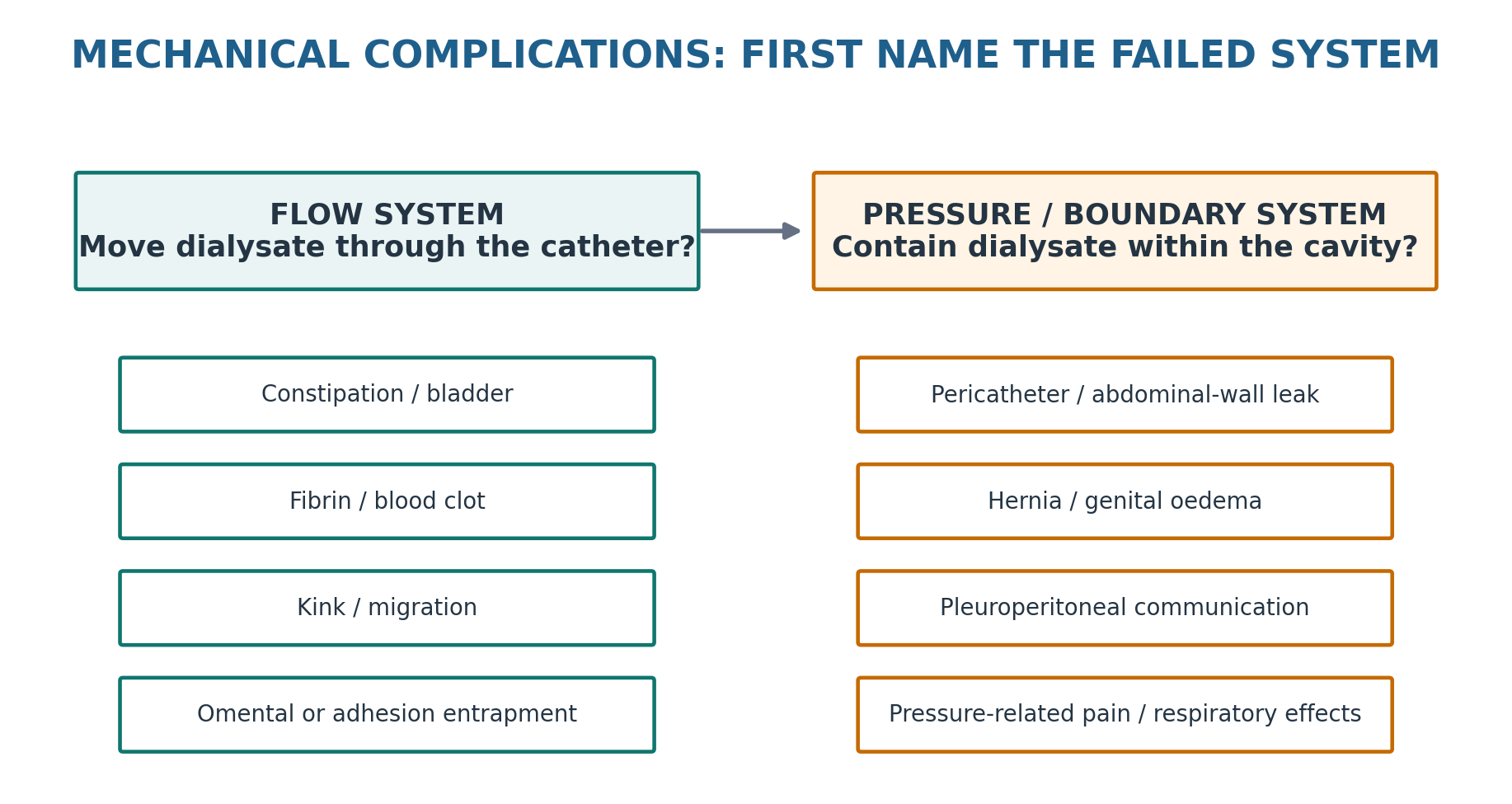
| MASTER PRINCIPLE Mechanical complications are not one list of unrelated events. Think FLOW and PRESSURE. Flow failure asks whether dialysate can move through the catheter. Pressure/boundary failure asks whether dialysate remains within the intended compartment and whether the patient tolerates the intraperitoneal volume. Name the failed system before changing the prescription or abandoning PD. |
|---|
0. One-page chapter map
Table 12.1 — The eight decisions that govern mechanical-complication care.
| Decision | Core question | Bedside output |
|---|---|---|
| 1. Threat | Is there bowel compromise, a strangulated hernia, severe respiratory compromise or another emergency? | Urgent surgical / hospital pathway when needed |
| 2. Phenotype | Is this primarily flow failure, a leak, a hernia, pleural transfer, pain or free air? | Named mechanical syndrome |
| 3. Catheter | Is dysfunction outflow-predominant or two-way? | Mechanism shortlist |
| 4. Pressure | Is fill volume, posture, straining or a fresh access track increasing mechanical stress? | Pressure-driver assessment |
| 5. Localise | Which bedside test or image will identify the site of failure? | X-ray / ultrasound / CT peritoneography / pleural studies as appropriate |
| 6. Correct | Is there a reversible bowel, bladder, fibrin, connector or prescription problem? | Least-invasive correction |
| 7. Rescue | Does the patient need radiologic manipulation, laparoscopy, hernia repair or thoracic intervention? | Definitive structural plan |
| 8. Verify | Is PD now reliable, comfortable and sustainable? | Function + symptom + delivery reassessment |
Learning outcomes
Distinguish catheter-flow dysfunction from pressure/boundary complications at the first bedside assessment.
Use the direction of flow failure—outflow-predominant versus two-way—to narrow the mechanism.
Recognise constipation as the most common reversible cause of PD catheter outflow dysfunction and assess urinary retention when clinically relevant.
Distinguish intraluminal fibrin/blood-clot obstruction from catheter kink, tip migration, omental wrapping and adhesions.
Apply a stepwise catheter-rescue strategy that progresses from reversible and non-invasive causes to radiologic or laparoscopic intervention.
Recognise early and late dialysate leaks and select imaging according to the suspected anatomical pathway.
Assess hernias for reducibility and emergency features, and use pressure-conscious PD around repair.
Diagnose PD hydrothorax without relying on an invented universal pleural-fluid glucose cutoff.
Distinguish volume overload from diaphragmatic splinting and pleuroperitoneal leak in a breathless PD patient.
Troubleshoot infusion pain, drain pain and pneumoperitoneum without masking surgical or infectious disease.
Use current ISPD/GREX activity guidance rather than advising every patient to exercise with an empty abdomen.
Preserve PD when a mechanical complication is correctable, while recognising when definitive repair or modality transition is safer.
| EVIDENCE POSTURE The current adult ISPD access guideline listed by ISPD remains the 2019 update, which recommends a two-week break-in when possible, low-volume recumbent PD for urgent starts, and a conservative-to-invasive sequence for catheter dysfunction. The 2023 SAGES update adds contemporary GRADE surgical guidance but repeatedly rates certainty as very low. The 2024 CJASN mechanical-complications review and 2023 AJKD Core Curriculum provide current clinical synthesis. Therefore this chapter states firm guideline thresholds only where they exist and deliberately avoids universal pressure cutoffs, fixed post-repair restart intervals, or a single diagnostic pleural-glucose threshold unsupported by evidence. [1–7] |
|---|
1. Core concept: mechanical complications are flow failure or boundary failure
Mechanical complications interrupt PD because either dialysate cannot move reliably through the access system or because intraperitoneal fluid and pressure act outside their intended space. These mechanisms overlap: a hernia can alter drainage, constipation can displace a catheter, and a prescription that is technically adequate for clearance can be mechanically intolerable. The first bedside task is therefore classification, not intensification. [1,3,4]
Table 12.2 — The two-system model.
| System | Typical failures | First clinical question |
|---|---|---|
| Flow | Constipation, urinary retention, fibrin/blood clot, kink, migration, omental wrap, adhesions | Can dialysate enter and leave through the catheter normally? |
| Pressure / boundary | Pericatheter leak, abdominal-wall/genital leak, hernia, hydrothorax, splinting, pressure-related pain | Is dialysate staying in the intended compartment and is the volume mechanically tolerated? |
| BEDSIDE TRANSLATION “Low drain volume” is not a diagnosis. Before adding exchanges or hypertonic solution, decide whether the missing volume is trapped behind a flow problem, has escaped through a leak, or reflects true ultrafiltration physiology. Cross-reference Chapter 8 when the question is membrane UF rather than mechanical loss. |
|---|
2. Intra-abdominal pressure: fill volume is also a mechanical dose
Instilled dialysate increases intra-abdominal pressure (IAP). Pressure varies with fill volume, body habitus, abdominal-wall compliance, posture, cough/strain and individual anatomy. Upright posture generally raises pressure relative to supine treatment. A fresh catheter tract or pre-existing weak point may fail under a pressure that another patient tolerates easily. No single universally validated “safe IAP” threshold should therefore be used as a bedside mandate. [1,3,4]
Table 12.3 — Pressure drivers and bedside consequences.
| Driver | Mechanical effect | Prescription consequence |
|---|---|---|
| Higher fill volume | Greater abdominal-wall and diaphragmatic load | Reduce volume when pressure symptoms/leak risk outweigh clearance gain |
| Upright dwell | Often higher IAP than supine dwell | Supine APD can be a pressure-sparing architecture |
| Fresh catheter tract | Incomplete tissue sealing | Traditional break-in preferred when possible; urgent start requires low-volume recumbent strategy |
| Constipation / straining | Transiently increases pressure and can compress/displace catheter | Treat bowel dysfunction as both flow and pressure prevention |
| Cough / heavy lifting | Brief high-pressure spikes | Individualise activity and address persistent cough/strain |
| Hernia / congenital communication | Provides a low-resistance escape pathway | Pressure reduction alone may palliate; structural repair treats the defect |
| PRESSURE DISCIPLINE Do not chase a clearance number with progressively larger fills when the patient develops reflux, pain, a leak, hernia symptoms or respiratory compromise. The mechanically tolerated prescription is part of adequate PD. |
|---|
3. First assessment: classify the phenotype before ordering a rescue procedure
Mechanical presentations are usually recognisable from a small number of patterns: poor drain, poor fill plus poor drain, new swelling/bulge, new pleural effusion, pain during a specific phase of exchange, or free intraperitoneal air. Threat assessment comes first because strangulated hernia, bowel perforation and severe respiratory compromise do not belong in an outpatient troubleshooting loop.
Table 12.4 — Presentation to first hypothesis.
| Presentation | Think first | Immediate discriminator |
|---|---|---|
| Fills normally, drains slowly/incompletely | Constipation, tip migration, omental/adhesion entrapment | Bowel history + plain abdominal radiograph |
| Poor inflow and poor outflow | Kink, intraluminal fibrin/blood clot, connector/tubing problem | Inspect system; image catheter; consider irrigation |
| Abdominal-wall/genital oedema or exit-site fluid | Dialysate leak | Timing after insertion + exam + localising imaging if needed |
| New reducible bulge | Hernia | Standing/strain examination; reducibility |
| Tender irreducible bulge +/- vomiting/obstruction | Incarceration/strangulation | Urgent surgical assessment |
| New unilateral pleural effusion | Pleuroperitoneal communication | Pleural studies + simultaneous serum glucose; imaging if uncertain |
| Pain only during inflow/drain | Flow mechanics / position / solution factors | Phase-specific history + catheter/bowel assessment |
| Free subdiaphragmatic air | Technique-related air versus perforation | Symptoms/peritoneal signs + effluent studies + imaging |
| THREAT GATE Mechanical explanations must never delay emergency assessment of a rigid/focal abdomen, bowel obstruction, tender irreducible hernia, sepsis, haemodynamic instability or severe respiratory compromise. |
|---|
4. Catheter flow dysfunction: use the direction of failure
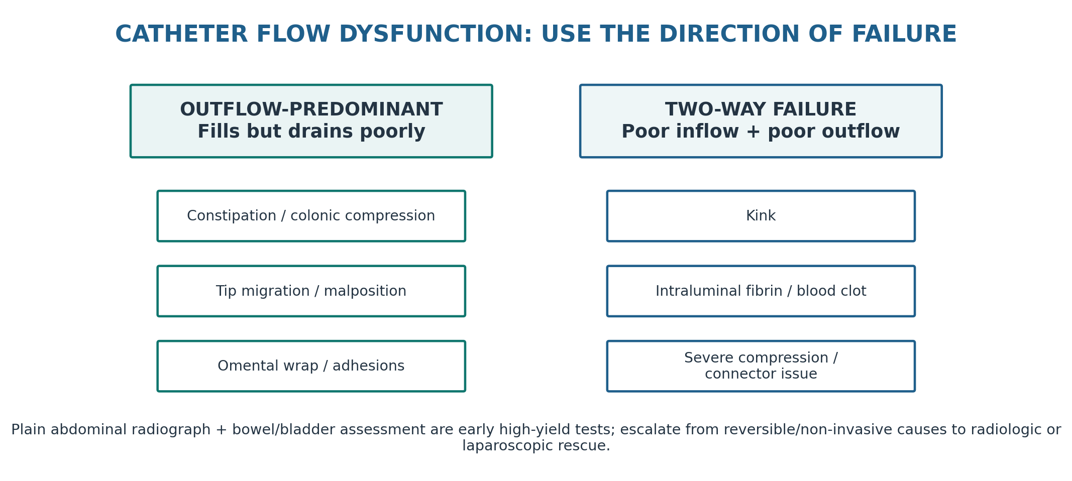
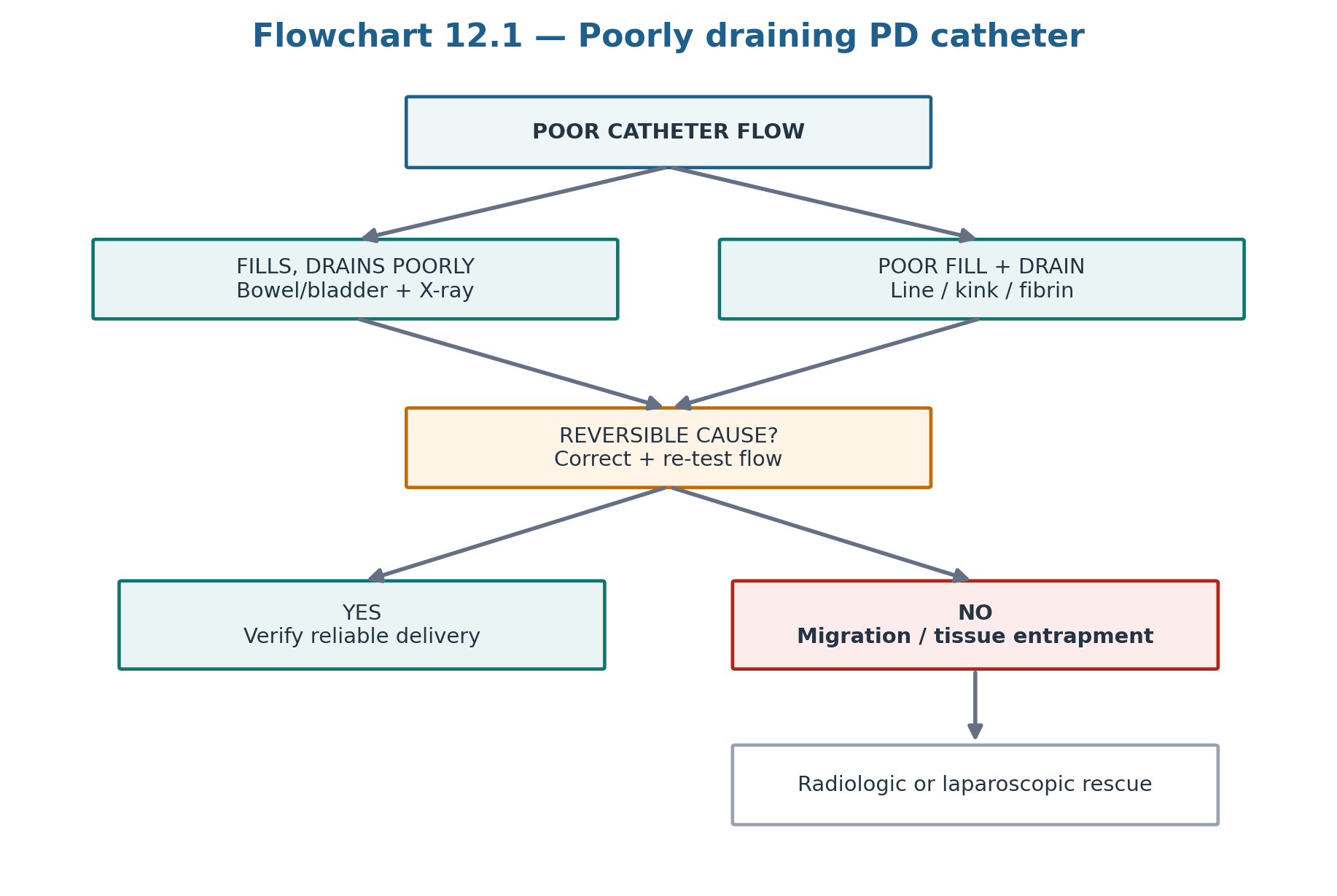
ISPD recommends approaching catheter dysfunction in a logical sequence from conservative/non-invasive causes toward more invasive rescue. Most flow dysfunction presents as outflow failure. The most common cause is constipation; urinary retention is less common but can similarly compress the catheter. Kink and intraluminal fibrin or blood clot more often produce two-way obstruction. A plain abdominal radiograph is a high-yield early test because it can show faecal loading, tip migration and obvious kink. [1]
Table 12.5 — Flow phenotype to mechanism.
| Flow pattern | Mechanisms to prioritise | First actions |
|---|---|---|
| Outflow-predominant | Constipation, bladder distension, tip migration, omental/adhesion entrapment | Bowel/bladder assessment; plain radiograph; correct reversible cause |
| Two-way poor flow | Kink, fibrin/blood clot, severe compression, connector/tubing problem | Inspect line; radiograph; saline irrigation when appropriate |
| Intermittent positional failure | Tip position, omental contact, constipation, posture-dependent kink | Compare positions; radiograph; bowel correction |
| Sudden failure after previously normal function | Constipation, fibrin/clot, migration | Treat reversible cause before invasive rescue |
| Recurrent failure after manipulation | Persistent anatomic cause | Avoid endless temporary manipulation; definitive access review |
5. Constipation and bladder distension: the reversible causes to find first
Constipation is the most common cause of PD catheter outflow dysfunction in the ISPD access guideline. A distended colon can compress the drainage holes or displace the catheter tip. The clinical clue is often a change from previously reliable drainage accompanied by reduced bowel frequency, hard stools or radiographic faecal loading. Treat constipation deliberately before assuming catheter failure. [1,3,4]
Table 12.6 — Bowel and bladder troubleshooting.
| Finding | Mechanism | Direction |
|---|---|---|
| New constipation + poor drain | Colonic distension compresses/displaces catheter | Use an effective bowel regimen and reassess drainage |
| Radiograph shows faecal loading | Supports mechanical bowel contribution | Treat bowel burden before invasive access procedure |
| Suprapubic fullness / urinary symptoms | Bladder distension can compress catheter | Assess retention and relieve according to clinical pathway |
| Flow normalises after bowel treatment | Reversible extrinsic cause confirmed | Build prevention into routine PD care |
| Flow remains poor despite correction | Another catheter mechanism exists | Proceed to position/kink/intraluminal/tissue-entrapment pathway |
| CLINICAL PEARL A bowel regimen is part of PD access maintenance. It is not merely symptom treatment. Constipation can simultaneously impair drainage, raise IAP and worsen pain. |
|---|
6. Intraluminal obstruction: fibrin and blood are a catheter problem, not a membrane problem
When constipation, bladder distension and obvious catheter malposition are excluded, intraluminal fibrin or blood clot becomes important—particularly with two-way flow impairment or visible fibrin. ISPD describes brisk saline irrigation first; if obstruction remains and clot is suspected, a fibrinolytic such as tissue plasminogen activator can be used according to centre protocol. Exact dose, dwell and compatibility should not be improvised from memory. [1]
Table 12.7 — Intraluminal obstruction reasoning.
| Clue | Interpretation | Action direction |
|---|---|---|
| Visible fibrin + two-way poor flow | Intraluminal obstruction likely | Check for peritonitis/bleeding context; saline irrigation |
| Recent bloody effluent + abrupt blockage | Blood clot possible | Irrigation; centre-protocol fibrinolytic if appropriate |
| No response to irrigation/fibrinolytic | Structural problem or organised obstruction | Image and escalate |
| Repeated fibrin formation | Underlying inflammatory driver possible | Assess for peritonitis/other cause rather than repeated rescue alone |
| SAFETY BOUNDARY Do not place an exact fibrinolytic dose in a teaching algorithm unless it is verified against the current local PD access/pharmacy protocol. Catheter material, concentration, dwell, contraindications and bleeding risk matter. |
|---|
7. Migration, kink, omental wrap and adhesions: know when anatomy must be rescued
After reversible extrinsic causes and intraluminal obstruction are addressed, persistent dysfunction usually reflects catheter position or tissue interaction. Cephalad migration can impair drainage; a kink may be intramural or intraperitoneal; omentum or adhesions can trap the tip or side holes. Plain radiography identifies many position problems, while CT or contrast studies can clarify selected cases. [1,3]
Radiologic guidewire manipulation can restore position without general anaesthesia but success is variable and recurrence is common when the underlying anatomic problem remains. Laparoscopy provides direct diagnosis and allows cause-specific treatment such as adhesiolysis, omental management or catheter repositioning. SAGES 2023 makes a conditional recommendation for either operative or non-operative salvage, reflecting very low-certainty comparative evidence; local expertise, patient preference, procedural burden and previous failure should determine the route. [2]
Table 12.8 — Access rescue: match the procedure to the problem.
| Problem | Potential rescue | Why it may fail |
|---|---|---|
| Simple migration without fixed entrapment | Radiologic manipulation or laparoscopic repositioning | Catheter memory / recurrence |
| Omental wrap | Laparoscopic release +/- omental procedure | Repeated manipulation does not remove tissue cause |
| Adhesion entrapment | Laparoscopic adhesiolysis | Extensive adhesions may limit durable function |
| Kink | Revision or replacement depending location | Manipulation may not correct fixed geometry |
| Repeated failed salvage | Definitive replacement may be more patient-centred | Procedural burden can exceed benefit |
| DO NOT AUTOMATE A malfunctioning catheter does not automatically need replacement—and repeated radiologic manipulations are not automatically “less invasive” from the patient’s perspective. Choose the rescue that is most likely to correct the named anatomical cause durably. |
|---|
8. Early pericatheter leak and urgent-start PD
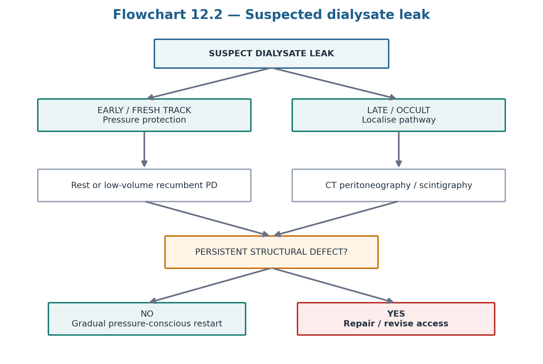
Dialysate leak is escape of dialysis solution from the peritoneal cavity. ISPD commonly classifies early versus late leaks around 30 days after catheter insertion. Early leaks arise before tissue ingrowth and wound healing have formed a secure boundary. Risk is influenced by insertion technique, timing of PD start, fill volume and abdominal-wall stress. [1,3,4]
ISPD recommends delaying PD initiation for approximately 2 weeks after catheter placement when possible (1B). When urgent start is clinically necessary within that interval, low-volume intermittent exchanges in the recumbent position with the abdomen dry during ambulatory periods are recommended (1C). The Cochrane review found that urgent-start PD may increase dialysate leak compared with conventional start, but the evidence is low/very low certainty. [1,8]
Table 12.9 — Early leak phenotype.
| Finding | Interpretation | Management direction |
|---|---|---|
| Fluid around exit/wound soon after start | Pericatheter track leak | Reduce IAP; consider PD rest or low-volume recumbent strategy |
| Abdominal-wall oedema after fresh catheter | Occult tissue-plane leak | Stop pressure escalation; localise if persistent/uncertain |
| Leak during urgent start | Fresh tract + treatment pressure | Use protected urgent-start protocol; cross-reference Chapter 6 |
| Persistent or dramatic early leak | Structural/wound failure possible | Access-team/surgical review rather than repeated pressure challenge |
| CROSS-REFERENCE Chapter 6 covers urgent-start prescription architecture. This chapter covers what to do when the pressure barrier fails. |
|---|
9. Late leaks: localise the pathway rather than assuming ultrafiltration failure
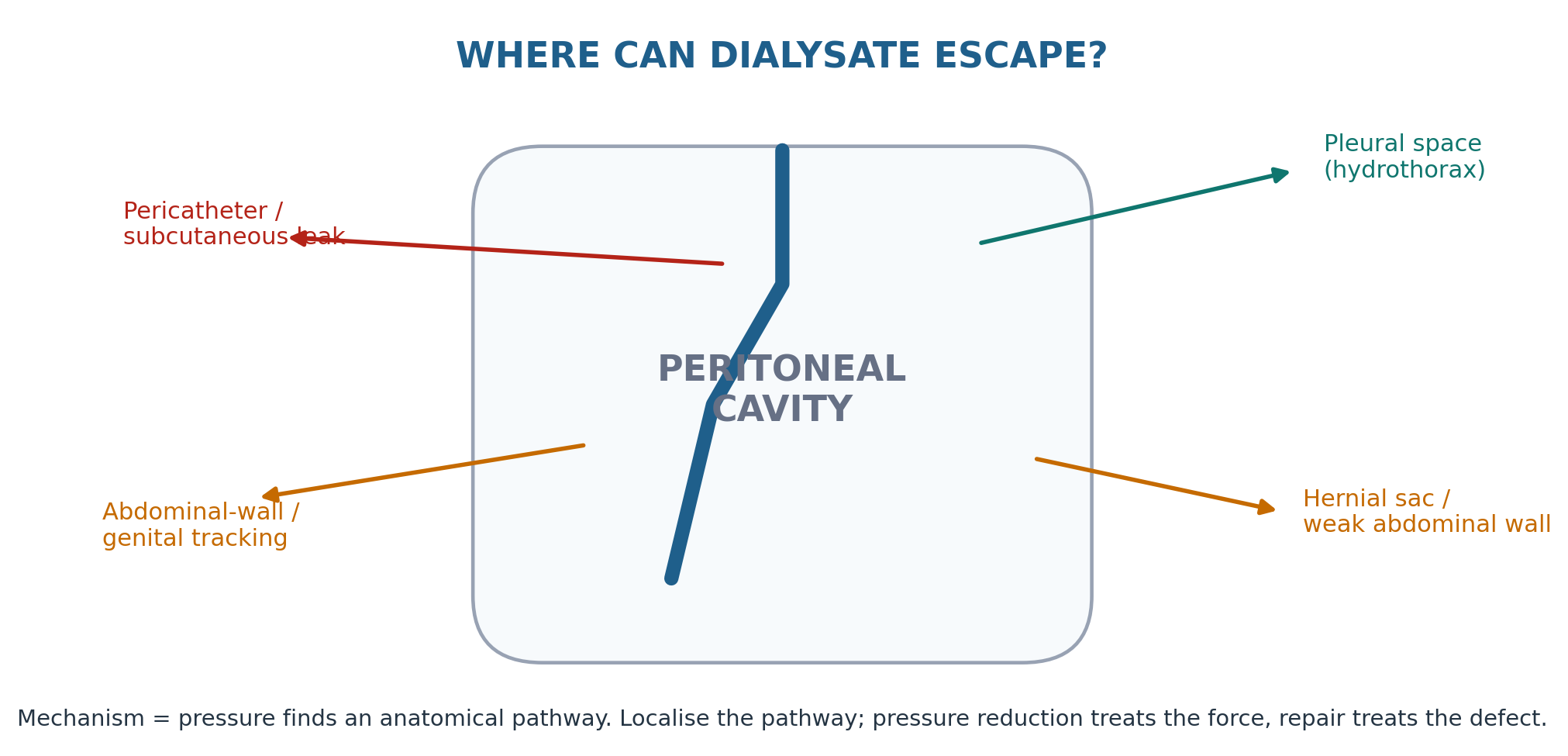
Late leaks may occur through an abdominal-wall defect, hernia, persistent catheter track, retroperitoneal pathway, patent processus vaginalis or an occult infected tunnel. Presentations include abdominal-wall oedema, genital swelling, an apparent new “mass,” unexpected weight gain or reduced net ultrafiltration. [1,3,4]
ISPD recommends CT peritoneography or peritoneal scintigraphy when a boundary leak is suspected and requires localisation (1A). Ultrasound can help with superficial pericatheter collections or pseudohernia. If the leak is associated with tunnel infection, it is an infectious source-control problem and Chapter 11 applies. [1]
Table 12.10 — Late leak patterns.
| Pattern | Likely pathway | Best next question |
|---|---|---|
| Scrotal/labial oedema | Patent processus vaginalis / inguinal communication | Is there an inguinal defect requiring repair? |
| Focal abdominal-wall swelling | Wall defect / pericatheter pseudohernia | Does imaging show dialysate outside the cavity? |
| Generalised wall oedema + low apparent UF | Occult extraperitoneal leak | Is “lost UF” actually dialysate tracking? |
| Leak + tunnel inflammation | Catheter-related infection | Enter Chapter 11 source-control pathway |
| Recurrent leak after rest alone | Uncorrected structural communication | Localise and repair rather than repeat rest indefinitely |
10. Hernias: distinguish reducible anatomy from surgical emergency
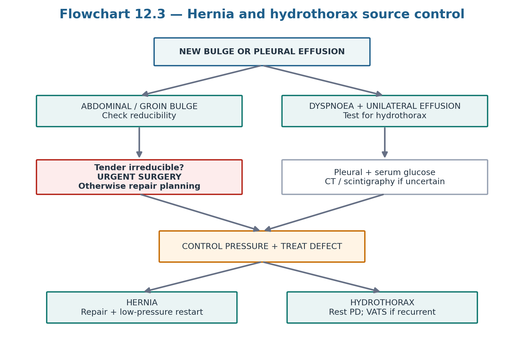
PD increases mechanical load on the abdominal wall and can make pre-existing defects clinically evident. Umbilical, inguinal, incisional and other ventral hernias may present as a bulge that enlarges with standing or straining. A new reducible hernia is usually a repair-planning problem; a tender irreducible hernia, particularly with vomiting, bowel obstruction or peritonism, requires urgent surgical assessment for incarceration or strangulation.
The 2023 SAGES guideline conditionally favours staged hernia repair followed by PD catheter insertion over simultaneous repair and catheter insertion in adults when medically possible, but the certainty is very low. For established PD patients needing hernia repair, no universal evidence-based number of “days off PD” applies to every repair. Postoperative dialysis should be individualised with the surgeon and PD team, commonly using lower-volume supine therapy when the repair and clinical state permit. [2,9]
Table 12.11 — Hernia decision matrix.
| Phenotype | Priority | PD direction |
|---|---|---|
| Small reducible, asymptomatic/mild symptoms | Elective surgical review | Pressure-conscious prescription while awaiting plan |
| Enlarging or symptomatic reducible hernia | Timely repair | Avoid unnecessary high upright fills |
| Tender irreducible hernia | Emergency surgical assessment | Do not continue routine exchanges while delaying evaluation |
| Post-repair, stable and surgeon permits PD | Protect repair | Low-volume supine restart with gradual titration |
| Recurrent hernia | Reassess anatomy + pressure drivers | Definitive surgical/access plan rather than repeated temporary reductions |
| NO INVENTED RESTART CLOCK Evidence does not support one fixed restart interval for every hernia repair. Type of repair, tissue quality, urgency, wound status, residual kidney function and dialysis need all modify the plan. |
|---|
11. Hydrothorax: a pleural effusion can be dialysate
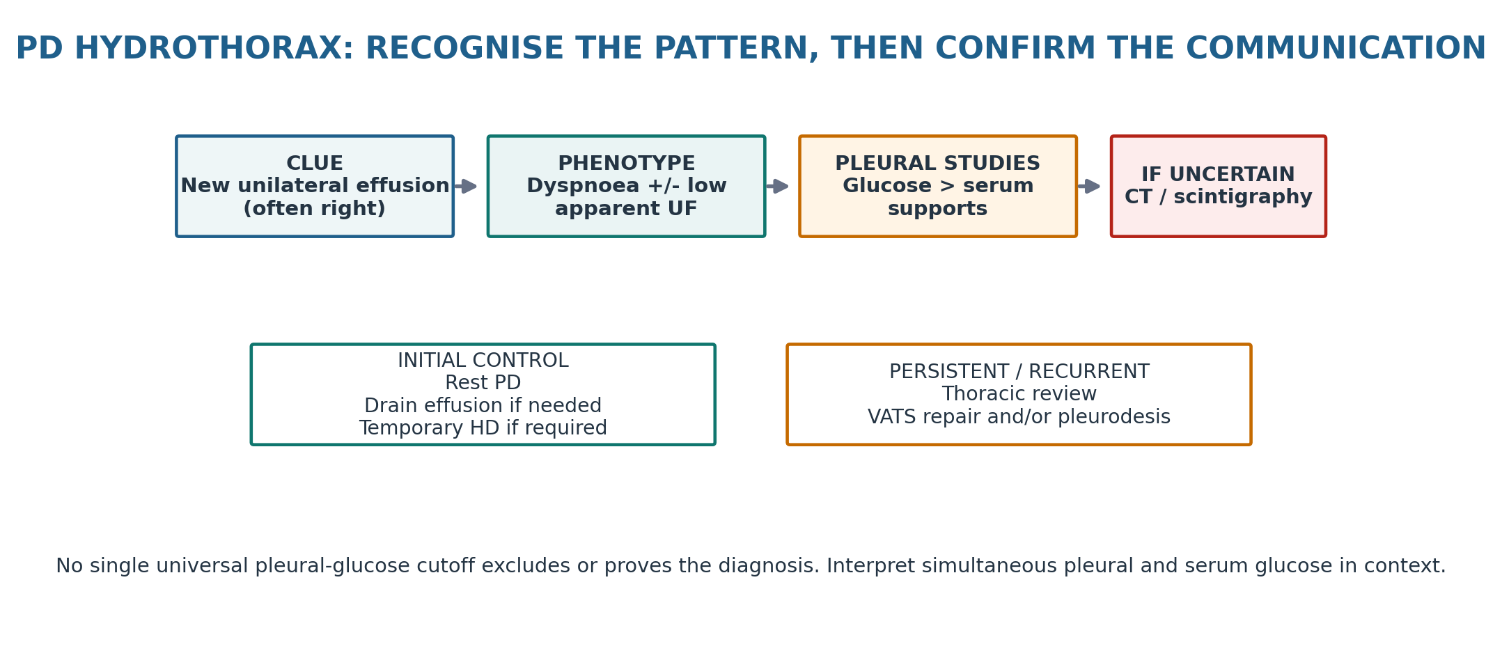
Pleuroperitoneal communication allows dialysate to cross a diaphragmatic defect into the pleural space. Hydrothorax is usually unilateral and predominantly right-sided. Patients may have dyspnoea, reduced net drain volume or may occasionally be minimally symptomatic. Chest imaging often shows a unilateral effusion without the bilateral pulmonary congestion pattern expected from simple fluid overload. [1,3,10,11]
Thoracentesis commonly shows a transudative effusion with pleural glucose higher than simultaneous serum glucose. In a review of verified cases, pleural-fluid-to-serum glucose ratios were >1, but the absolute gradient varied widely; therefore a low gradient does not exclude the diagnosis and no single universal cutoff should be treated as definitive. When biochemical findings are equivocal or anatomical confirmation is needed, CT peritoneography or radionuclide scintigraphy can demonstrate the communication. [10]
Confirmed symptomatic hydrothorax usually requires PD interruption/rest and drainage of the pleural space when clinically indicated, with temporary haemodialysis if required. Persistent or recurrent communication should prompt thoracic surgical assessment; video-assisted thoracoscopic repair and/or pleurodesis can allow many patients to return to PD. [1,11]
Table 12.12 — Hydrothorax versus ordinary volume overload.
| Feature | PD hydrothorax | Volume overload / cardiac effusion |
|---|---|---|
| Distribution | Often unilateral, usually right | Often bilateral but variable |
| Pulmonary congestion | May be absent despite large effusion | May accompany systemic/pulmonary congestion |
| Apparent PD UF | May fall as dialysate enters pleura | May be genuinely insufficient relative to intake |
| Pleural glucose | Often exceeds simultaneous serum | Usually not dialysate-like |
| Definitive question | Is there pleuroperitoneal communication? | Is sodium/water balance positive? |
| DIAGNOSTIC DISCIPLINE Do not label every pleural effusion “PD hydrothorax,” and do not dismiss hydrothorax because the pleural glucose gradient is not dramatically high. The diagnosis is a pattern plus confirmation of communication. |
|---|
12. Respiratory mechanics and back pain: a full abdomen changes biomechanics
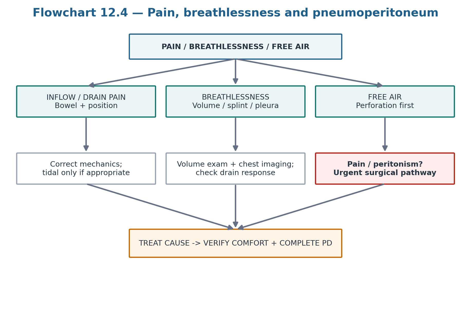
Intraperitoneal dialysate elevates the diaphragm and alters trunk mechanics. Some patients develop breathlessness from diaphragmatic splinting without pleural transfer; others experience reflux, early satiety or back discomfort from increased abdominal load and lumbar postural change. Symptoms that improve promptly after drainage suggest a mechanical volume effect, but cardiopulmonary disease and true volume overload must still be excluded. [3,4]
Table 12.13 — Mechanical symptom versus competing diagnosis.
| Symptom | Mechanical clue | Do not miss |
|---|---|---|
| Breathlessness with full abdomen, improves after drain | Diaphragmatic splinting / fill intolerance | Pulmonary oedema, hydrothorax, infection, cardiac disease |
| Back pain worse during larger fills | Postural/mechanical load | Independent spinal/neurologic disease |
| Reflux/fullness | High intraperitoneal volume/pressure | GI pathology if persistent or atypical |
| Poor sleep on APD | Fill/drain discomfort or alarms | Chapter 9 delivery/prescription mismatch |
| PRESCRIPTION LEVER For pressure-related symptoms, smaller ambulatory fills, greater use of supine treatment and a prescription that preserves the required dialysis goal with lower mechanical burden may be more effective than simply “tolerating” discomfort. |
|---|
13. Infusion pain and drain pain: the phase of the exchange is diagnostic
Pain that occurs only during inflow has a different differential from pain that appears as the abdomen empties. Inflow pain can reflect cold solution, rapid jet/position effects, solution characteristics or catheter contact. Drain pain is classically related to negative pressure and catheter tip contact with pelvic structures, and it may be worsened by constipation or malposition. Persistent pain deserves an anatomical diagnosis rather than chronic analgesia alone. [3,4]
Table 12.14 — Phase-specific PD pain.
| Pain timing | Think first | Corrective direction |
|---|---|---|
| During inflow | Solution temperature, flow jet, catheter position, irritant solution | Check warming/process, inflow mechanics and position; exclude peritonitis if symptoms are not phase-limited |
| Near end of drain | Negative pressure / pelvic catheter contact | Check constipation and catheter position; adjust drain strategy |
| Repeated cycler drain alarms + pain | Drain mechanics or malposition | Treat cause; consider tidal PD only after correctable causes excluded |
| Pain throughout dwell | Not typical isolated mechanical drain pain | Evaluate peritonitis, leak, pressure intolerance, visceral pathology |
| CROSS-REFERENCE Chapter 9 covers tidal PD as a prescription tool. Tidal therapy should not be used to hide a kink, migration, severe constipation or another treatable mechanical lesion. |
|---|
14. Pneumoperitoneum: free air is commoner in PD, but perforation still comes first
Small amounts of intraperitoneal air can be introduced during PD connections, incomplete bag priming or exchanges and may be detected incidentally. Contemporary reviews report wide prevalence estimates. However, the presence of a PD catheter does not neutralise the usual danger of free air: bowel perforation must be considered first when there is abdominal pain, peritoneal irritation, systemic illness or an atypical clinical course. [3]
Table 12.15 — Pneumoperitoneum reasoning.
| Context | Interpretation | Action |
|---|---|---|
| Asymptomatic, small incidental free air | Technique-related air possible | Review connection/priming technique and observe clinically |
| Free air + abdominal pain | Perforation or peritonitis must be excluded | Urgent clinical assessment; effluent studies; imaging/surgical review as indicated |
| Recurrent benign free air | Repeated air entry during exchanges | Observe technique and retrain |
| Shoulder-tip discomfort after exchanges | Intraperitoneal air can irritate diaphragm | Remove technique source; do not dismiss if abdominal findings concerning |
| BOARD TRAP “PD patients often have free air” is not permission to ignore a surgical abdomen. The safe sequence is perforation first, technique second. |
|---|
15. External catheter damage and connector failure
External catheter cracks, cuts or connector damage create two simultaneous problems: loss of mechanical integrity and potential contamination. If a break is identified, the system should be clamped proximal to the defect where feasible and the patient should contact the PD programme immediately. Whether a damaged segment can be repaired/spliced or requires catheter replacement depends on the location and remaining usable tubing. [1]
If the system was open or contaminated, manage the event using the wet-contamination/peritonitis-prevention pathway in Chapter 10. If the damage involves the tunnel/exit site or infection, Chapter 11 applies. Mechanical repair never replaces contamination assessment.
Table 12.16 — Catheter damage: two parallel decisions.
| Decision | Question | Output |
|---|---|---|
| Mechanical | Can the external segment be safely repaired without compromising catheter integrity? | Repair/splice versus replacement |
| Contamination | Was the closed system breached or contaminated fluid able to enter? | Dry/wet contamination pathway |
| Infection | Is the exit/tunnel inflamed or draining? | Chapter 11 evaluation |
| Delivery | Can PD be performed reliably after repair? | Functional verification before routine use |
16. Prevention: pressure management without unnecessary restriction
Mechanical-complication prevention begins before dialysis starts: appropriate catheter placement, inspection and repair planning for abdominal-wall defects, constipation prevention, a protected break-in when possible, and a prescription that respects pressure tolerance. SAGES 2023 conditionally favours traditional over urgent start where medically possible; ISPD recommends a two-week break-in where circumstances allow. [1,2]
Exercise advice should be specific rather than restrictive. ISPD/GREX practice recommendations state that the abdomen does not need to be drained for activities that do not substantially raise IAP, such as walking, hiking or jogging, unless fullness is uncomfortable. For higher-IAP activities such as weightlifting or jumping, draining the abdomen before exercise is advised. Evidence quality is limited, so activity is individualised. [7]
Table 12.17 — Mechanical prevention bundle.
| Intervention | Why it matters | Practice translation |
|---|---|---|
| Break-in when possible | Allows tract/wound healing | Traditional start preferred where clinically feasible |
| Protected urgent start | Reduces pressure on fresh access | Low-volume recumbent exchanges; dry ambulatory periods |
| Bowel regularity | Prevents compression, displacement and straining | Routine constipation prevention/treatment |
| Hernia awareness | Identifies weak points before emergency | Examine new bulges; repair appropriately |
| Pressure-conscious fills | Reduces leak/hernia/symptom burden | Do not increase fill beyond mechanical tolerance |
| Exercise-specific abdomen strategy | Avoids unnecessary sedentary restriction | Drain before high-IAP activity; not required for routine low-IAP activity |
| Technique competence | Reduces introduced air and line damage | Observe exchanges when recurrent air/mechanical events occur |
| ACTIVITY RULE Do not tell every PD patient to remain inactive or exercise only with an empty abdomen. Preserve life participation while identifying activities that materially raise IAP and modifying the prescription around them. |
|---|
17. Keeping PD going: correct the cause, then rebuild a sustainable prescription
Many mechanical complications are correctable and do not require permanent conversion to haemodialysis. The goal is not to preserve PD at any cost; it is to preserve a preferred and effective modality when the mechanical problem can be corrected safely. A successful rescue restores reliable flow, contains dialysate within the intended cavity, avoids recurrent pain or respiratory compromise, and permits the prescribed treatment to be delivered consistently. [1–4]
Table 12.18 — Mechanical rescue hierarchy.
| Level | Examples | Decision principle |
|---|---|---|
| 1. Reversible bedside cause | Constipation, bladder distension, connector issue, air-entry technique | Fix before invasive procedure |
| 2. Intraluminal rescue | Saline irrigation; protocol-based fibrinolytic | Use when clot/fibrin is plausible |
| 3. Diagnostic localisation | X-ray, ultrasound, CT peritoneography, pleural studies | Choose the test that answers the anatomical question |
| 4. Minimally invasive salvage | Radiologic manipulation | Useful when anatomy is amenable and durable success plausible |
| 5. Operative repair | Laparoscopic catheter rescue, hernia repair, VATS diaphragmatic repair | Treat structural cause directly |
| 6. Planned modality transition | Irreparable/recurrent mechanical failure or patient preference | Shared decision, not reflex failure |
| VERIFY After every mechanical intervention, document: inflow, outflow, net UF interpretation, symptoms, leaks/bulges, prescription delivery and patient burden. A technically “patent” catheter that repeatedly interrupts therapy is not fully rescued. |
|---|
18. Major clinical algorithms
19. Retention tables: pattern recognition
Table 12.19 — If you see this, think this first.
| Finding | First hypothesis | Immediate action |
|---|---|---|
| Fills well, drains poorly + constipation | Colonic compression/displacement | Treat constipation; radiograph if needed |
| Poor inflow + poor outflow | Kink or intraluminal obstruction | Inspect tubing; image; consider irrigation |
| Sudden low UF + abdominal-wall oedema | Dialysate leak | Stop pressure escalation; localise if needed |
| New scrotal/labial oedema | Inguinal communication / patent processus | Surgical/anatomical assessment |
| Tender irreducible hernia | Incarceration/strangulation | Urgent surgery |
| Right pleural effusion + no pulmonary congestion | PD hydrothorax | Pleural/serum glucose + confirm communication |
| Breathlessness only with full abdomen | Splinting / pressure intolerance | Drain/reduce volume; still exclude cardiac/pulmonary disease |
| End-drain pelvic pain | Negative-pressure tip contact | Check constipation/position; adjust drain strategy |
| Free air + benign exam | Technique air possible | Review priming/connection |
| Free air + focal abdominal pain | Perforation/peritonitis | Emergency evaluation |
Table 12.20 — What not to confuse.
| Do not confuse | With | Correction |
|---|---|---|
| Low drain volume | Intrinsic UF failure | Check flow and leak before membrane diagnosis |
| Constipation | Minor unrelated symptom | It is the commonest reversible outflow cause |
| Radiographic migration | Automatic need for replacement | Choose radiologic vs laparoscopic rescue based on cause/context |
| Early leak | Automatic permanent HD transfer | Pressure-sparing rest/modified PD may permit recovery |
| Reducible hernia | Strangulated hernia | Tender irreducible/obstructive phenotype is emergency |
| Unilateral effusion | Automatic heart failure | Consider pleuroperitoneal communication |
| Pleural glucose gradient <50 mg/dL | Hydrothorax excluded | No universal exclusion threshold |
| Drain pain | Reason for analgesics only | Find catheter/bowel/negative-pressure cause |
| Pneumoperitoneum in PD | Automatically benign | Perforation must be excluded when clinically concerning |
| Exercise on PD | Always requires empty abdomen | Drain mainly for higher-IAP activity; individualise |
20. Clinical pearls
1. Poor drain + constipation is a bowel problem until proved otherwise.
2. Outflow-only versus two-way failure is a rapid mechanism filter.
3. A plain abdominal radiograph can answer three early questions at once: faecal load, catheter position and kink.
4. Do not use fibrinolytic therapy to treat a catheter that is visibly kinked or displaced.
5. When repeated manipulation fails, ask what fixed anatomical cause has not been corrected.
6. A leak can masquerade as ultrafiltration failure because dialysate is lost into tissue rather than the drain bag.
7. Supine low-volume PD is a pressure-sparing tool, not a universal substitute for definitive repair.
8. A tender irreducible hernia is a surgical problem before it is a dialysis-prescription problem.
9. Right-sided pleural effusion without pulmonary congestion should trigger the hydrothorax question.
10. Pleural glucose greater than simultaneous serum supports hydrothorax; a modest gradient does not exclude it.
11. Breathlessness relieved by draining can reflect mechanical splinting rather than total-body fluid overload.
12. Infusion pain and drain pain have different mechanisms—ask exactly when the pain occurs.
13. Tidal PD may reduce drain pain, but only after constipation and catheter malposition have been addressed.
14. Free air can be technique-related; a surgical abdomen cannot.
15. The best mechanical rescue is the least burdensome intervention likely to correct the named cause durably.
21. Common pitfalls — and the correction
Table 12.21 — High-frequency errors in mechanical PD care.
| Pitfall | Why it fails | Correction |
|---|---|---|
| Increasing glucose concentration for low drain volume | Treats presumed UF when flow/leak may be cause | Verify mechanics first |
| Skipping bowel history | Misses commonest reversible outflow problem | Ask and treat constipation early |
| Repeated tPA without imaging | Cannot correct migration/kink/omentum | Match rescue to mechanism |
| Repeated radiologic manipulation after recurrent failure | May create procedural burden without durability | Escalate to definitive anatomical review |
| Calling early leak “technique failure” | Many heal with pressure protection | Use protected rest/low-volume recumbent strategy when safe |
| Managing recurrent leak with rest alone | Structural defect remains | Localise and repair |
| Waiting on a tender irreducible hernia | Risks bowel compromise | Urgent surgical pathway |
| Using one pleural-glucose cutoff as absolute | Diagnostic gradients vary | Interpret ratio/context and image if uncertain |
| Calling all PD breathlessness overload | Misses splinting/hydrothorax | Examine volume state + pleural/pressure phenotype |
| Treating recurrent drain pain only with tidal settings | Masks constipation or malposition | Correct cause before prescription workaround |
| Assuming free air is from PD | Can miss perforation | Threat assessment first |
| Blanket advice to avoid exercise | Reduces life participation without evidence | Use activity-specific IAP guidance |
22. Mini-cases: decisions, not trivia
Case 1 — The constipated catheter
A stable CAPD patient who previously drained normally develops slow, incomplete outflow over 3 days. Inflow is normal. He has not opened his bowels for 4 days.
| BEST NEXT STEP The phenotype is outflow-predominant and constipation is the leading reversible cause. Treat bowel dysfunction and reassess catheter drainage before arranging invasive catheter salvage. |
|---|
Case 2 — Two-way obstruction after bloody effluent
A patient develops poor inflow and poor outflow after an episode of blood-stained dialysate. The catheter position is unchanged on radiograph and there is no major faecal loading.
| BEST NEXT STEP Intraluminal blood/fibrin is plausible. Use the centre access protocol: saline irrigation first, then a verified fibrinolytic strategy if appropriate. Do not invent the dose or skip bleeding/peritonitis assessment. |
|---|
Case 3 — The catheter that keeps migrating
A catheter is successfully repositioned radiologically twice but again migrates and drains intermittently.
| BEST NEXT STEP Temporary repositioning has not corrected the durable anatomical problem. Discuss laparoscopic diagnosis/rescue or replacement rather than an indefinite cycle of manipulations. |
|---|
Case 4 — Urgent-start wall oedema
Five days after catheter insertion, a patient on low-volume urgent-start PD develops new oedema around the lower abdominal wall and unexpectedly poor net drain volume.
| BEST NEXT STEP Treat this as a suspected early dialysate leak. Reduce/stop intraperitoneal pressure according to clinical dialysis need, keep the patient recumbent for any protected exchanges, and involve the access team if the leak is significant or persistent. |
|---|
Case 5 — A new groin bulge
A patient notices a reducible groin bulge that enlarges when standing. There is no pain, vomiting or obstruction.
| BEST NEXT STEP This is an elective hernia phenotype, not an emergency. Arrange surgical repair planning and use a pressure-conscious PD prescription while avoiding unnecessary interruption. |
|---|
Case 6 — The “heart-failure” effusion
A euvolaemic PD patient develops progressive dyspnoea and a large right pleural effusion without pulmonary congestion. Net PD UF appears to have fallen.
| BEST NEXT STEP Suspect pleuroperitoneal communication. Sample pleural fluid with simultaneous serum glucose and use CT peritoneography/scintigraphy if confirmation remains uncertain. Do not assume a low absolute glucose gradient excludes hydrothorax. |
|---|
Case 7 — Cycler pain at every final drain
APD is complete except for severe pelvic pain during the final part of each drain. There is no abdominal pain during the dwell and effluent is clear.
| BEST NEXT STEP Think negative-pressure catheter contact, constipation or malposition. Correct reversible causes first; a tidal strategy can then be considered if anatomy is acceptable and pain persists. |
|---|
Case 8 — Free air and abdominal pain
A PD patient has subdiaphragmatic free air on chest radiograph and new focal abdominal tenderness.
| BEST NEXT STEP Do not label this technique-related pneumoperitoneum. Treat it as possible perforation/surgical disease: urgent examination, effluent studies and imaging/surgical assessment as appropriate. |
|---|
23. Active recall
MUST MEMORIZE
Table 12.22 — Core facts.
| Prompt | Answer |
|---|---|
| Two mechanical systems? | Flow failure and pressure/boundary failure |
| Most common cause of outflow dysfunction? | Constipation |
| Outflow-only failure suggests? | Constipation, migration, omental/adhesion entrapment |
| Two-way failure suggests? | Kink or intraluminal fibrin/blood clot, among other causes |
| Early high-yield image for poor flow? | Plain abdominal radiograph |
| Break-in when possible? | Approximately 2 weeks after catheter insertion |
| Urgent-start pressure strategy? | Low-volume recumbent exchanges; dry when ambulatory |
| Imaging for occult leak? | CT peritoneography or peritoneal scintigraphy |
| Emergency hernia phenotype? | Tender irreducible +/- bowel obstruction/peritonism |
| Typical hydrothorax side? | Usually right-sided |
| Does low pleural glucose gradient exclude hydrothorax? | No |
| Pleural/serum glucose pattern supporting hydrothorax? | Pleural glucose greater than simultaneous serum supports communication in context |
| Persistent hydrothorax after rest? | Thoracic surgical repair/pleurodesis pathway |
| Drain pain: first correctable causes? | Constipation and catheter position/mechanics |
| Pneumoperitoneum with pain? | Exclude perforation/peritonitis before benign technique explanation |
| Exercise: empty abdomen for walking/jogging? | Not routinely required unless uncomfortable |
| Exercise: empty abdomen for high-IAP weightlifting/jumping? | Recommended by ISPD/GREX practice points |
| Mechanical success after rescue? | Reliable flow, no leak, tolerable symptoms and complete delivered PD |
USE AS REFERENCE
Exact fibrinolytic agent/dose/dwell and contraindications.
Contrast agent and local CT-peritoneography protocol.
Perioperative antibiotic and anticoagulation protocols for access surgery.
Exact postoperative PD restart plan after individual hernia/abdominal/thoracic surgery.
Radiologic manipulation technique and sedation requirements.
Pleurodesis agent selection and VATS procedural approach.
24. Flashcards: spaced repetition
1. Q: Poor outflow + constipation? A: Treat constipation before declaring catheter failure.
2. Q: Poor inflow + outflow? A: Think kink or intraluminal obstruction; inspect/image.
3. Q: First image for catheter dysfunction? A: Plain abdominal radiograph.
4. Q: Visible fibrin + poor flow? A: Irrigation, then protocol-based fibrinolytic if appropriate.
5. Q: Migration after repeated manipulation? A: Consider definitive laparoscopic rescue/replacement.
6. Q: Early leak mechanism? A: Fresh tract + intraperitoneal pressure before full healing.
7. Q: Preferred break-in when possible? A: About 2 weeks.
8. Q: Urgent-start posture? A: Recumbent, low-volume; dry ambulatory period.
9. Q: Genital oedema? A: Think inguinal communication/patent processus vaginalis.
10. Q: Occult leak imaging? A: CT peritoneography or peritoneal scintigraphy.
11. Q: Tender irreducible hernia? A: Emergency surgical assessment.
12. Q: Hydrothorax classic pattern? A: New usually right-sided pleural effusion, dyspnoea, reduced apparent UF.
13. Q: Pleural glucose rule? A: Higher than simultaneous serum supports; no universal cutoff excludes.
14. Q: Uncertain hydrothorax? A: Demonstrate communication with CT peritoneography/scintigraphy.
15. Q: Persistent/recurrent hydrothorax? A: VATS repair and/or pleurodesis assessment.
16. Q: Breathlessness relieved by drain? A: Think diaphragmatic splinting/fill intolerance; still exclude overload/pleural disease.
17. Q: Drain pain? A: Negative-pressure tip contact, constipation or malposition.
18. Q: Tidal PD role? A: Symptom strategy after correctable mechanical causes excluded.
19. Q: Free air without symptoms? A: Technique-related air possible; review priming/connections.
20. Q: Free air with focal pain? A: Exclude perforation urgently.
21. Q: Exercise walking? A: Abdomen need not routinely be empty.
22. Q: High-IAP lifting/jumping? A: Drain abdomen before activity.
23. Q: Mechanical prevention bowel principle? A: Maintain regular bowel function.
24. Q: End point of rescue? A: Reliable, comfortable, sustainable delivered PD.
25. Rapid differential / troubleshooting
Table 12.23 — Mechanical troubleshooting from problem to action.
| Problem | Differential | First actions |
|---|---|---|
| Slow drain only | Constipation, migration, omentum/adhesions | Bowel review/treatment; plain radiograph |
| Poor fill and drain | Kink, fibrin/clot, connector problem | Inspect system; radiograph; irrigation if appropriate |
| Intermittent positional drain | Migration/omentum/constipation | Position test; bowel correction; image |
| New wall swelling + low UF | Leak | Reduce pressure; localise if persistent |
| Genital oedema | Patent processus/inguinal leak | CT peritoneography/surgical review |
| New reducible hernia | Abdominal-wall defect | Surgical plan + pressure-conscious PD |
| Tender irreducible hernia | Incarceration/strangulation | Emergency surgery |
| Dyspnoea + right effusion | Hydrothorax | Pleural studies + simultaneous serum glucose; confirm communication |
| Dyspnoea only when full | Splinting, high fill, overload | Drain response + volume/cardiopulmonary assessment |
| Pain during inflow | Solution/flow/position | Review warming, flow and catheter position |
| Pain at final drain | Negative pressure, constipation, malposition | Treat constipation; assess position; consider tidal if appropriate |
| Incidental free air | Technique air | Observe exchange/priming; retrain |
| Free air + abdominal signs | Perforation/peritonitis | Urgent diagnostic/surgical pathway |
| External catheter crack | Mechanical breach + contamination | Clamp proximal; contact unit; contamination pathway |
| Repeated mechanical events | Persistent anatomy/process problem | Multidisciplinary access CQI review |
26. Final revision sheet
| CORE CONCEPT Mechanical PD failure is solved fastest by naming the system first. FLOW failure requires a sequence from bowel/bladder and intraluminal causes to position/tissue rescue. PRESSURE/BOUNDARY failure requires reduction of mechanical stress, localisation of the escape pathway and repair of the anatomical weak point when it persists. |
|---|
Table 12.24 — One-minute revision.
| Domain | Must remember |
|---|---|
| Flow | Outflow-only versus two-way narrows mechanism |
| Constipation | Commonest reversible outflow cause |
| Imaging | Plain radiograph early for flow; CT peritoneography/scintigraphy for leaks |
| Intraluminal obstruction | Irrigation -> verified local fibrinolytic protocol when appropriate |
| Migration/omentum/adhesions | Radiologic or laparoscopic rescue based on anatomy/context |
| Break-in | ~2 weeks when possible |
| Urgent start | Low-volume recumbent; dry when ambulatory |
| Leak | Think pressure + anatomical pathway; do not call it UF failure |
| Hernia | Tender irreducible = emergency |
| Hydrothorax | Usually right; pleural glucose > serum supports, but no absolute cutoff |
| Pain | Phase of exchange is diagnostic |
| Free air | Perforation first when symptomatic |
| Exercise | No blanket empty-abdomen rule; drain for high-IAP activity |
| Goal | Correct cause and preserve sustainable PD where safe |
| TEN TAKE-HOME RULES 1) Start with FLOW versus PRESSURE. 2) Constipation is the commonest reversible outflow cause. 3) Use flow direction to narrow the differential. 4) Do not use fibrinolytic therapy for an anatomical kink/migration. 5) Protect a fresh catheter with a break-in when possible and low-volume recumbent urgent start when not. 6) A leak can masquerade as UF failure. 7) Tender irreducible hernia is an emergency. 8) Right pleural effusion without congestion is hydrothorax until assessed; no single glucose cutoff is absolute. 9) Pain and free air require mechanism-specific threat assessment. 10) Preserve PD when the defect is correctable, not when repeated rescue only prolongs failure. |
|---|
Table 12.25 — One-minute bedside synthesis.
| If you see... | Think... | Do now... |
|---|---|---|
| Poor drain + constipation | Extrinsic outflow obstruction | Treat bowel cause |
| Poor inflow + outflow | Kink/fibrin/clot | Inspect + image +/- irrigation |
| Wall oedema + low UF | Leak | Pressure control + localise |
| Tender irreducible bulge | Strangulated/incarcerated hernia | Urgent surgery |
| Right effusion + dyspnoea | Pleuroperitoneal communication | Pleural/serum glucose + confirmation |
| Pain only at drain | Negative-pressure mechanics | Bowel/position assessment |
| Free air + focal pain | Possible perforation | Urgent evaluation |
| Recurrent failure after temporary rescue | Uncorrected anatomy | Definitive access review |
| FINAL MENTAL MODEL Threat -> FLOW or PRESSURE -> reversible cause -> localise anatomy -> least-burdensome durable rescue -> pressure-conscious PD restart -> verify reliable delivery. |
|---|
Rapid oral viva
Explain the difference between outflow-predominant and two-way catheter dysfunction.
Why is constipation the first reversible cause to address?
When is a fibrinolytic reasonable and when is it the wrong intervention?
Compare radiologic manipulation with laparoscopic catheter rescue.
Define the protected urgent-start strategy and explain why it reduces leak risk.
How do you recognise an occult late dialysate leak?
What makes a hernia an emergency?
Diagnose hydrothorax without relying on a single glucose threshold.
Distinguish diaphragmatic splinting, hydrothorax and volume overload.
Differentiate infusion pain, drain pain and pain that should trigger peritonitis/surgical evaluation.
How should pneumoperitoneum be approached in a PD patient?
Give current ISPD/GREX advice about exercise with a full or empty abdomen.
| SAFETY BOUNDARY This chapter teaches clinical reasoning and guideline architecture. Exact fibrinolytic dosing, contrast-peritoneography technique, procedural sedation, perioperative antibiotic/anticoagulation management, timing of PD reintroduction after a specific operation and details of thoracic or abdominal surgery require the current local access, radiology, surgical and pharmacy protocols. Surgical abdomen, bowel compromise and respiratory instability supersede routine PD troubleshooting. |
|---|
27. Selected authoritative references
1. Crabtree JH, Shrestha BM, Chow KM, et al. Creating and Maintaining Optimal Peritoneal Dialysis Access in the Adult Patient: 2019 Update. Perit Dial Int. 2019;39(5):414–436. https://doi.org/10.3747/pdi.2018.00232. PMID: 31028108.
2. Haggerty SP, Kumar SS, Collings AT, et al. SAGES peritoneal dialysis access guideline update 2023. Surg Endosc. 2024;38(1):1–23. https://doi.org/10.1007/s00464-023-10550-8. PMID: 37989887.
3. Cheng XBJ, Bargman J. Complications of Peritoneal Dialysis Part I: Mechanical Complications. Clin J Am Soc Nephrol. 2024;19(6):784–790. https://doi.org/10.2215/CJN.0000000000000417. PMID: 38190178.
4. Khan SF. Updates on Infectious and Other Complications in Peritoneal Dialysis: Core Curriculum 2023. Am J Kidney Dis. 2023;82(4):481–490. https://doi.org/10.1053/j.ajkd.2023.03.011. PMID: 37436349.
5. Brown EA, Blake PG, Boudville N, et al. International Society for Peritoneal Dialysis practice recommendations: Prescribing high-quality goal-directed peritoneal dialysis. Perit Dial Int. 2020;40(3):244–253. https://doi.org/10.1177/0896860819895364. PMID: 32063219.
6. Briggs VR, Jacques RM, Fotheringham J, Maheswaran R, Campbell M, Wilkie ME. Catheter insertion techniques for improving catheter function and clinical outcomes in peritoneal dialysis patients. Cochrane Database Syst Rev. 2023;2(2):CD012478. https://doi.org/10.1002/14651858.CD012478.pub2. PMID: 36810986.
7. Bennett PN, Bohm C, Harasemiw O, et al. Physical activity and exercise in peritoneal dialysis: International Society for Peritoneal Dialysis and the Global Renal Exercise Network practice recommendations. Perit Dial Int. 2022;42(1):8–24. https://doi.org/10.1177/08968608211055290. PMID: 34743628.
8. Htay H, Johnson DW, Craig JC, Teixeira-Pinto A, Hawley CM, Cho Y. Urgent-start peritoneal dialysis versus conventional-start peritoneal dialysis for people with chronic kidney disease. Cochrane Database Syst Rev. 2020;12(12):CD012913. https://doi.org/10.1002/14651858.CD012913.pub2. PMID: 33320346.
9. Lew SQ, Collins A. When end-stage kidney disease complicates abdominal surgery. Semin Dial. 2020;33(3):270–278. https://doi.org/10.1111/sdi.12872. PMID: 32277515.
10. Momenin N, Colletti PM, Kaptein EM. Low pleural fluid-to-serum glucose gradient indicates pleuroperitoneal communication in peritoneal dialysis patients: presentation of two cases and a review of the literature. Nephrol Dial Transplant. 2012;27(3):1212–1219. https://doi.org/10.1093/ndt/gfr393. PMID: 21771760.
11. Chow KM, Szeto CC, Li PKT. Management options for hydrothorax complicating peritoneal dialysis. Semin Dial. 2003;16(5):389–394. https://doi.org/10.1046/j.1525-139x.2003.16080.x. PMID: 12969393.
12. Kim JK, Lolas M, Keefe DT, et al. Omental Procedures During Peritoneal Dialysis Insertion: A Systematic Review and Meta-Analysis. World J Surg. 2022;46(5):1183–1195. https://doi.org/10.1007/s00268-021-06413-9. PMID: 35076821.
13. Leblanc M, Ouimet D, Pichette V. Dialysate leaks in peritoneal dialysis. Semin Dial. 2001;14(1):50–54. https://doi.org/10.1046/j.1525-139x.2001.00014.x.
14. Prischl FC, Muhr T, Seiringer EM, et al. Magnetic resonance imaging of the peritoneal cavity among peritoneal dialysis patients, using the dialysate as contrast medium. J Am Soc Nephrol. 2002;13(1):197–203. https://doi.org/10.1681/ASN.V131197.
15. Prasad N, Agarwal V, Mishra P, et al. Modalities of Diagnosis and Management of Peritoneal Dialysis-related Hydrothorax Including Videothoracoscopy-assisted Repair: A Single-center Experience. Indian J Nephrol. 2021;31(6):562–567. https://doi.org/10.4103/ijn.IJN_101_20. PMID: 35068768.
16. Auguste BL, Bargman JM. Peritoneal Dialysis Prescription and Adequacy in Clinical Practice: Core Curriculum 2023. Am J Kidney Dis. 2023;81(1):100–109. https://doi.org/10.1053/j.ajkd.2022.07.004. PMID: 36208963.