Applied Nephrology Master Series
Chapter 13
Noninfectious and Metabolic Complications
Abnormal Effluent | Hemoperitoneum | Eosinophilia | Chylous Effluent | Glucose Load | Icodextrin Safety | Hypokalemia | Surveillance
| CHAPTER MISSION Turn an abnormal bag or a drifting metabolic profile into a mechanism-based diagnosis: exclude infectious and surgical emergencies before labelling effluent “noninfectious”; identify hemoperitoneum, eosinophilic inflammation, chylous effluent and chemical reactions; treat glucose-containing dialysate as a systemic metabolic exposure; correct potassium problems by cause; use glucose-sparing strategies without sacrificing sodium-volume control; and recognise which findings belong to the later membrane-failure and nutrition chapters. |
|---|
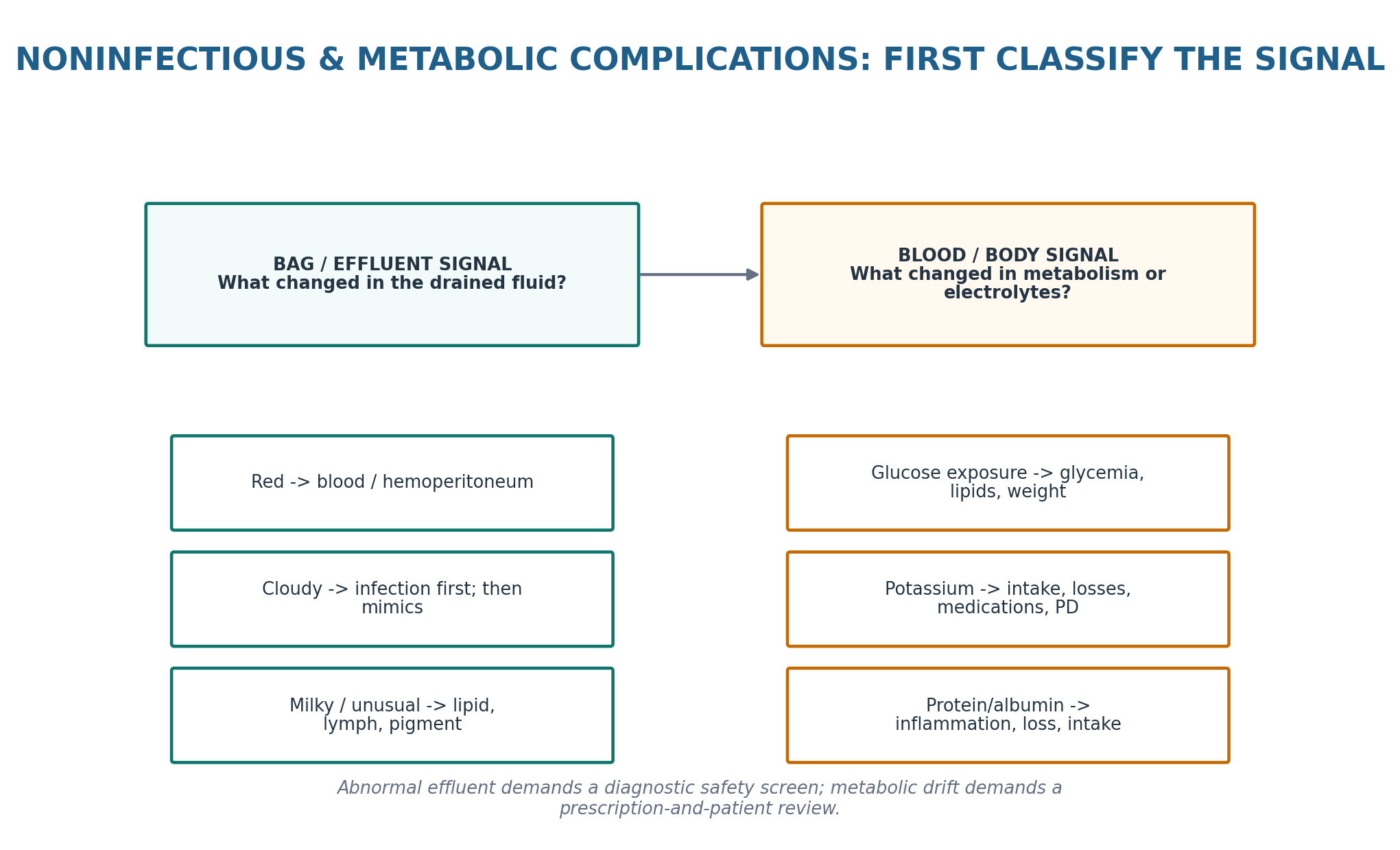
| MASTER PRINCIPLE Think BAG and BODY. An abnormal bag is a diagnostic problem until infection, bleeding and surgical disease are safely addressed. A metabolic abnormality is a whole-patient and prescription problem until intake, comorbidity, medications, residual kidney function, volume status and dialysate exposure are integrated. Never trade fluid control for a prettier metabolic number. |
|---|
0. One-page chapter map
Table 13.1 — The eight decisions that govern noninfectious and metabolic-complication care.
| Decision | Core question | Bedside output |
|---|---|---|
| 1. Threat | Is there sepsis, surgical abdomen, major bleeding, hemodynamic instability or another emergency? | Urgent hospital / surgical / peritonitis pathway when needed |
| 2. Effluent | Is the bag red, cloudy, milky, fibrin-rich or unusually coloured? | Named visual phenotype |
| 3. Cells / chemistry | Are RBCs, PMNs, eosinophils, triglyceride or another analyte explaining the appearance? | Objective effluent classification |
| 4. Source | Gynecologic, traumatic, allergic/chemical, lymphatic, medication-related or intra-abdominal? | Mechanism/source hypothesis |
| 5. Metabolic dose | How much glucose-containing dialysate is being used and why? | Prescription exposure review |
| 6. Potassium | Is low potassium due to intake, GI loss, medication, insulin shifts, residual diuresis or PD context? | Cause-directed potassium plan |
| 7. Correct | Which trigger, solution or prescription lever can be changed without compromising volume/clearance? | Targeted intervention |
| 8. Verify | Did effluent, symptoms, glucose profile, weight/volume and electrolytes improve? | Reassessment + next trigger |
Learning outcomes
Distinguish noninfectious effluent abnormalities from PD-associated peritonitis without delaying appropriate infectious evaluation.
Use effluent colour, cellular differential, timing and clinical severity to classify hemoperitoneum, eosinophilic inflammation and chylous effluent.
Recognise when bloody effluent is compatible with a benign gynecologic phenomenon and when it signals intra-abdominal hemorrhage or surgical disease.
Apply the conventional literature definition of eosinophilic peritonitis cautiously and understand that infection can coexist with peritoneal eosinophilia.
Approach milky effluent through triglyceride/lymphatic and medication pathways rather than treating appearance alone.
Explain why glucose-containing PD solution functions as both an osmotic prescription and a systemic metabolic exposure.
Interpret HbA1c cautiously in dialysis and use directly measured glucose or CGM-derived information when HbA1c and clinical glucose patterns disagree.
Use icodextrin and other glucose-sparing strategies as targeted prescription tools while protecting sodium-volume control and observing product-specific safety warnings.
Recognise hypokalemia as a common PD vulnerability signal, identify reversible causes and distinguish trial targets from guideline recommendations.
Interpret hypoalbuminemia and peritoneal protein loss without reducing nutrition assessment to a single serum albumin value.
Cross-reference Chapter 14 for long-term membrane failure/EPS and Chapter 15 for detailed protein-energy wasting and nutrition management.
| EVIDENCE POSTURE Current ISPD adult guidance does not provide one dedicated guideline for all noninfectious/metabolic complications. The chapter therefore integrates the 2020 ISPD goal-directed prescription recommendations, the 2022 ISPD peritonitis guideline for safe exclusion of infection, the 2021 membrane-dysfunction recommendations where relevant, KDIGO 2022 diabetes guidance (the 2026 update remains a public-review draft, not final guidance), randomized glucose-sparing and potassium-supplementation trials, the 2024 hypokalemia meta-analysis, current icodextrin product safety information, and high-quality reviews of abnormal PD effluent. Evidence for eosinophilic, chylous and many unusual-effluent syndromes is mainly observational or case-based. [1–16] |
|---|
1. Core concept: abnormal effluent and metabolic drift are different PD signals
Noninfectious PD complications in this chapter fall into two clinically distinct groups. The first becomes visible in the drained bag: blood, unusual cloudiness, milkiness, fibrin or another colour change. The second evolves in the patient: hyperglycemia, glycemic variability, weight/adiposity change, dyslipidemia, hypokalemia and the consequences of protein loss. Both are PD-specific clues, but they should not be solved with the same reflex. [1,2,8]
Table 13.2 — Two-signal model.
| Signal | What changed | First question |
|---|---|---|
| Effluent | Colour, clarity, cells or composition of drained dialysate | Is this infection, bleeding, lymph/lipid, inflammation, medication/pigment or surgical disease? |
| Metabolic | Glucose, lipids, body composition, potassium or protein balance | Is PD contributing, and what non-PD drivers coexist? |
| BEDSIDE TRANSLATION Cloudy fluid is a peritonitis problem until appropriately evaluated; a high glucose or low potassium value is not “from PD” until intake, medications, illness, residual kidney function, volume and prescription are reviewed. |
|---|
2. The safety screen: benign mimics are diagnoses of exclusion when the phenotype is dangerous
The chapter starts where Chapter 10 left off: abnormal effluent can be noninfectious, but infectious peritonitis remains the first safety concern when the fluid is cloudy or the patient has abdominal pain. Severe focal pain, guarding, hemodynamic instability, rapid hemoglobin fall, pregnancy-related concern, recent abdominal procedure, bile-stained fluid or systemic toxicity move the patient out of a routine “effluent mimic” pathway. [2,8–10]
Table 13.3 — Red flags that bypass routine noninfectious troubleshooting.
| Finding | Why it matters | Immediate direction |
|---|---|---|
| Fever / PMN-predominant cloudy effluent | Infectious peritonitis remains likely | Chapter 10 sampling + treatment pathway |
| Rigid/focal abdomen, rebound, ileus | Possible bowel or surgical disease | Hospital imaging/surgical assessment |
| Hemodynamic instability or major Hb drop | Clinically important hemorrhage | Resuscitation + bleeding-source evaluation |
| Pregnancy possible + pain/blood | Ectopic pregnancy or gynecologic emergency possible | Pregnancy testing + urgent gynecologic pathway |
| Green/bilious or feculent-appearing fluid | Enteric/biliary communication possible | Urgent intra-abdominal evaluation |
| DO NOT MISCLASSIFY “Noninfectious” describes the final mechanism, not the first assumption. The safe sequence is exclude dangerous infection/bleeding/surgical disease, then narrow the mimic. |
|---|
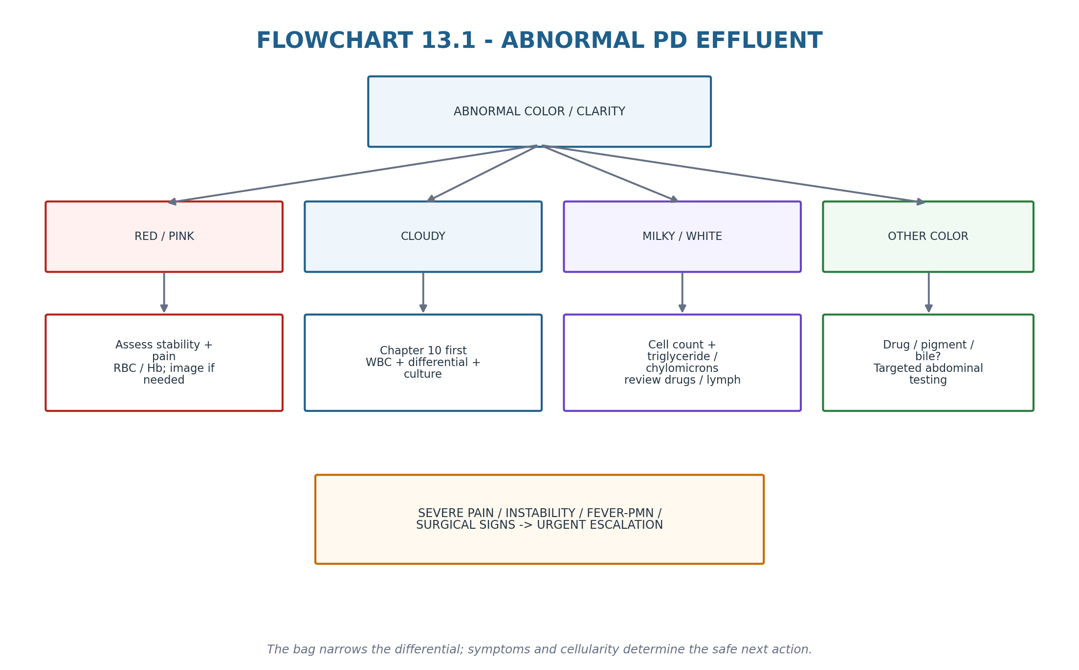
3. Abnormal effluent: colour narrows the differential, cellularity changes the probability
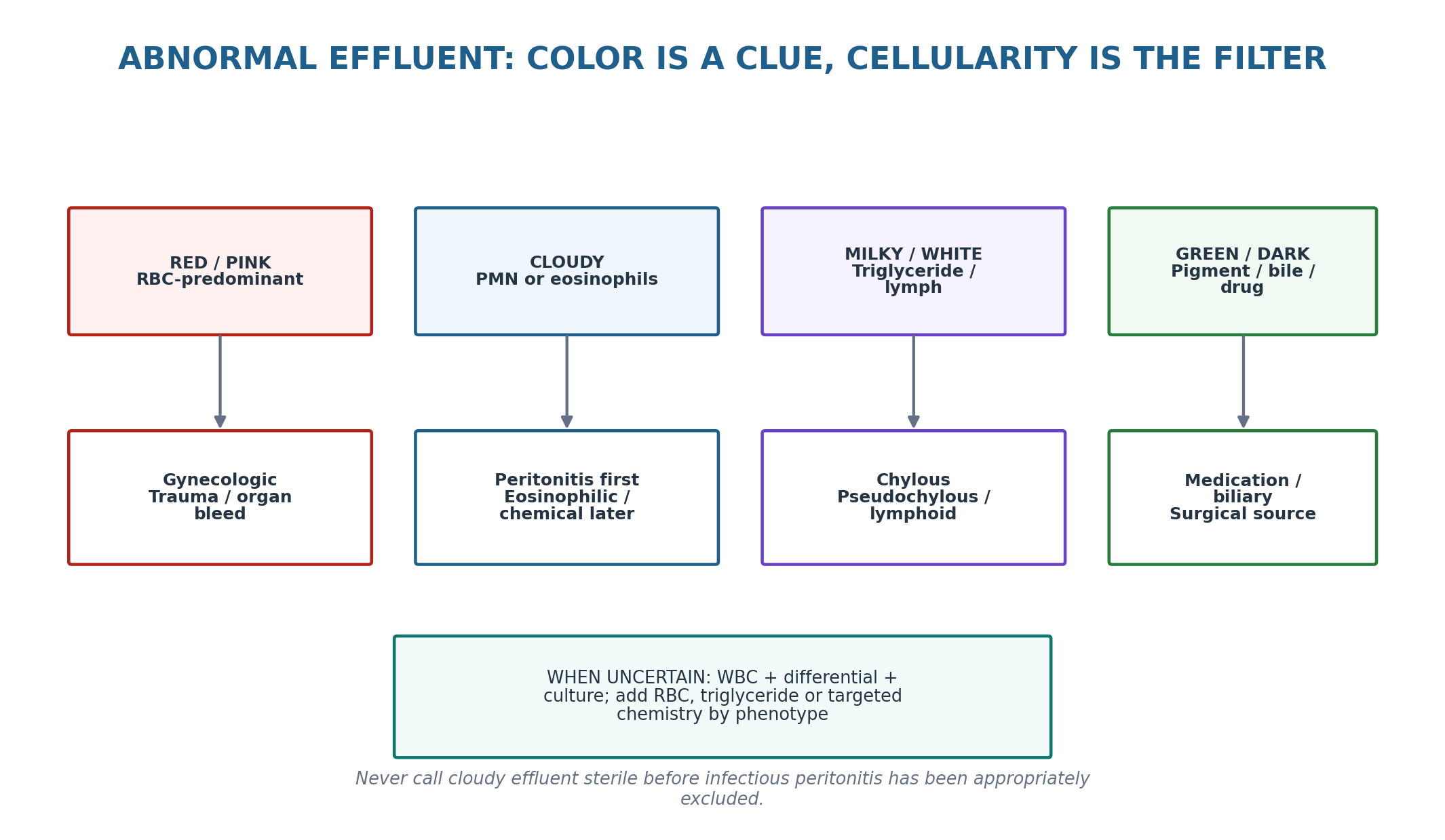
A transparent bag is the normal visual baseline. When appearance changes, ask four questions: what colour is it, is it truly turbid or simply pigmented, what cells dominate, and what happened immediately before the change? Effluent WBC/differential and culture are central whenever peritonitis remains plausible; RBC count, triglyceride/chylomicron testing or targeted chemistry is added according to phenotype. [2,8]
Table 13.4 — Effluent phenotype to first diagnostic move.
| Appearance | Common noninfectious hypotheses | Do not miss |
|---|---|---|
| Pink / red | Menstruation/ovulation, catheter or postoperative trauma, anticoagulation-associated bleeding | Ectopic pregnancy, ruptured cyst, solid-organ/GI bleeding, severe hemorrhage |
| Cloudy | Eosinophilic reaction, chemical inflammation, fibrin/cellular debris | Bacterial/fungal/NTM peritonitis, enteric source |
| Milky / white | Chyloperitoneum, pseudochylous or lymph-rich fluid | Peritonitis, malignancy/lymphatic obstruction, pancreatitis, drug-associated chylous effluent |
| Green / dark / unusual | Medication/pigment, bile or blood breakdown | Biliary leak, bowel pathology, surgical abdomen |
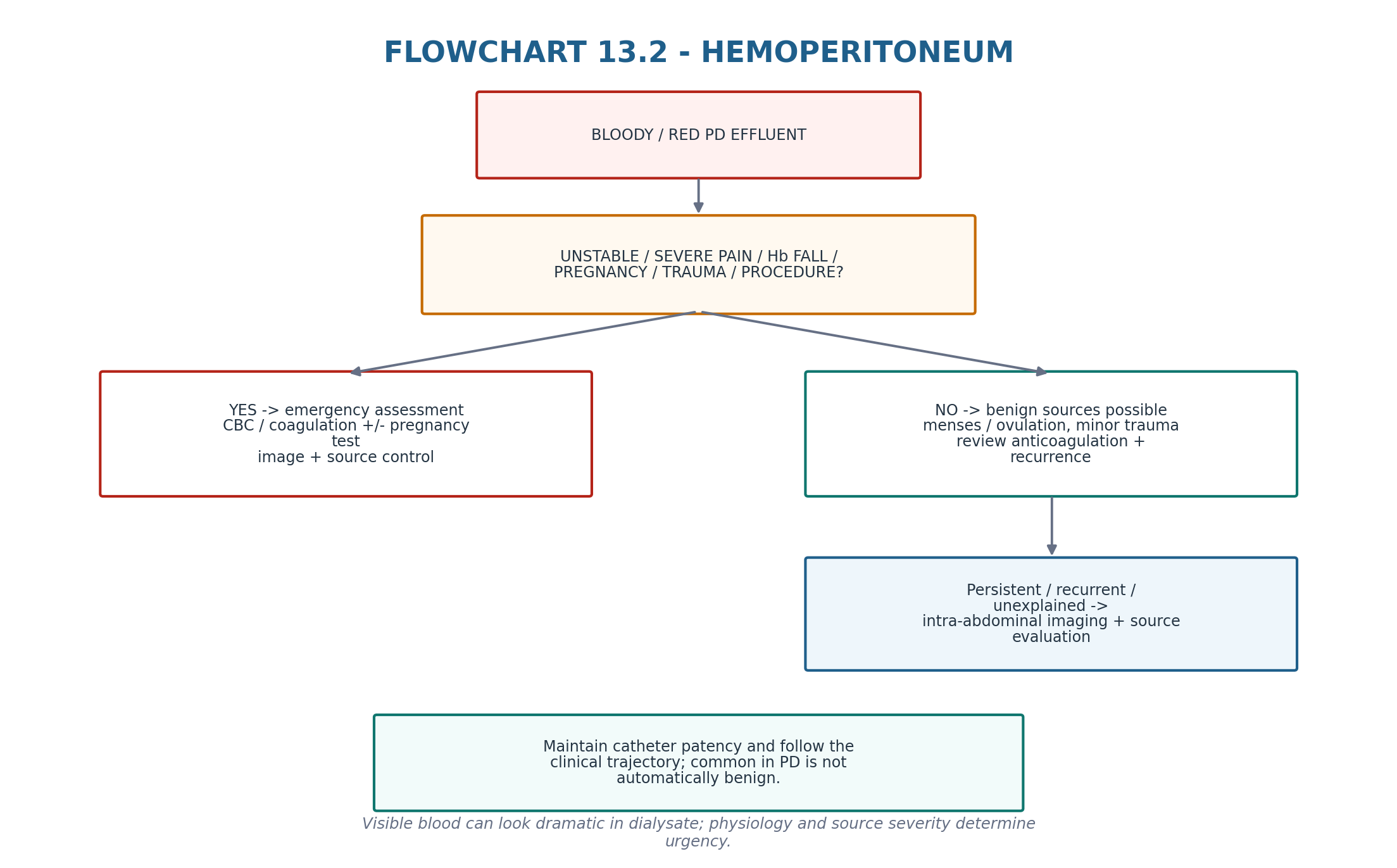
4. Hemoperitoneum: dramatic colour does not equal severe bleeding, but severity must be proved
PD provides a “window” into the peritoneal cavity, so very small quantities of blood can colour a large volume of dialysate. Historical series show that many episodes are benign, particularly menstruation/ovulation-related bleeding, yet the same presentation can arise from gynecologic emergencies, recent procedures, trauma, cyst rupture, solid-organ or gastrointestinal disease, malignancy and long-vintage peritoneal pathology. [9,10]
Table 13.5 — Hemoperitoneum: phenotype to action.
| Pattern | Interpretation | Action direction |
|---|---|---|
| Mild pink effluent, stable, cyclical timing | Gynecologic source common | Confirm pattern; observe trajectory; maintain catheter patency |
| Recent catheter manipulation/procedure | Traumatic source possible | Review timing, coagulation/anticoagulation, clinical stability |
| Persistent/recurrent unexplained bleeding | Underlying intra-abdominal pathology more likely | CBC/coagulation + targeted imaging/gynecologic or surgical assessment |
| Severe pain, instability, rapid Hb fall | Major hemorrhage/surgical disease | Emergency pathway; PD-specific troubleshooting is secondary |
| CLINICAL PEARL The apparent amount of blood in a PD bag is visually amplified by dialysate. Use hemodynamics, hemoglobin trajectory, pain and source—not colour intensity alone—to judge severity. |
|---|
5. Eosinophilic peritonitis: an inflammatory mimic, not permission to ignore infection
Eosinophilic peritonitis is a noninfectious inflammatory phenotype most often described early after catheter insertion or PD initiation and has been linked to exposure to air, catheter/solution materials, drugs and hypersensitivity-type reactions. A commonly used literature definition is >100 eosinophils/mL in effluent with eosinophils >10% of total effluent WBC; this is a conventional research/clinical definition rather than a new ISPD diagnostic criterion. [11,12]
Crucially, peritoneal eosinophilia can occur during infectious peritonitis. Therefore the presence of eosinophils does not safely cancel the Chapter 10 infectious pathway when symptoms, neutrophilia or culture findings support infection. [2,11,12]
Table 13.6 — Eosinophilic phenotype.
| Feature | Supports eosinophilic reaction | Raises concern for infection/other disease |
|---|---|---|
| Timing | Early after catheter/PD exposure or new material/drug | Late abrupt episode without exposure explanation |
| Symptoms | Often mild or asymptomatic cloudy bag | Fever, marked pain, systemic toxicity |
| Differential | Eosinophil-predominant effluent | PMN predominance or evolving neutrophilia |
| Culture | Repeatedly negative | Positive bacterial/fungal/NTM culture |
| Course | Often self-limited after trigger removal | Persistent/worsening despite trigger removal |
| DIAGNOSTIC DISCIPLINE Do not prolong antibiotics indefinitely for a culture-negative eosinophil-predominant, clinically well patient after infection has been properly reassessed—but do not declare eosinophilic peritonitis from eosinophils alone. |
|---|
6. Chemical and sterile peritoneal inflammation: reconstruct the exposure
Chemical/sterile peritoneal inflammation is uncommon and is usually inferred from a temporal relationship to a dialysate, disinfectant, medication, connector contamination, solution lot or another intraperitoneal exposure after infectious causes have been appropriately investigated. The syndrome can resemble peritonitis clinically and cytologically; the key clinical task is exposure reconstruction rather than simply assigning the label “culture negative.” [2,8]
Table 13.7 — Exposure reconstruction.
| Question | What to ask | Why it matters |
|---|---|---|
| Solution | New brand/batch, icodextrin/amino-acid exposure, warming or storage abnormality? | Identifies solution-related trigger |
| Medication | What was injected into the bag and how was the port disinfected? | Drug/chemical contamination can inflame the peritoneum |
| Connection | Was disinfectant or another agent introduced into the system? | Technique-related chemical exposure |
| Cluster | Are other patients using the same product affected? | Signals product/process event requiring unit escalation |
| CQI SIGNAL More than one similar sterile inflammatory episode in a unit is not a “rare patient reaction” until solution, storage, preparation and process factors are investigated. |
|---|
7. Milky or white effluent: identify lipid/lymph rather than guessing from appearance
Milky effluent can reflect chyloperitoneum—triglyceride-rich lymph within the peritoneal cavity—but visual appearance alone cannot separate chylous fluid from infection, cholesterol-rich pseudochylous fluid or lymph-rich variants. General chylous-ascites literature often uses elevated fluid triglyceride and/or chylomicrons to establish the diagnosis; PD-specific interpretation should account for dialysate dilution and local laboratory methods rather than imposing one universal cutoff. [8,13]
PD-specific causes include catheter/surgical lymphatic injury, malignancy or lymphatic obstruction, pancreatitis and medication-associated chylous effluent. Dihydropyridine calcium-channel blockers have a well-described reversible association in PD case literature and systematic review. [14]
Table 13.8 — Milky effluent reasoning.
| Clue | Think | Next step |
|---|---|---|
| Acellular/low-cell milky bag | Lipid/lymph rather than acute inflammatory peritonitis | Effluent triglyceride ± chylomicron/targeted lipid testing |
| Milky bag + abdominal pain/high WBC | Peritonitis still possible | Culture/cell differential first |
| New dihydropyridine CCB exposure | Drug-associated chyloperitoneum possible | Medication review; clinician-directed withdrawal/substitution |
| Persistent unexplained chyle | Lymphatic obstruction, malignancy, pancreatitis, cirrhosis/other disease | Targeted imaging and systemic work-up |
8. Fibrin and unusual colour: a clue to inflammation or another source, not a diagnosis
Fibrin strands may occur after catheter insertion, bleeding, peritonitis or other peritoneal inflammation and can contribute to catheter obstruction. Their management therefore belongs partly to the mechanical-access pathway in Chapter 12. The diagnostic mistake is to interpret fibrin as proof of infection—or to focus on fibrin clearance while missing the inflammatory source. [2]
Unusual orange, green, yellow, purple or black effluent has been reported with blood breakdown, medications, rhabdomyolysis, bile and other pigments. These are low-frequency phenotypes in which history and targeted testing dominate; green/bilious or feculent appearances should lower the threshold for urgent intra-abdominal evaluation. [8]
| CROSS-REFERENCE For fibrin causing inflow/outflow obstruction, return to Chapter 12. For cloudy effluent with infectious probability, return to Chapter 10. This chapter owns the noninfectious differential—not duplicated access or infection treatment. |
|---|
9. Glucose absorption: the osmotic prescription becomes a systemic metabolic exposure
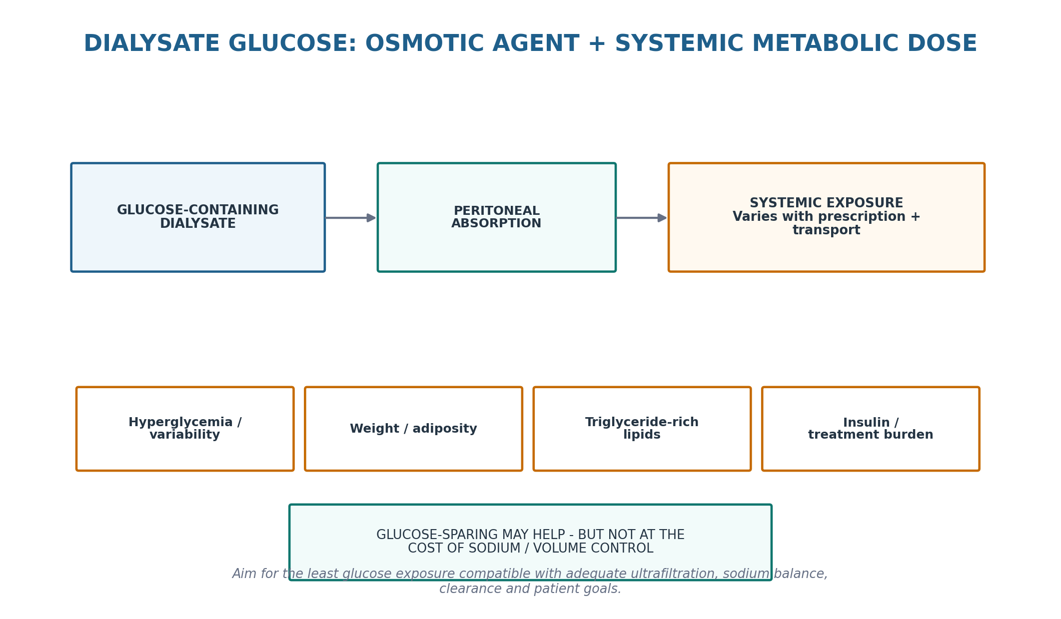
Glucose is an effective and familiar crystalloid osmotic agent, but a clinically meaningful fraction is absorbed across the peritoneum. The absorbed amount varies with glucose concentration, dwell duration, transport characteristics and prescription architecture. The consequences can include higher glycemic exposure, insulin requirements, weight/adiposity gain and a triglyceride-rich lipid pattern, although the strength of evidence linking glucose absorption itself to hard cardiovascular outcomes remains incomplete. [1,4–6,16–18]
Table 13.9 — Glucose exposure: benefit, cost and safeguard.
| Prescription feature | Potential benefit | Metabolic cost / safeguard |
|---|---|---|
| Higher dextrose concentration | Greater crystalloid osmotic force | More glucose absorption; use only when clinically needed |
| Repeated hypertonic rescue | Can restore short-term UF | Signals unresolved sodium/volume or membrane problem; Chapter 8 review |
| Long glucose dwell | Solute contact time | Glucose gradient dissipates; fast transport may lose UF |
| Icodextrin long dwell | Sustained colloid UF with less glucose exposure | Product-specific safety/monitor compatibility required |
| Amino-acid solution | Glucose-sparing/nutritional osmotic option in selected settings | Not a generic metabolic solution; nutrition/acid-base and product limits apply |
| PRESCRIPTION RULE The goal is not “zero glucose.” It is the least glucose exposure that still achieves safe ultrafiltration, sodium balance, clearance, tolerability and patient goals. |
|---|
10. Dysglycemia: measure the patient’s glucose exposure, not just HbA1c
Diabetes management in PD is complicated by glucose absorption from dialysate and by the reduced reliability of HbA1c in advanced CKD and dialysis. KDIGO 2022 states that HbA1c remains a monitoring tool but its accuracy and precision decline in dialysis; CGM-derived glucose management information can be used when HbA1c does not match directly measured glucose or clinical symptoms. [3,4]
Prescription changes can alter glucose exposure rapidly. A patient who shifts from repeated dextrose to a glucose-sparing regimen may need active review of insulin or other glucose-lowering therapy to avoid hypoglycemia, while increased hypertonic dextrose use during volume stress can raise glucose requirements. Diabetes-specific drug selection and targets are deferred to Chapter 16 and current diabetes/dialysis guidance. [3,4]
Table 13.10 — Glycemic assessment in PD.
| Signal | Interpretation | Clinical use |
|---|---|---|
| HbA1c | Long-term marker with lower reliability in dialysis | Use trend; question discordance |
| SMBG pattern | Direct capillary pattern around exchanges/meals | Shows immediate prescription-related glycemia |
| CGM / GMI | Captures variability and hypoglycemia when compatible with device/product | Useful when HbA1c is discordant |
| Prescription glucose review | Identifies change in dialysate carbohydrate exposure | Links dialysis changes to glucose changes |
| MEASUREMENT HUMILITY Do not intensify glucose-lowering therapy solely because an HbA1c looks high—or relax therapy solely because it looks low—when the dialysis context, anemia/ESA exposure and directly measured glucose tell a different story. |
|---|
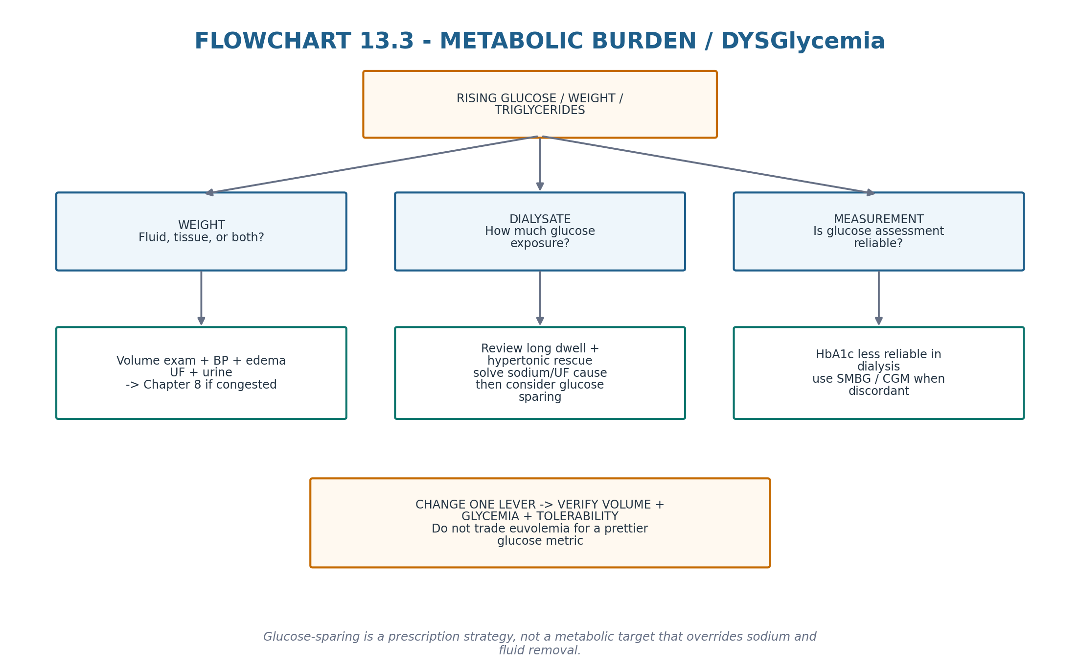
11. Glucose-sparing prescriptions: metabolic improvement can create a volume problem
The IMPENDIA/EDEN randomized program showed that a glucose-sparing regimen using icodextrin and amino-acid solution improved HbA1c and several lipid measures in patients with diabetes, but the intervention group had more serious adverse events and deaths, including events compatible with extracellular fluid expansion. The lesson is not that glucose sparing is unsafe; it is that metabolic endpoints cannot outrank volume control. [5]
Table 13.11 — Glucose-sparing decision.
| Question | Good reason to consider | Reason to slow down |
|---|---|---|
| Long-dwell UF problem? | Icodextrin can improve sustained long-dwell UF | Do not substitute it for an adequate full prescription |
| High glucose burden? | Reduce unnecessary hypertonic exposure | First solve sodium intake, leak, adherence or membrane mismatch |
| Glycemia worsening after stronger bags? | Prescription is contributing | Adjust dialysis and diabetes plan together |
| Weight rising? | Could reflect glucose calories | Exclude fluid overload before calling it adiposity |
| TRIAL LESSON IMPENDIA/EDEN improved metabolic markers but warned against “metabolic optimisation” that inadvertently under-treats extracellular volume. Every glucose-sparing change gets a volume reassessment. |
|---|
12. Icodextrin safety: the glucose-meter interaction is a never-event risk
Icodextrin is metabolised to oligosaccharides including maltose. Current product information warns that GDH-PQQ, glucose-dye-oxidoreductase and some GDH-FAD glucose-monitoring methods can produce falsely elevated glucose readings. This has caused inappropriate insulin administration or failure to recognise hypoglycemia. Falsely elevated readings may persist for up to approximately 2 weeks after stopping icodextrin. [15]
Compatibility of continuous glucose monitors and other devices is product-specific and should be checked with the current device manufacturer rather than assumed. This safety information must travel with the patient across emergency departments, hospital wards and procedures. [15]
Table 13.12 — Icodextrin safety checklist.
| Risk | Failure mode | Safeguard |
|---|---|---|
| Non-glucose-specific meter | Maltose interpreted as glucose | Use a compatible glucose-specific method |
| Hospital transfer | Staff unaware of icodextrin | Flag icodextrin prominently in medication/dialysis handover |
| Recent discontinuation | Interference can persist | Maintain warning after stopping according to product information |
| CGM use | Compatibility may vary by device | Verify manufacturer compatibility |
| Rash / hypersensitivity | Known adverse reaction in some patients | Assess temporal relation and alternative causes; discontinue if clinically required |
| SAFETY ALERT A falsely high bedside glucose in an icodextrin-treated patient can lead to insulin treatment of a patient who is actually hypoglycemic. This is a device–drug interaction, not a minor laboratory nuisance. |
|---|
13. Weight gain: separate fluid, lean tissue and adiposity before changing the prescription
Weight gain after PD initiation is not synonymous with glucose-induced fat gain. Fluid status, return of appetite after uremia, changes in lean mass, reduced activity, insulin exposure and absorbed dialysate glucose can all contribute. The clinical task is to separate extracellular fluid expansion from tissue gain because the treatments are opposite. [1,17]
Table 13.13 — Weight gain differential.
| Pattern | Likely driver | Direction |
|---|---|---|
| Weight ↑ + edema/BP/low UF | Fluid overload | Chapter 8 sodium-volume assessment |
| Weight ↑ without congestion + higher glucose exposure | Adiposity/metabolic gain possible | Review glucose burden, diet/activity; Chapter 16 obesity context |
| Weight ↑ with improved intake/function | Recovery of tissue mass possible | Assess body composition/nutrition rather than assuming harm |
| Weight loss + low albumin/appetite/inflammation | PEW or illness possible | Chapter 15 nutrition/PEW pathway |
| DO NOT CONFUSE A falling scale weight can represent harmful volume depletion or PEW; a rising weight can represent fluid overload or nutritional recovery. Interpret the phenotype before celebrating or treating the number. |
|---|
14. Dyslipidemia: common metabolic phenotype, uncertain PD-specific outcome target
PD commonly produces or amplifies hypertriglyceridemia, VLDL/remnant abnormalities and other atherogenic lipid changes through insulin resistance, glucose absorption and kidney-failure metabolism. Contemporary reviews also explore possible links between dyslipidemia and peritoneal membrane outcomes, but causality and PD-specific treatment targets remain uncertain. [16–18]
The practical response is to avoid assuming that stronger glucose-based dialysis is metabolically neutral, assess cardiovascular risk in the wider CKD/dialysis context, and follow current lipid guidance rather than inventing a PD-specific lipid target. Changes in glucose exposure may modestly improve triglyceride/VLDL indices, but hard-outcome benefit from glucose sparing has not been established. [5,16]
| EVIDENCE CALIBRATION Metabolic plausibility and improved lipid biomarkers are not equivalent to proven cardiovascular benefit. Use lipid results for global risk management, not as a reason to compromise dialysis adequacy or volume. |
|---|
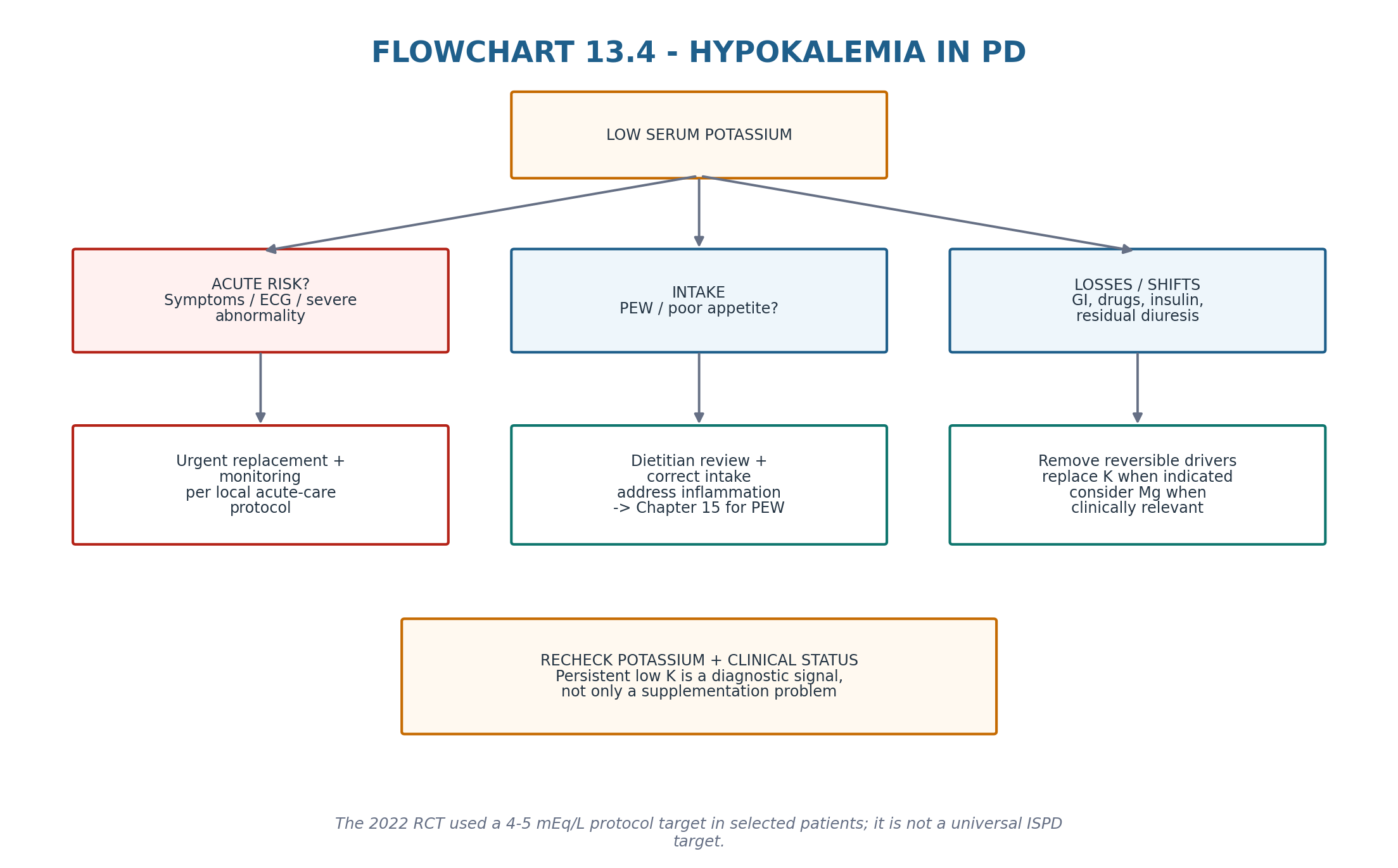
15. Hypokalemia: a common PD vulnerability signal
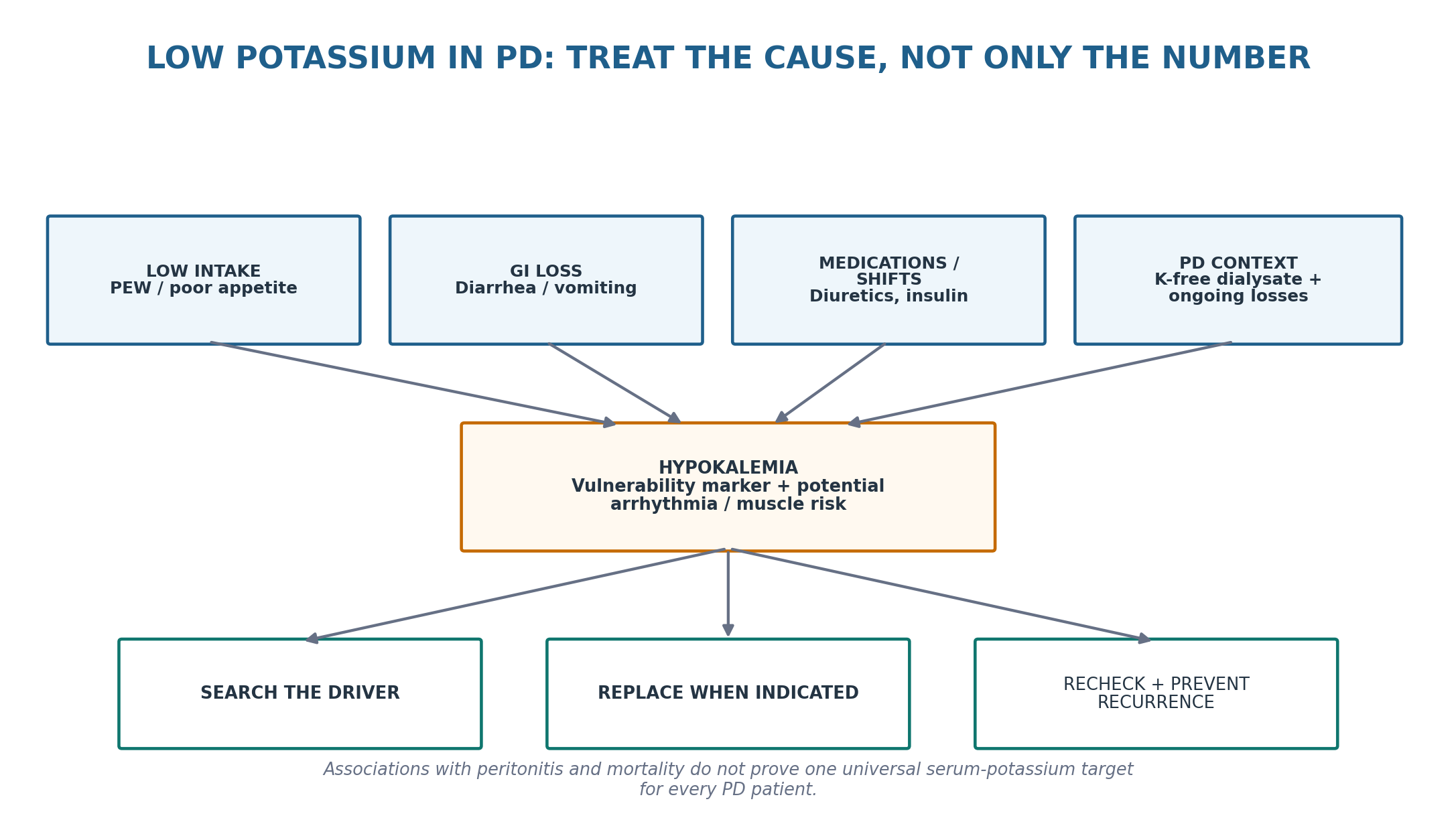
ISPD 2020 goal-directed recommendations identify hypokalemia as associated with poor nutritional intake and adverse outcomes including peritonitis, and state that dietary and/or oral potassium supplementation should be considered. The 2024 systematic review/meta-analysis found hypokalemia common and associated with higher all-cause mortality, cardiovascular mortality and peritonitis, but certainty was low to very low and causality cannot be assumed. [1,6]
A 2022 multicentre randomized trial enrolled PD patients with recurrent/time-averaged potassium <3.5 mEq/L and compared protocol-based oral potassium supplementation targeting 4–5 mEq/L with reactive supplementation below 3.5 mEq/L. Protocol treatment increased serum potassium and reduced first-peritonitis hazard, but the trial was open-label and not designed to establish 4–5 mEq/L as a universal ISPD target. Hyperkalemia occurred in a small number of protocol-treated patients. [7]
Table 13.14 — Hypokalemia: cause before replacement.
| Driver | Clue | Corrective direction |
|---|---|---|
| Low intake / PEW | Poor appetite, low intake, frailty, weight loss | Dietitian/nutrition assessment; Chapter 15 |
| GI loss | Diarrhea, vomiting, laxative excess | Treat cause + replace |
| Diuretics / residual urine | Ongoing kaliuresis | Review medication and volume plan |
| Insulin / glucose shifts | Temporal glucose-insulin changes | Coordinate glycemic plan |
| Potassium-free dialysate + low intake | Persistent tendency in PD | Replacement strategy per local protocol |
| Coexisting magnesium deficit | Refractory replacement in relevant context | Check/correct when clinically indicated |
| THRESHOLD DISCIPLINE The 4–5 mEq/L range was the intervention target of one RCT in selected hypokalemic PD patients. Present it as trial evidence—not as a universal guideline target for every PD patient. |
|---|
16. Protein loss and hypoalbuminemia: albumin is a risk signal, not a nutrition diagnosis
PD causes ongoing peritoneal protein losses, which can increase during peritonitis and coexist with poor intake, inflammation, volume expansion and comorbidity. ISPD notes that hypoalbuminemia is more common in PD than HD and is associated with protein-energy wasting and peritoneal protein loss. [1]
Serum albumin is therefore clinically important but nonspecific. A low value should trigger assessment of inflammation/infection, intake, gastrointestinal disease, liver disease, urinary loss when relevant, volume state and dialysis-related protein loss. Detailed dietary protein prescription, PEW diagnosis, body-composition assessment and amino-acid solution use belong to Chapter 15. [1]
Table 13.15 — Low albumin: do not collapse the differential.
| Possible contributor | Clue | Next direction |
|---|---|---|
| Inflammation/peritonitis | CRP/illness, recent infection | Treat inflammatory driver |
| Volume expansion | Edema, overhydration | Chapter 8 volume pathway |
| Poor intake / PEW | Weight/muscle/appetite decline | Chapter 15 nutrition assessment |
| Peritoneal protein loss | Long PD exposure, inflammatory episodes | Interpret with whole clinical picture |
| Hepatic/GI/other disease | Context-specific signs | Targeted systemic evaluation |
17. Integrated surveillance: make metabolic burden visible without duplicating later chapters
A high-quality PD review should make noninfectious and metabolic complications visible before they become technique-threatening. The minimum useful pattern is longitudinal: ask about abnormal effluent, review glucose-containing solution use and rescue hypertonicity, inspect glucose data in patients with diabetes, separate weight from volume, trend potassium, lipids and albumin in context, and link every abnormality to a named mechanism and reassessment plan. [1,3,4]
Table 13.16 — Integrated review dashboard.
| Domain | Ask / measure | Cross-reference |
|---|---|---|
| Effluent | Any blood, persistent cloudiness, milkiness, fibrin or unusual colour? | Chapter 10 if infection plausible; Chapter 12 if flow obstruction |
| Glucose exposure | Which solutions/concentrations and why? | Chapter 8 volume; Chapter 9 prescription optimisation |
| Glycemia | HbA1c trend + direct glucose/CGM when discordant | Chapter 16 diabetes-specific management |
| Weight / volume | Fluid versus tissue gain? | Chapter 8 volume; Chapter 16 obesity/frailty |
| Potassium | Trend + intake/GI/medication cause | Chapter 15 if PEW/poor intake |
| Albumin / nutrition | Inflammation, intake, fluid, protein loss | Chapter 15 |
| Membrane warning | Rising glucose requirement / transport change | Chapter 14 membrane failure/EPS |
| SERIES CONTINUITY Chapter 13 teaches recognition and immediate management of noninfectious/metabolic signals. Chapter 14 owns long-term membrane failure and EPS; Chapter 15 owns detailed nutrition/PEW; Chapter 16 owns diabetes, obesity, frailty and other special-population prescriptions. |
|---|
18. Major clinical algorithms
19. Retention tables: pattern recognition
Table 13.17 — If you see this, think this first.
| Finding | First hypothesis | Immediate action |
|---|---|---|
| Pink/red bag, stable, cyclical | Gynecologic hemoperitoneum possible | Confirm pattern; assess severity/source |
| Red bag + severe pain/instability | Major intra-abdominal bleeding | Emergency evaluation |
| Cloudy + fever/PMN predominance | Infectious peritonitis | Chapter 10 pathway |
| Cloudy early after PD + eosinophil predominance + cultures negative | Eosinophilic reaction possible | Reassess infection and exposures |
| Milky, low-cell effluent | Chylous/lymphatic fluid | Triglyceride/chylomicron + medication review |
| New milky effluent after dihydropyridine CCB | Drug-associated chyloperitoneum possible | Medication causality review |
| Glucose worsens after stronger dextrose | Dialysate glucose burden | Review prescription + diabetes therapy |
| Weight up without edema | Adiposity/tissue gain possible | Separate body composition from fluid |
| Persistent low K | Intake/GI/medication/PD vulnerability | Cause search + replacement plan |
| Low albumin | Inflammation/volume/intake/protein loss | Do not diagnose PEW from albumin alone |
Table 13.18 — What not to confuse.
| Do not confuse | With | Correction |
|---|---|---|
| Cloudy bag | Automatically noninfectious because culture was once negative | Repeat full clinical/cellular assessment |
| Eosinophils in effluent | Proof of sterile eosinophilic peritonitis | Infection can coexist with eosinophilia |
| Red dialysate | Major hemorrhage by appearance alone | Use physiology/source severity |
| Milky effluent | Peritonitis automatically | Check cells and lipid/lymph chemistry |
| Glucose sparing | Permission to under-treat volume | Verify UF/sodium and euvolemia |
| HbA1c | Perfect glycemia measure in dialysis | Use direct glucose/CGM if discordant |
| Weight gain | Always fat gain | Exclude extracellular-volume expansion |
| Trial potassium target | Universal guideline target | Present RCT evidence in context |
| Low albumin | Synonym for malnutrition | Inflammation/volume/loss also matter |
20. Clinical pearls
1. The bag is a diagnostic window: colour gets attention; cells and clinical context make the diagnosis.
2. Cloudy effluent remains infectious until appropriately evaluated—even when a noninfectious mimic is plausible.
3. A tiny amount of intraperitoneal blood can make a dramatic red bag; severity comes from hemodynamics, hemoglobin and source.
4. Menstruation/ovulation is common in hemoperitoneum, but persistent or severe bleeding deserves a broader intra-abdominal search.
5. Eosinophilic peritonitis is often early and benign, but eosinophils do not exclude infection.
6. A “culture-negative peritonitis” that stays eosinophil-predominant should trigger an exposure and differential-count review.
7. Milky effluent can be medication-related; always review recent calcium-channel blocker changes.
8. Fibrin is an inflammatory/access clue, not a stand-alone infectious diagnosis.
9. Dialysate glucose is a metabolic dose as well as an osmotic prescription.
10. The correct glucose-sparing prescription is the one that preserves fluid and sodium control.
11. HbA1c is less reliable in dialysis; discordant direct glucose data deserve more weight.
12. Icodextrin glucose-meter incompatibility is a high-consequence medication-device safety issue.
13. Weight change in PD must be split into fluid, fat, lean tissue and nutritional recovery.
14. Persistent hypokalemia is frequently a marker of poor intake or illness—not merely dialysate potassium loss.
15. Serum albumin is a prognostic risk marker, not a one-test diagnosis of malnutrition.
21. Common pitfalls — and the correction
Table 13.19 — High-frequency errors.
| Pitfall | Why it fails | Correction |
|---|---|---|
| Calling cloudy culture-negative fluid “sterile” too early | Misses infection or surgical source | Use Chapter 10 criteria/trajectory first |
| Treating eosinophils as proof of allergy | Infection can coexist | Integrate symptoms, PMN, culture and timing |
| Ignoring severe hemoperitoneum because PD blood is often benign | Misses life-threatening bleeding | Use severity/source red flags |
| Giving antibiotics for every milky bag | Chyle/lymph can mimic cloudiness | Cell count/culture + lipid testing |
| Using stronger dextrose for every weight gain | May worsen metabolic burden and miss sodium/leak/UF mechanism | Re-enter Chapter 8 volume reasoning |
| Chasing lower glucose exposure while reducing UF | Creates extracellular volume expansion | Verify volume after glucose-sparing change |
| Trusting an incompatible glucose meter with icodextrin | Can cause lethal insulin error | Use verified glucose-specific device |
| Assuming every kilogram gained is adipose tissue | Fluid and recovery confound | Phenotype weight change |
| Replacing potassium without looking for poor intake/diarrhea | Recurrence continues | Treat cause + replace |
| Calling low albumin “malnutrition” | Albumin is inflammation/volume sensitive | Comprehensive Chapter 15 assessment |
22. Mini-cases: decisions, not trivia
Case 1 — Pink effluent on a predictable cycle
A stable premenopausal patient has painless pink dialysate around mid-cycle, normal BP, no hemoglobin fall and spontaneous clearing.
| BEST NEXT STEP A benign gynecologic source is plausible. Confirm recurrence pattern and clinical stability rather than launching an emergency pathway solely because the bag looks dramatic; persistent or changing bleeding still requires investigation. |
|---|
Case 2 — Red bag with pain and dizziness
A patient presents with bright-red effluent, new focal abdominal pain, dizziness and a falling hemoglobin after an abdominal procedure.
| BEST NEXT STEP Treat this as clinically important intra-abdominal hemorrhage until proved otherwise. Resuscitation, imaging and surgical/procedural source evaluation take priority over routine PD troubleshooting. |
|---|
Case 3 — Cloudy fluid, eosinophils, negative cultures
Two weeks after PD initiation, the effluent becomes cloudy. The patient is well, cultures remain negative and the differential is strongly eosinophilic rather than neutrophilic.
| BEST NEXT STEP Reassess infectious probability and recent exposures, then consider eosinophilic peritoneal inflammation. Do not keep broadening antibiotics indefinitely if the phenotype remains noninfectious—but do not use eosinophilia alone to cancel the peritonitis safety screen. |
|---|
Case 4 — Milky effluent after a medication change
A patient develops painless milky dialysate shortly after a new dihydropyridine calcium-channel blocker. Cell count is low and cultures are negative.
| BEST NEXT STEP Test for a chylous/lipid phenotype and review the medication as a possible cause. Drug-associated chyloperitoneum is well described in PD and may resolve after clinician-directed withdrawal/substitution. |
|---|
Case 5 — Better HbA1c, worse edema
A diabetic APD patient is moved to a glucose-sparing regimen. Glucose indices improve, but weight, BP and edema increase.
| BEST NEXT STEP Do not celebrate the metabolic endpoint. Reassess net UF, sodium removal, residual urine and the entire volume prescription; glucose sparing must not compromise euvolemia. |
|---|
Case 6 — Icodextrin and an unexpectedly high bedside glucose
An emergency department gives insulin for a very high point-of-care glucose in a patient using icodextrin, but the patient is confused and diaphoretic.
| BEST NEXT STEP Immediately verify that the meter is compatible/glucose-specific and confirm glucose using an appropriate method. Icodextrin metabolites can cause falsely elevated readings with incompatible systems and dangerous insulin treatment. |
|---|
Case 7 — Persistent potassium 3.2–3.4 mmol/L
A PD patient repeatedly has low potassium, poor appetite and recent diarrhea. There is no acute ECG instability.
| BEST NEXT STEP Treat the cause and the potassium together: correct GI loss and poor intake, review medications and replace potassium according to local protocol. Do not infer a universal 4–5 mmol/L target from the supplementation RCT. |
|---|
Case 8 — Low albumin after peritonitis
Albumin falls after a severe peritonitis episode while the patient remains edematous and anorexic.
| BEST NEXT STEP Do not diagnose isolated protein deficiency. Inflammation, increased peritoneal protein loss, dilutional volume expansion and poor intake can coexist. Treat the acute drivers and enter the Chapter 15 nutrition/PEW pathway once clinically stable. |
|---|
23. Active recall
MUST MEMORIZE
Table 13.20 — Core facts.
| Prompt | Answer |
|---|---|
| First rule for cloudy effluent? | Peritonitis must be appropriately excluded/treated according to Chapter 10. |
| Most common benign hemoperitoneum source in menstruating patients? | Menstruation/ovulation-related bleeding. |
| What makes hemoperitoneum urgent? | Instability, severe/focal pain, Hb fall, pregnancy/trauma/procedure or other high-risk source. |
| Conventional eosinophilic peritonitis definition? | >100 eosinophils/mL and >10% of effluent WBC in commonly cited literature; not an ISPD peritonitis criterion. |
| Do eosinophils exclude infection? | No. |
| Milky low-cell effluent suggests? | Chylous/lymphatic or lipid-rich fluid; test rather than judge by appearance. |
| Drug class associated with chylous PD effluent? | Dihydropyridine calcium-channel blockers in case literature/systematic review. |
| Why is dialysate glucose a metabolic dose? | A fraction is absorbed systemically and can affect glycemia, insulin, weight and lipids. |
| HbA1c in dialysis? | Useful but less reliable; direct glucose/CGM can resolve discordance. |
| Key glucose-sparing safeguard? | Verify sodium-volume control after the prescription change. |
| Icodextrin meter danger? | Some GDH-PQQ/GDO and some GDH-FAD methods read maltose as glucose. |
| How long may icodextrin meter interference persist after stopping? | Product information warns falsely high readings may occur for up to about 2 weeks. |
| Weight gain in PD: first distinction? | Fluid versus tissue gain. |
| ISPD potassium message? | Hypokalemia is associated with poor intake/adverse outcomes; dietary/oral supplementation should be considered. |
| RCT potassium target? | 4–5 mEq/L in selected hypokalemic patients—trial target, not universal guideline target. |
| Low albumin equals malnutrition? | No; inflammation, fluid status and peritoneal protein loss also matter. |
| Detailed EPS content belongs where? | Chapter 14. |
| Detailed PEW/nutrition content belongs where? | Chapter 15. |
USE AS REFERENCE
Exact acute potassium-replacement doses and ECG-monitoring thresholds.
Patient-specific glycemic targets and dialysis-compatible diabetes drug selection.
Current glucose-meter/CGM compatibility with icodextrin.
Laboratory-specific chylous-effluent triglyceride/chylomicron interpretation.
Investigation of recurrent hemoperitoneum according to gynecologic, surgical and malignancy context.
Solution- or lot-specific investigation of suspected chemical peritonitis.
24. Flashcards: spaced repetition
1. Q: Cloudy bag: first assumption? A: Infectious peritonitis until appropriately evaluated.
2. Q: Red bag: first severity question? A: Is the patient unstable, in severe pain, or losing hemoglobin?
3. Q: Common benign cause of hemoperitoneum? A: Menstruation/ovulation-related bleeding.
4. Q: Eosinophilic peritonitis typical timing? A: Often early after catheter insertion/PD exposure.
5. Q: Can infection cause peritoneal eosinophilia? A: Yes.
6. Q: Milky effluent: first lab direction? A: Cell count/culture if indicated plus triglyceride/chylous assessment.
7. Q: CCB class linked to chyloperitoneum? A: Dihydropyridine calcium-channel blockers.
8. Q: Fibrin means infection? A: No; it is an inflammation/bleeding/access clue.
9. Q: Why can PD worsen glycemia? A: Systemic absorption of dialysate glucose.
10. Q: HbA1c reliability in dialysis? A: Reduced compared with earlier CKD; use direct glucose/CGM if discordant.
11. Q: Glucose-sparing trade-off to watch? A: Extracellular volume expansion/insufficient UF.
12. Q: Icodextrin safety phrase? A: Use a compatible glucose-specific measurement method.
13. Q: Why can incompatible meters read high? A: Maltose/oligosaccharides interfere with some enzymatic methods.
14. Q: How long after stopping icodextrin can interference persist? A: Up to about 2 weeks per current product information.
15. Q: Weight gain first split? A: Fluid versus tissue.
16. Q: Typical PD dyslipidemia pattern? A: Often triglyceride/VLDL-rich, though individual patterns vary.
17. Q: Persistent low potassium: common hidden driver? A: Poor intake/PEW or GI loss, often with medication/PD contributions.
18. Q: Does hypokalemia cause peritonitis? A: Association exists; causality is not proved.
19. Q: What did the potassium RCT show? A: Protocol supplementation raised potassium and reduced first-peritonitis hazard in selected patients.
20. Q: Universal potassium target from that RCT? A: No.
21. Q: Low albumin: why nonspecific? A: Inflammation, volume expansion, intake and peritoneal loss all affect it.
22. Q: Membrane failure / EPS chapter? A: Chapter 14.
23. Q: Nutrition / PEW chapter? A: Chapter 15.
24. Q: Final mental model? A: Safety screen → classify effluent/body signal → name mechanism → change one lever → verify.
25. Rapid differential / troubleshooting
Table 13.21 — Noninfectious/metabolic troubleshooting from problem to action.
| Problem | Differential | First actions |
|---|---|---|
| Pink/red effluent, stable | Menstruation/ovulation, minor trauma, anticoagulation | Assess severity, timing, Hb; observe if convincingly benign |
| Red effluent, pain/instability | Major hemorrhage, ectopic pregnancy, organ/GI source | Emergency evaluation + imaging/source control |
| Cloudy, culture negative | Early treated infection, eosinophilic/chemical inflammation, unusual organism | Repeat WBC/differential/culture; exposure review; special studies if needed |
| Milky effluent | Chyle, pseudochyle, lymph-rich fluid, peritonitis | Cell count/culture + triglyceride/chylous testing + medication review |
| Fibrin strands | Postoperative/bleeding/inflammation/peritonitis | Identify source; Chapter 12 if catheter obstruction |
| Rising glucose after hypertonic bags | Dialysate glucose absorption | Review why hypertonic therapy is needed; coordinate diabetes plan |
| Better glucose after glucose-sparing but edema worsens | Under-ultrafiltration / sodium-volume problem | Return to Chapter 8 volume assessment |
| High bedside glucose on icodextrin with hypoglycemic symptoms | Meter interference | Use compatible confirmatory glucose method urgently |
| Weight gain | Fluid, adiposity, nutritional recovery | Volume phenotype + body-composition/nutrition context |
| Persistent hypokalemia | Poor intake, GI loss, diuretics, insulin, PD context | Cause-directed review + replacement + recheck |
| Low albumin | Inflammation, dilution, PEW, peritoneal loss, systemic disease | Treat drivers; Chapter 15 for comprehensive nutrition |
26. Final revision sheet
| CORE CONCEPT Noninfectious and metabolic PD complications are solved by two linked mental models: abnormal EFFLUENT requires a safety-first source diagnosis; metabolic drift requires a WHOLE-PATIENT + PRESCRIPTION diagnosis. The prescription is never metabolically neutral, but metabolic improvement must not compromise fluid, sodium or dialysis goals. |
|---|
Table 13.22 — One-minute revision.
| Domain | Must remember |
|---|---|
| Cloudy effluent | Chapter 10 first; mimics come after safe infectious assessment. |
| Hemoperitoneum | Colour can look dramatic; urgency comes from physiology and source. |
| Eosinophilic inflammation | Often early/self-limited; conventional >100/mL and >10% definition; infection can coexist. |
| Milky effluent | Think triglyceride/lymph + drugs; do not diagnose from colour alone. |
| Fibrin / unusual colour | Clue to inflammation, bleeding, pigment or intra-abdominal source. |
| Glucose burden | Dialysate glucose is absorbed and affects systemic metabolism. |
| Glycemic monitoring | HbA1c less reliable in dialysis; direct glucose/CGM helps discordance. |
| Glucose sparing | Can improve metabolic markers; verify UF/sodium/volume. |
| Icodextrin | Use compatible glucose measurement; interference may persist ~2 weeks after stopping. |
| Weight | Separate fluid from tissue. |
| Hypokalemia | Common, multifactorial, associated with adverse outcomes; treat cause + replace when indicated. |
| Albumin | Risk marker, not a stand-alone nutrition diagnosis. |
TEN TAKE-HOME RULES
1) Cloudy effluent is infectious until appropriately assessed. 2) Red fluid is common but not automatically benign. 3) Eosinophils do not exclude infection. 4) Milky fluid needs lipid/lymph testing, not visual diagnosis. 5) Dialysate glucose is a systemic metabolic dose. 6) HbA1c has reduced reliability in dialysis. 7) Glucose sparing never outranks euvolemia. 8) Icodextrin requires glucose-meter compatibility. 9) Persistent hypokalemia requires a cause search; one RCT target is not a universal guideline target. 10) Low albumin triggers a whole-patient assessment and the Chapter 15 nutrition pathway.
Table 13.23 — One-minute bedside synthesis.
| If you see… | Think… | Do now… |
|---|---|---|
| Cloudy + pain | Peritonitis | Effluent studies + Chapter 10 treatment logic |
| Pink + stable cyclical pattern | Benign gynecologic source possible | Confirm pattern and stability |
| Red + unstable | Major hemorrhage | Emergency/source evaluation |
| Cloudy + eosinophil-predominant + negative cultures | Eosinophilic/chemical mimic possible | Reassess infection + exposures |
| Milky low-cell bag | Chyle/lymph | Triglyceride/chylous work-up + medication review |
| Glucose rises after stronger bags | Dialysate metabolic load | Review prescription + diabetes plan |
| Glucose-sparing + edema | Volume trade-off | Reassess UF/sodium/volume |
| Icodextrin + implausible glucose | Meter interaction | Confirm with compatible method |
| Persistent low K | Whole-patient vulnerability | Cause + replacement + recheck |
| Low albumin | Inflammation/volume/intake/loss | Comprehensive assessment; Chapter 15 |
| FINAL MENTAL MODEL Threat → bag/body phenotype → cells/chemistry → source/mechanism → targeted change → verify volume + symptoms + laboratory trajectory → cross-reference membrane/nutrition/special-population chapters when the problem belongs there. |
|---|
Rapid oral viva
Explain why a culture-negative cloudy bag is not automatically a noninfectious complication.
Classify hemoperitoneum into benign-pattern versus emergency-pattern presentations.
Define eosinophilic peritonitis using the conventional literature definition and explain its limitation.
Build the differential for milky PD effluent and describe the role of medication review.
Explain why dialysate glucose must be treated as a systemic metabolic exposure.
Explain when HbA1c becomes misleading in dialysis and how directly measured glucose can help.
Describe the IMPENDIA/EDEN lesson about glucose sparing and extracellular volume.
Explain the icodextrin glucose-meter interaction and how long the warning remains relevant after discontinuation.
Build a cause-based approach to recurrent hypokalemia and explain why 4–5 mEq/L is not a universal PD target.
Explain why serum albumin cannot diagnose PEW by itself.
| SAFETY BOUNDARY This chapter teaches recognition and decision architecture. Acute hemorrhage, surgical abdomen, severe electrolyte disturbance, symptomatic hypoglycemia/hyperglycemia and sepsis require current acute-care protocols. Exact potassium replacement, diabetes-drug dosing, glycemic targets, icodextrin/CGM compatibility, chylous-fluid laboratory cutoffs, and treatment of eosinophilic/chemical peritonitis should use current local protocols, product information and specialist input. |
|---|
27. Selected authoritative references
1. Brown EA, Blake PG, Boudville N, et al. International Society for Peritoneal Dialysis practice recommendations: Prescribing high-quality goal-directed peritoneal dialysis. Perit Dial Int. 2020;40(3):244–253. https://doi.org/10.1177/0896860819895364.
2. Li PKT, Chow KM, Cho Y, et al. ISPD peritonitis guideline recommendations: 2022 update on prevention and treatment. Perit Dial Int. 2022;42(2):110–153. https://doi.org/10.1177/08968608221080586. PMID: 35264029.
3. Kidney Disease: Improving Global Outcomes (KDIGO) Diabetes Work Group. KDIGO 2022 Clinical Practice Guideline for Diabetes Management in Chronic Kidney Disease. Kidney Int. 2022;102(5S):S1–S127.
4. Wijewickrama P, Williams J, Bain S, et al. Narrative Review of Glycemic Management in People With Diabetes on Peritoneal Dialysis. Kidney Int Rep. 2023;8(4):700–714. https://doi.org/10.1016/j.ekir.2023.01.040. PMID: 37069983.
5. Li PKT, Culleton BF, Ariza A, et al. Randomized, controlled trial of glucose-sparing peritoneal dialysis in diabetic patients. J Am Soc Nephrol. 2013;24(11):1889–1900. https://doi.org/10.1681/ASN.2012100987. PMID: 23949801.
6. Yang C, Hu X, Ling X, et al. Hypokalemia in Peritoneal Dialysis: A Systematic Review and Meta-analysis of Prevalence, Treatment, and Outcomes. Kidney Med. 2024;6(12):100923. https://doi.org/10.1016/j.xkme.2024.100923. PMID: 39634334.
7. Pichitporn W, Kanjanabuch T, Phannajit J, et al. Efficacy of Potassium Supplementation in Hypokalemic Patients Receiving Peritoneal Dialysis: A Randomized Controlled Trial. Am J Kidney Dis. 2022;80(5):580–588.e1. https://doi.org/10.1053/j.ajkd.2022.03.013. PMID: 35597332.
8. Dossin T, Goffin E. When the color of peritoneal dialysis effluent can be used as a diagnostic tool. Semin Dial. 2019;32(1):72–79. https://doi.org/10.1111/sdi.12740. PMID: 30032485.
9. Lew SQ. Hemoperitoneum: bloody peritoneal dialysate in ESRD patients receiving peritoneal dialysis. Perit Dial Int. 2007;27(3):226–233. PMID: 17468466.
10. Greenberg A, Bernardini J, Piraino BM, Johnston JR, Perlmutter JA. Hemoperitoneum complicating chronic peritoneal dialysis: single-center experience and literature review. Am J Kidney Dis. 1992;19(3):252–256. https://doi.org/10.1016/S0272-6386(13)80006-6. PMID: 1553970.
11. Zhang Q, Xia Y, Zhang M, Jiang C. Peritoneal dialysis related eosinophilic peritonitis: a case report and review of the literature. BMC Nephrol. 2023;24(1):10. https://doi.org/10.1186/s12882-022-03027-8. PMID: 36635670.
12. Uzun Kenan B, Büyükkaragöz B, Leventoğlu E, Bakkaloğlu SA. Eosinophilic peritonitis in children undergoing maintenance peritoneal dialysis: A case report and literature review. Semin Dial. 2022;35(6):548–555. https://doi.org/10.1111/sdi.13113. PMID: 35788998.
13. Bhardwaj R, Vaziri H, Gautam A, et al. Chylous Ascites: A Review of Pathogenesis, Diagnosis and Treatment. J Clin Transl Hepatol. 2018;6(1):105–113. https://doi.org/10.14218/JCTH.2017.00035. PMID: 29577037.
14. Chen YC, et al. Calcium Channel Blocker-Related Chylous Ascites: A Systematic Review and Meta-Analysis. Medicina (Kaunas). 2019;55(4):93. PMID: 30959848.
15. EXTRANEAL (icodextrin) peritoneal dialysis solution. Current U.S. prescribing information / DailyMed. Safety warning on glucose-monitor interference. Accessed 3 September 2026.
16. Stepanova N. Dyslipidemia in Peritoneal Dialysis: Implications for Peritoneal Membrane Function and Patient Outcomes. Biomedicines. 2024;12(10):2377. https://doi.org/10.3390/biomedicines12102377. PMID: 39457689.
17. Wang J, Zhao J, Li L, et al. Association Between Peritoneal Glucose Absorption, Lipid Metabolism, and Cardiovascular Disease Risk in Nondiabetic Patients on Peritoneal Dialysis. J Ren Nutr. 2025;35(1):196–206. https://doi.org/10.1053/j.jrn.2024.05.005. PMID: 38851308.
18. Holmes CJ. Adverse effects of systemic glucose absorption with peritoneal dialysis: how good is the evidence? Curr Opin Nephrol Hypertens. 2013;22(6):635–642. PMID: 24076559.
19. Morelle J, Stachowska-Pietka J, Öberg C, et al. ISPD recommendations for the evaluation of peritoneal membrane dysfunction in adults: classification, measurement, interpretation and rationale for intervention. Perit Dial Int. 2021;41(4):352–372. https://doi.org/10.1177/0896860820982218.
| SOURCE NOTE Guideline status and key contemporary evidence were checked 3 September 2026. The ISPD guideline repository continues to list the 2020 high-quality prescription, 2021 membrane-dysfunction and 2022 peritonitis guidance as current relevant adult standards. KDIGO has a 2026 Diabetes in CKD public-review draft, but the 2022 guideline remains the final published KDIGO standard used here. Exact acute electrolyte replacement, diabetes therapy, glucose-monitor compatibility, rare-effluent diagnostic cutoffs and product-specific solution instructions remain governed by current local protocols, laboratory methods and manufacturer information. |
|---|