Applied Nephrology Master Series
Chapter 14
Long-Term Peritoneal Membrane Failure and Encapsulating Peritoneal Sclerosis
Membrane Trajectory | Acquired Ultrafiltration Failure | Long-Term Risk | EPS | CT Diagnosis | Nutrition | Medical Therapy | Surgery
| CHAPTER MISSION Track the peritoneal membrane as a changing organ: recognise acquired transport and ultrafiltration failure before volume control collapses; separate long-term membrane dysfunction from encapsulating peritoneal sclerosis; discuss EPS risk without imposing an arbitrary PD “time limit”; recognise the post-PD risk window; diagnose EPS from the clinical–radiologic syndrome; and organise nutrition, medical therapy and expert surgery around the dominant disease phenotype. |
|---|
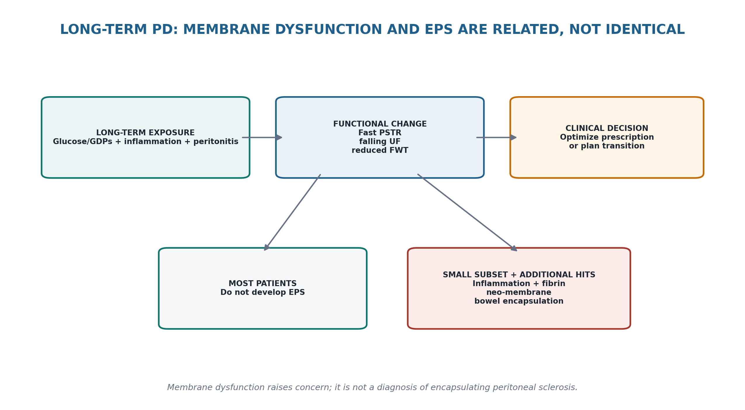
| MASTER PRINCIPLE Follow the TRAJECTORY, not the CLOCK. Long PD duration raises EPS risk, but there is no evidence-based duration at which every patient should stop PD. The decisive questions are whether membrane function is deteriorating, whether clinical goals remain achievable, whether new gastrointestinal symptoms suggest EPS, and what alternative kidney-replacement options fit the person. |
|---|
0. One-page chapter map
Table 14.1 — The eight decisions that govern long-term membrane and EPS care.
| Decision | Core question | Bedside output |
|---|---|---|
| 1. Trajectory | Is membrane function stable, adapting, or progressively deteriorating? | Named functional trajectory |
| 2. Mechanism | Is the problem fast PSTR, low osmotic conductance, non-membrane fluid loss, or mixed? | Mechanism of low UF |
| 3. Risk | Does long PD exposure plus membrane/inflammatory history meaningfully raise EPS concern? | Individualized risk discussion |
| 4. Symptoms | Are there recurrent obstructive, inflammatory or nutritional gastrointestinal signals? | EPS suspicion level |
| 5. Imaging | Does CT show bowel encapsulation/tethering and characteristic peritoneal abnormalities? | Clinical–radiologic synthesis |
| 6. Support | Is there intestinal failure, malnutrition, sepsis or another immediate threat? | Nutrition + acute-care plan |
| 7. Treat | Is the phenotype inflammatory, fibrotic/obstructive, or mixed? | Medical / surgical strategy |
| 8. Transition | Can PD remain safe, or is planned modality change required? | Shared modality plan + follow-up |
Learning outcomes
Distinguish ordinary long-term peritoneal remodeling, acquired membrane dysfunction and true encapsulating peritoneal sclerosis (EPS).
Interpret longitudinal PET, ultrafiltration capacity and sodium-dip/free-water-transport information without confusing a transport abnormality with EPS itself.
Apply the 2021 ISPD definitions for low UF capacity and intrinsic membrane dysfunction when clinically indicated.
Explain why prolonged PD exposure increases EPS risk while avoiding a universal time-based rule for stopping PD.
Use the 2017 ISPD position that EPS risk should be discussed during long-term PD and that continuation versus transition should be individualized through shared decision-making.
Recognise that EPS frequently presents after PD has been discontinued, including after transition to haemodialysis or kidney transplantation.
Recognise recurrent bowel-obstruction symptoms, inflammatory features and nutritional decline as the core clinical signals of EPS.
Use CT as the principal imaging test in suspected EPS and interpret a pattern of peritoneal thickening/enhancement/calcification, bowel tethering/encapsulation, loculated fluid and obstruction rather than relying on one sign.
Treat nutrition and intestinal-failure support as central disease-modifying care while recognising that corticosteroid and tamoxifen evidence is observational and phenotype dependent.
Know when recurrent or fixed obstruction requires referral to an experienced EPS surgical centre for enterolysis/peritonectomy.
Avoid unvalidated routine CT or biomarker screening of asymptomatic long-term PD patients.
Cross-reference Chapter 8 for UF failure, Chapter 9 for prescription optimization and Chapter 15 for detailed protein-energy wasting/nutrition management.
| EVIDENCE POSTURE The current ISPD guideline repository still lists the 2017 position paper on length of time on PD and EPS as the dedicated EPS guidance. It concludes that EPS is rare, risk increases with longer PD exposure, discussion is reasonable around years 3–4, and there is insufficient evidence for a single maximum PD duration. The 2021 ISPD membrane-dysfunction recommendations provide current functional thresholds and explicitly state that acquired intrinsic membrane injury during prolonged PD should trigger discussion of EPS risk and modality transition. Diagnostic, pharmacologic and surgical EPS evidence remains dominated by observational cohorts, registries and expert-centre experience; no randomized trial establishes corticosteroid, tamoxifen or surgical timing as a universal standard. [1–8] |
|---|
1. Core concept: membrane failure and EPS are related—but not the same disease
Long-term PD changes the peritoneal membrane. Some patients develop faster small-solute transport, reduced glucose-driven ultrafiltration, diminished free-water transport or a progressively less efficient membrane. These functional changes matter because they can threaten sodium-volume control and increase glucose exposure. They are not, by themselves, encapsulating peritoneal sclerosis. EPS is a distinct clinical syndrome in which a fibrotic/neo-membranous process encapsulates bowel and produces recurrent or progressive intestinal obstruction, inflammation and nutritional failure. [1–4]
Table 14.2 — Do not collapse these three entities into one.
| Entity | What it means | What it does NOT mean |
|---|---|---|
| Long-term membrane remodeling | Structural and functional change with prolonged PD exposure | Inevitable technique failure or EPS |
| Acquired membrane dysfunction | Measured transport/UF abnormality developing over time | A diagnosis of EPS |
| EPS | Clinical syndrome of bowel encapsulation/obstruction with characteristic imaging/context | Any long PD vintage, calcification or fast transporter status |
| BEDSIDE TRANSLATION A declining sodium dip or worsening UF can identify an injured membrane that deserves a modality-risk discussion. It cannot diagnose the fibrous bowel-cocoon syndrome of EPS. |
|---|
2. Long-term membrane remodeling: repeated injury changes both vessels and interstitium
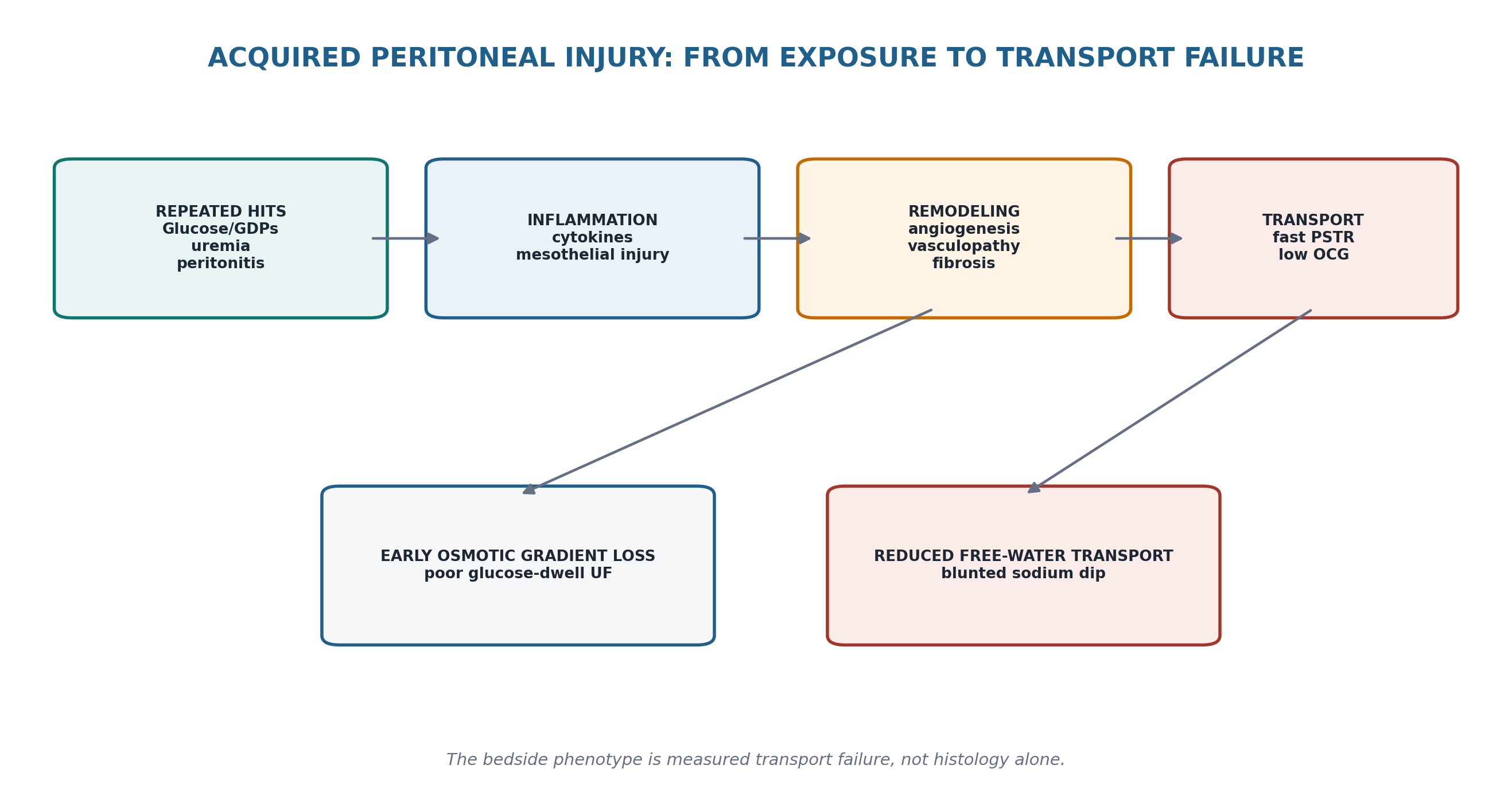
The pathobiology is not a simple linear “mesothelium becomes scar” story. Long-term exposure to glucose and glucose-degradation products, episodes of peritoneal inflammation, uremic biology, angiogenesis and vasculopathy all interact. Contemporary Japanese pathologic work also emphasizes that established EPS may involve formation of a fibrin-rich neo-membrane over an injured peritoneal surface rather than merely an extreme degree of ordinary peritoneal sclerosis. [4,9]
Table 14.3 — Mechanism → functional consequence → clinical signal.
| Mechanism | Functional consequence | Bedside signal |
|---|---|---|
| Inflammation / increased vascular surface area | Faster PSTR; rapid glucose absorption | Long glucose dwells lose osmotic force early |
| Angiogenesis / vasculopathy | Altered effective capillary exchange | Transport phenotype changes over time |
| Interstitial fibrosis | Lower osmotic conductance to glucose | Low UF despite strong glucose gradient |
| Reduced effective free-water transport | Blunted early sodium dilution | Small/absent 1-h sodium dip |
| Persistent fibrin/inflammatory response in susceptible patient | Neo-membrane, adhesions, encapsulation | Obstructive EPS phenotype |
| MECHANISM DISCIPLINE Peritoneal fibrosis is biologically important, but neither fibrosis nor calcification alone is synonymous with EPS. EPS is defined clinically by the bowel-encapsulating syndrome. |
|---|
3. Functional surveillance: measure change when the clinical trajectory changes
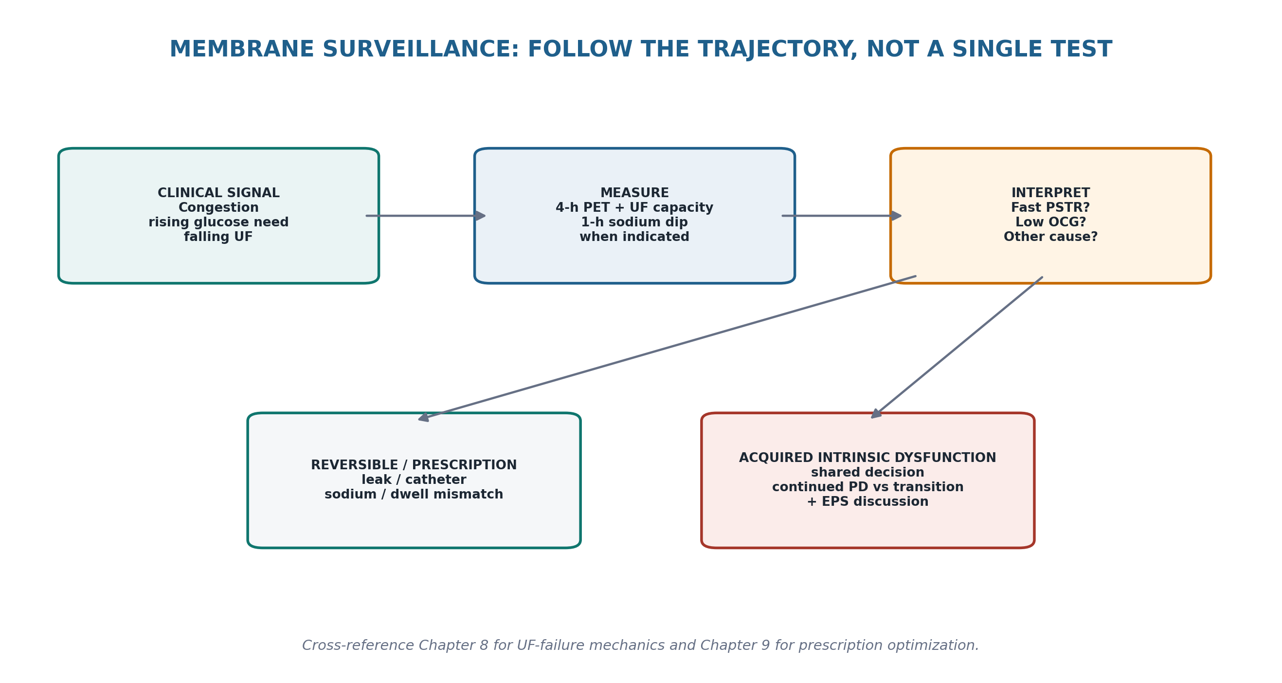
ISPD recommends characterizing PSTR with a 4-hour PET early in PD and subsequently when clinically indicated. Long-term surveillance becomes particularly important when a previously stable prescription no longer controls volume, when glucose exposure rises, when daily UF falls, or when membrane behavior appears to have changed. [2]
Table 14.4 — Long-term membrane surveillance signals.
| Signal | What to measure/check | Interpretive caution |
|---|---|---|
| Falling net UF | Delivered prescription, drain completeness, leaks, daily UF, PET UF capacity | Low UF is not automatically membrane failure |
| Increasing hypertonic glucose use | Sodium intake, RKF trend, PSTR, long-dwell strategy | Prescription escalation can mask the mechanism |
| New fast PSTR | Inflammation/peritonitis history, PET trend | Fast PSTR can be acquired or transient |
| Blunted sodium dip | High-glucose PET with 1-h sodium measurement | Reduced FWT can reflect fibrosis but is not EPS-specific |
| Congestion despite adherence | Total sodium/water balance + membrane testing | Chapter 8 differential still applies |
| SERIAL RULE A change from the patient’s own previous membrane behavior is often more clinically informative than a one-time transporter label. |
|---|
4. Acquired intrinsic ultrafiltration insufficiency: when the membrane itself is failing
The 2021 ISPD membrane recommendations define low UF capacity as a valuable screening phenotype. Insufficient UF should be suspected when net UF from a 4-hour PET is <400 mL using 3.86% glucose/4.25% dextrose or <100 mL using 2.27% glucose/2.5% dextrose, and/or when daily UF cannot maintain adequate fluid status. These thresholds identify low UF capacity; they do not by themselves specify the mechanism. [2]
When intrinsic membrane dysfunction is suspected, ISPD recommends supplementing the 4-hour high-glucose PET with a 1-hour sodium dip. A sodium dip ≤5 mmol/L and/or sodium sieving ratio ≤0.03 at 1 hour indicates UF insufficiency due to impaired free-water/osmotic conductance physiology. Acquired intrinsic dysfunction developing after years on PD—particularly when residual kidney function is absent—should prompt discussion of the risks of continuing PD, including EPS, versus transition to another modality. [2]
Table 14.5 — ISPD functional anchors that belong in long-term PD review.
| Measure | Current ISPD anchor | Clinical meaning |
|---|---|---|
| 4-h UF, 3.86% glucose/4.25% dextrose | <400 mL | Low UF capacity; search mechanism |
| 4-h UF, 2.27% glucose/2.5% dextrose | <100 mL | Low UF capacity; search mechanism |
| 1-h sodium dip, high-glucose PET | ≤5 mmol/L | Supports intrinsic UF insufficiency |
| 1-h sodium sieving ratio | ≤0.03 | Supports intrinsic UF insufficiency |
| Daily UF unable to maintain fluid status | Clinical practice point | Low UF capacity may be clinically important even beyond test thresholds |
| DO NOT SHORT-CIRCUIT Before calling a membrane intrinsically failed, exclude catheter dysfunction, leak, incomplete drainage, excessive sodium/fluid intake, prescription mismatch and increased fluid absorption. Chapter 8 remains the UF-failure diagnostic pathway. |
|---|
5. EPS risk: duration matters—but there is no stopwatch rule
EPS is strongly associated with prolonged PD exposure, and incidence rises with PD vintage. The 2017 ISPD position paper emphasizes that EPS remains uncommon and that the majority of long-term PD patients do not develop it. The paper found insufficient evidence to support a single maximum safe duration of PD. It specifically argues against withholding or stopping PD solely because a clock threshold has been reached. [1]
A 2022 meta-analysis of 10 studies (12,595 participants) found associations with longer PD duration, higher transporter status, longer peritonitis duration, younger age at PD onset and glomerulonephritis history; the evidence is observational and does not create a validated individual prediction score. [5]
Table 14.6 — EPS risk factors: use for context, not deterministic prediction.
| Factor | Evidence signal | How to use it |
|---|---|---|
| Longer PD duration | Most consistent epidemiologic association | Raises vigilance; does not set an automatic stop date |
| Acquired fast PSTR / low FWT | Associated with long-term membrane injury and EPS risk in observational studies | Prompts functional review and shared decision |
| Prolonged/severe peritonitis burden | Associated in observational data | Strengthens concern when membrane function also deteriorates |
| PD discontinuation / post-transplant period | Many cases present after PD stops | Continue symptom vigilance after modality change |
| Younger age at PD onset / GN history | Meta-analysis associations | Context only; not actionable thresholds |
| RISK LANGUAGE Say “risk is higher” rather than “EPS is developing” unless the patient has the clinical–radiologic syndrome. |
|---|
6. When to discuss EPS and modality transition
The ISPD 2017 position paper recommends that EPS should be discussed with patients on long-term PD—not necessarily when PD starts, but reasonably around years 3–4—so that future choices are anticipated rather than presented during a crisis. The discussion should integrate prognosis, PD duration, dialysis quality, UF/volume control, peritonitis frequency, transplant access, HD risks and the patient’s quality-of-life priorities. [1]
Table 14.7 — The long-term PD shared-decision conversation.
| Domain | Question | Why it changes the decision |
|---|---|---|
| Membrane | Is function stable or progressively deteriorating? | Progressive intrinsic dysfunction may make continued PD less safe/effective |
| Clinical goals | Can sodium-volume and biochemical goals still be achieved? | Technique viability is clinical, not just historical |
| Peritonitis history | Is there repeated/prolonged inflammatory injury? | Adds membrane and EPS concern |
| Transplant | Is transplantation likely soon? | Competing pathway may dominate timing |
| HD feasibility | Vascular access, haemodynamic tolerance, home-HD options? | Alternative modality risk matters |
| Patient goals | What burden/trade-off is acceptable? | No evidence justifies a one-size-fits-all stop rule |
| SHARED DECISION RULE Do not use EPS fear to force an otherwise well patient off PD. Do not use patient preference to ignore progressive inability to achieve safe volume control. Both risk and benefit belong in the same conversation. |
|---|
7. After PD stops: the risk conversation is not over
EPS commonly becomes clinically apparent after PD has been discontinued, either after transfer to haemodialysis or following kidney transplantation. In a European multicentre study, post-transplant EPS and classical EPS were histologically similar; diagnosis occurred earlier after PD cessation in the post-transplant group. This reinforces an important practical rule: stopping PD does not instantly remove EPS risk. [6,10]
Table 14.8 — Post-PD vigilance.
| Situation | What to remember | Clinical response |
|---|---|---|
| Transferred to HD after long PD | EPS may present months or later after stopping | Educate about GI symptoms; investigate promptly |
| Kidney transplant after long PD | Post-transplant EPS is recognized | Do not attribute vomiting/weight loss automatically to drugs/infection |
| Acquired UF failure before stopping | Higher-risk membrane phenotype | Document trajectory in handover |
| Asymptomatic former PD patient | No validated routine CT/biomarker screening standard | Clinical vigilance rather than indiscriminate imaging |
| HANDOVER PEARL When a long-vintage PD patient transfers to transplant or HD, include “prior PD duration + membrane trajectory + EPS vigilance” in the receiving-team handover. |
|---|
8. The EPS clinical syndrome: think bowel obstruction, inflammation and nutritional decline
EPS most often presents as a gastrointestinal syndrome rather than as a dialysis-number abnormality. Common features include recurrent or progressive abdominal pain, anorexia, nausea, vomiting, early satiety, weight loss and episodes of partial or complete bowel obstruction. Hemoperitoneum, ascites or sterile inflammatory episodes may occur. Malnutrition is not merely a consequence; it is a major determinant of outcome. [3,4,7]
Table 14.9 — EPS bedside phenotypes.
| Phenotype | Typical clues | Urgency |
|---|---|---|
| Prodromal/inflammatory | Anorexia, weight loss, abdominal pain, inflammatory markers, ascites | Early specialist assessment |
| Intermittent obstruction | Recurrent vomiting/distension, episodic constipation/obstruction | CT + surgical/nutrition MDT |
| Fixed obstruction | Persistent vomiting, dilated bowel, inability to tolerate intake | Urgent intestinal-failure/surgical pathway |
| Complicated disease | Sepsis, ischemia, perforation, fistula, severe cachexia | Emergency surgical/critical-care pathway |
| CLINICAL TRIGGER In a current or former long-term PD patient, recurrent “subacute bowel obstruction” is EPS until the diagnosis has been seriously considered. |
|---|
9. Diagnosis: symptoms create the question; CT tests it
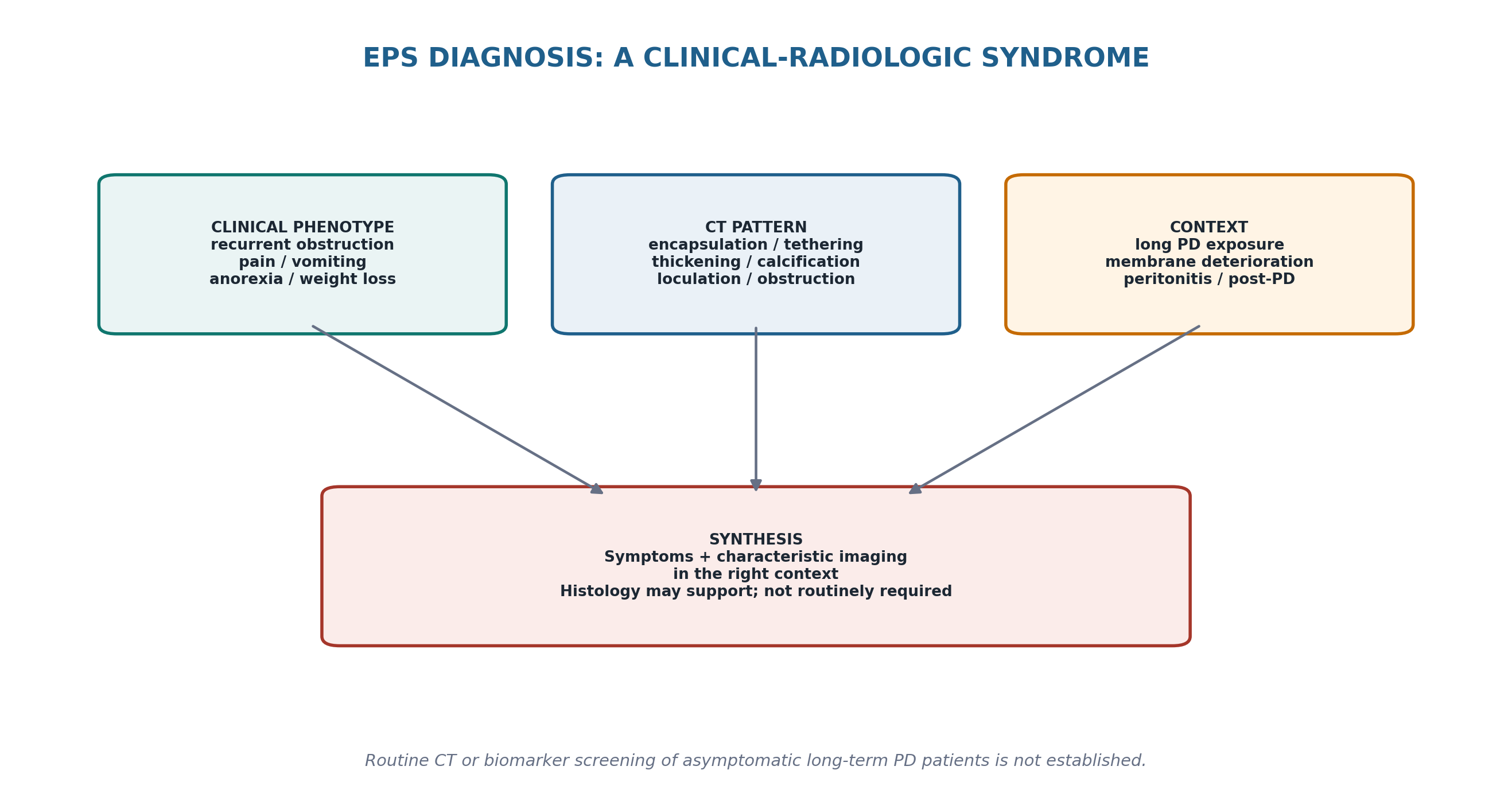
No blood test diagnoses EPS. Biomarkers such as CA-125, IL-6, PAI-1 and free-water transport have shown research signals in small case-control cohorts, but no validated screening strategy is recommended for routine asymptomatic surveillance. CT is the principal imaging modality when EPS is clinically suspected. [3,8,11]
Table 14.10 — Diagnostic sequence for suspected EPS.
| Step | Data required | Interpretation |
|---|---|---|
| 1. Clinical syndrome | Obstructive symptoms, weight loss, inflammatory features, prior PD history | Defines pre-test probability |
| 2. Safety screen | Sepsis, perforation, ischemia, severe dehydration/malnutrition | Determines urgent pathway |
| 3. CT abdomen/pelvis | Peritoneal + bowel + fluid findings | Confirms characteristic pattern / alternative diagnosis |
| 4. Multidisciplinary synthesis | Nephrology + radiology + surgery + nutrition | Determines treatment phenotype |
| 5. Histology/laparotomy | Only when obtained for clinical/surgical reasons or diagnosis remains uncertain | Supportive—not a routine screening test |
| SCREENING BOUNDARY A CT scan can support diagnosis in symptomatic patients. Routine serial CT of every long-term asymptomatic PD patient is not an established prevention strategy. |
|---|
10. CT pattern: diagnose the constellation, not one calcified line
Experienced radiologists identify EPS from a combination of findings. In the classic case-control study, peritoneal enhancement, thickening and calcification; adhesions/tethering of bowel loops; signs of obstruction; and loculated/septated fluid were more frequent in EPS than in long-term PD controls. Contemporary radiology reviews continue to regard CT as the imaging modality of choice. [8,12]
Table 14.11 — CT findings that support EPS.
| CT feature | What it represents | Important caution |
|---|---|---|
| Bowel tethering/clustering/encapsulation | Fibrous cocoon/adhesive process | Most meaningful when symptoms fit |
| Peritoneal thickening/enhancement | Inflammatory/fibrotic membrane change | Not specific alone |
| Peritoneal calcification | Chronic peritoneal injury | Can exist without clinical EPS |
| Loculated/septated ascites | Adhesive compartmentalization | Also occurs in infection/other disease |
| Bowel wall thickening/dilatation | Obstruction/inflammatory consequence | Assess ischemia/perforation when severe |
| Transition points / obstruction | Mechanical consequence of encasement | Drives surgical urgency |
| RADIOLOGY PEARL The highest-value CT question is not “Is there calcification?” It is “Is there a bowel-encapsulating adhesive pattern that explains this patient’s obstructive syndrome?” |
|---|
11. Differential diagnosis: not every long-vintage PD obstruction is EPS
Table 14.12 — Major EPS mimics.
| Alternative | Clues | How to separate |
|---|---|---|
| Adhesive small-bowel obstruction after surgery | Prior abdominal surgery; focal transition point | CT pattern lacks diffuse encapsulating process |
| Peritoneal carcinomatosis | Cancer history, nodularity, masses, ascites | Imaging/biopsy where indicated |
| Tuberculous peritonitis | Exposure/risk, fever, lymphadenopathy, ascites | Microbiology/histology; can itself cause sclerosing peritonitis |
| Active bacterial/fungal peritonitis | Cloudy effluent, PMN/culture pattern, systemic infection | Chapter 10 pathway |
| Mesenteric ischemia | Pain severity, lactate/vascular clues, acute deterioration | Emergency vascular/surgical assessment |
| Medication / transplant GI toxicity | Temporal drug relation, diarrhea rather than obstruction | Medication/infectious work-up |
| Malignancy-related obstruction | Mass/transition point/weight loss | CT + oncologic evaluation |
| DIAGNOSTIC HUMILITY EPS is uncommon. In a patient with acute focal peritonism, severe sepsis or a clear focal transition point, pursue the dangerous alternative diagnosis in parallel rather than forcing the EPS label. |
|---|
12. Immediate management when EPS is strongly suspected or diagnosed
Management begins with stabilization and removal of continuing peritoneal injury, not with a reflex drug prescription. Patients with established EPS generally require transition away from PD, correction of fluid/electrolyte abnormalities, early nutrition support and multidisciplinary assessment. Infection, ischemia and perforation must be excluded or treated. Exact catheter timing and replacement KRT planning depend on whether peritonitis, transplantation or other access issues coexist. [3,7]
Table 14.13 — First management bundle.
| Domain | Immediate task | Why |
|---|---|---|
| Kidney replacement | Plan non-PD KRT if PD is still ongoing | Avoid continued peritoneal exposure in established syndrome |
| Bowel | Assess degree of obstruction and decompression needs | Determines medical vs surgical urgency |
| Nutrition | Dietitian/intestinal-failure assessment early | Malnutrition strongly affects outcome |
| Fluid/electrolytes | Correct dehydration, K/Mg/P and acid–base abnormalities | Vomiting/poor intake create rapid deficits |
| Infection | Culture/image when indicated; treat sepsis | Infection can mimic/complicate EPS |
| Specialist referral | Experienced EPS surgical + nephrology centre | Outcome depends on multidisciplinary expertise |
| FIRST-DAY RULE Treat EPS as an intestinal-failure disease occurring in a kidney-replacement patient—not merely as “another PD complication.” |
|---|
13. Nutrition is core treatment—not supportive decoration
Reduced oral intake, vomiting, chronic inflammation and recurrent obstruction can produce severe protein-energy wasting and intestinal failure. Contemporary EPS reviews consistently identify malnutrition as a major driver of mortality. Enteral nutrition is preferred when the gut can be used safely; parenteral nutrition may be required when obstruction prevents adequate enteral intake and is particularly important for preoperative optimization in advanced disease. [3,7,13]
Table 14.14 — Nutrition reasoning in EPS.
| Problem | Clinical consequence | Management direction |
|---|---|---|
| Early satiety / intermittent obstruction | Falling intake before obvious cachexia | Early dietitian + energy/protein plan |
| Persistent vomiting / fixed obstruction | Inability to meet needs enterally | Intestinal-failure team; parenteral nutrition when appropriate |
| Severe weight loss / sarcopenia | Higher operative/infectious risk | Optimize before elective surgery when feasible |
| Refeeding after prolonged undernutrition | P/K/Mg shifts and fluid complications | Controlled refeeding + biochemical monitoring |
| Postoperative bowel recovery | Prolonged nutritional vulnerability | Continue enteral/parenteral support until intake adequate |
| CROSS-REFERENCE Chapter 15 develops protein-energy wasting, dietary assessment and metabolic health in PD. In EPS, the extra priority is intestinal-failure physiology and preoperative nutrition. |
|---|
14. Medical therapy: plausible biology, low-certainty evidence
No randomized controlled trial establishes a universal pharmacologic regimen for EPS. Corticosteroids have been used most often when inflammatory activity is prominent, based on case series and expert practice. Tamoxifen is used for its antifibrotic effects; a Dutch multicentre retrospective study associated tamoxifen exposure with lower mortality, but confounding and treatment-selection bias remain substantial. Other immunosuppressive or antifibrotic therapies lack sufficient evidence for routine use. [3,7,14]
Table 14.15 — Medical therapy evidence calibration.
| Therapy | Where it may fit | Evidence boundary / safety |
|---|---|---|
| Corticosteroids | Inflammatory phenotype after infection excluded | Observational evidence; infection, diabetes and catabolic risks |
| Tamoxifen | Fibrotic/mixed phenotype in selected patients | Observational evidence; thromboembolic/endometrial and other risks require individualized assessment |
| Combination therapy | Sometimes used when phenotype overlaps | No trial proves superiority |
| Other immunosuppression | Only selected specialist contexts / another indication | Not routine EPS standard |
| Routine prophylactic drug therapy in asymptomatic high-risk PD | Not established | No validated preventive pharmacologic standard |
| EVIDENCE CALIBRATION “Commonly used” does not mean “proven.” In EPS, pharmacologic treatment should be framed as specialist, phenotype-guided therapy supported mainly by observational evidence. |
|---|
15. Surgery: fixed obstruction is an anatomical problem
Advanced EPS with persistent or recurrent mechanical bowel obstruction often requires surgical enterolysis, with removal/dissection of the encapsulating fibrotic membrane and peritonectomy as appropriate. This is complex high-risk surgery: inadvertent enterotomy, fistula, sepsis, recurrent obstruction and reoperation are recognized complications. Outcomes are best reported from specialist centres with large accumulated experience. [7,15]
In the 24-year Japanese single-centre series, 243 patients underwent surgery and long-term survival improved compared with older historical expectations, supporting the modern principle that surgery should not be dismissed as futile. These results reflect exceptional expertise and should not be generalized to low-volume centres. [15]
Table 14.16 — When surgical referral becomes central.
| Clinical problem | Why surgery enters the pathway | Operational principle |
|---|---|---|
| Persistent complete or recurrent obstruction | Fixed mechanical encasement is unlikely to resolve with medication alone | Refer early to experienced EPS surgeon |
| Progressive nutritional failure from obstruction | Anatomy prevents adequate intake | Optimize nutrition before surgery when feasible |
| Recurrent admissions despite conservative therapy | Disease burden remains uncontrolled | Expert enterolysis assessment |
| Ischemia/perforation/acute abdomen | Life-threatening bowel complication | Emergency surgery; EPS expertise if possible |
| Mild inflammatory symptoms without fixed obstruction | Potential medical/nutritional window | Avoid premature high-risk surgery |
| SURGICAL PEARL The mistake is not only operating too early. It is also referring too late, after repeated obstruction has produced profound malnutrition and sepsis risk. |
|---|
16. Prevention and risk reduction: protect the membrane without promising prevention
No intervention has been proven in randomized trials to prevent EPS. The ISPD position paper nevertheless supports risk-reduction logic that aligns with high-quality PD practice: minimize unnecessary glucose exposure without compromising volume control; prevent and promptly treat peritonitis; monitor membrane function; use icodextrin and optimized dwell architecture when appropriate; and avoid continuing PD simply to preserve modality identity when the membrane can no longer achieve safe goals. [1,2]
Table 14.17 — Practical risk-reduction strategy.
| Strategy | Rationale | Do not overclaim |
|---|---|---|
| Minimize unnecessary hypertonic glucose | Reduces chronic metabolic/membrane exposure | Not proven to prevent EPS |
| Optimize long dwell with icodextrin when indicated | May reduce glucose burden and improve UF | Not an EPS prophylactic drug |
| Prevent peritonitis | Reduces repeated inflammatory hits | Not all peritonitis has equal EPS risk |
| Serial membrane review when clinically indicated | Detects acquired dysfunction | No validated EPS screening schedule |
| Shared modality transition when PD goals fail | Avoids unsafe prolonged exposure | No universal year-based stop rule |
| Use more biocompatible solutions where available | Observational/biological rationale; Japanese experience suggests lower severity/incidence | Not definitive causal trial evidence |
| PREVENTION LANGUAGE The correct promise is “reduce avoidable membrane stress and detect deterioration early,” not “this prevents EPS.” |
|---|
17. Long-term follow-up after diagnosis: measure recovery in patient-important terms
Treatment response should be assessed through symptom burden, obstructive events, oral/enteral tolerance, weight and muscle trajectory, inflammatory/infectious complications, need for parenteral nutrition, imaging when clinically useful and the ability to sustain the chosen kidney-replacement modality. CT appearance alone is not the therapeutic endpoint.
Table 14.18 — EPS follow-up dashboard.
| Domain | What to track | Escalation trigger |
|---|---|---|
| Obstruction | Vomiting, distension, admissions, bowel movements | Persistent/recurrent obstruction |
| Nutrition | Weight trajectory, intake, sarcopenia, micronutrients/electrolytes | Progressive nutritional failure |
| Inflammation/infection | Clinical sepsis/inflammation, catheter history if present | New infection or persistent inflammatory state |
| Treatment toxicity | Steroid/tamoxifen adverse effects where used | Risk exceeds likely benefit |
| Imaging | CT when symptoms/management require it | Progressive obstruction/complication |
| KRT | HD/transplant function, vascular access, volume control | Modality instability |
| OUTCOME RULE A patient who eats, gains strength and remains free of obstruction is improving even if the CT is not “normal.” |
|---|
18. Major clinical algorithms
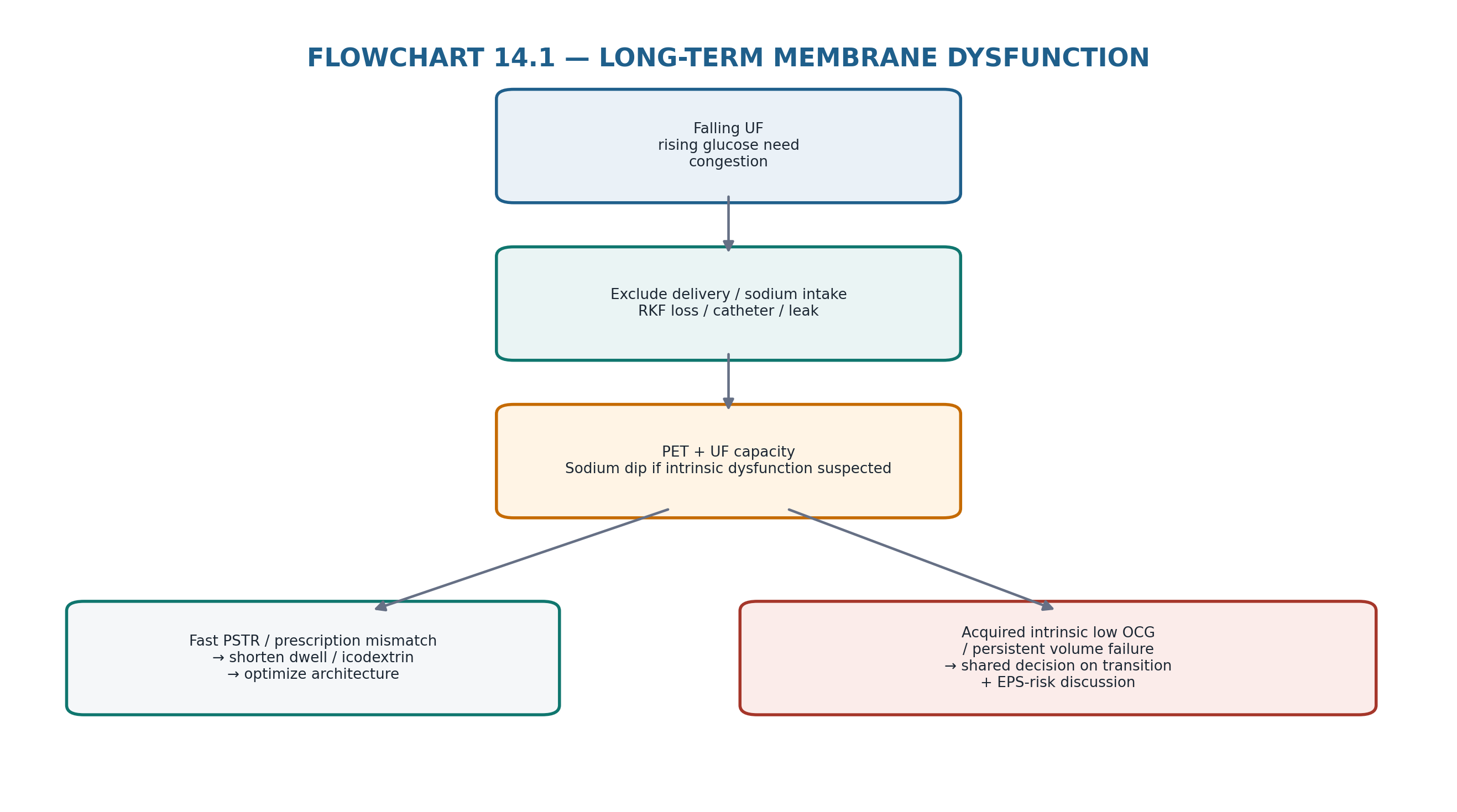
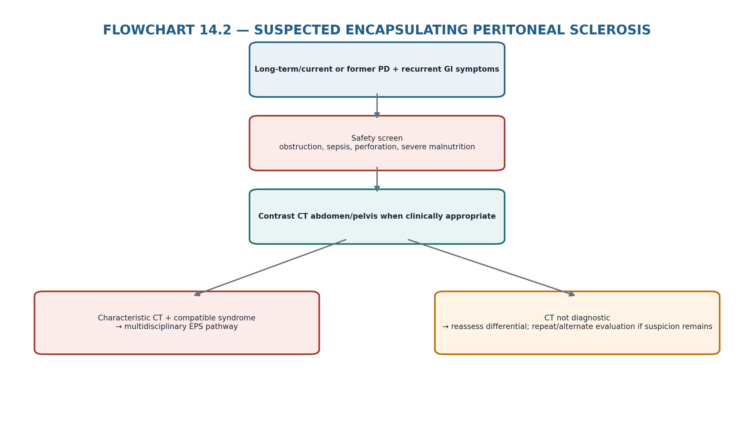
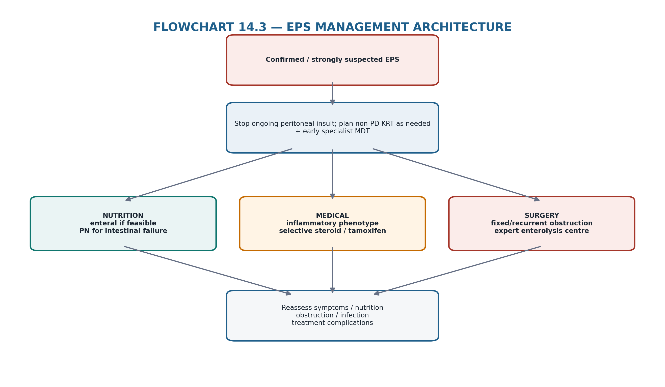
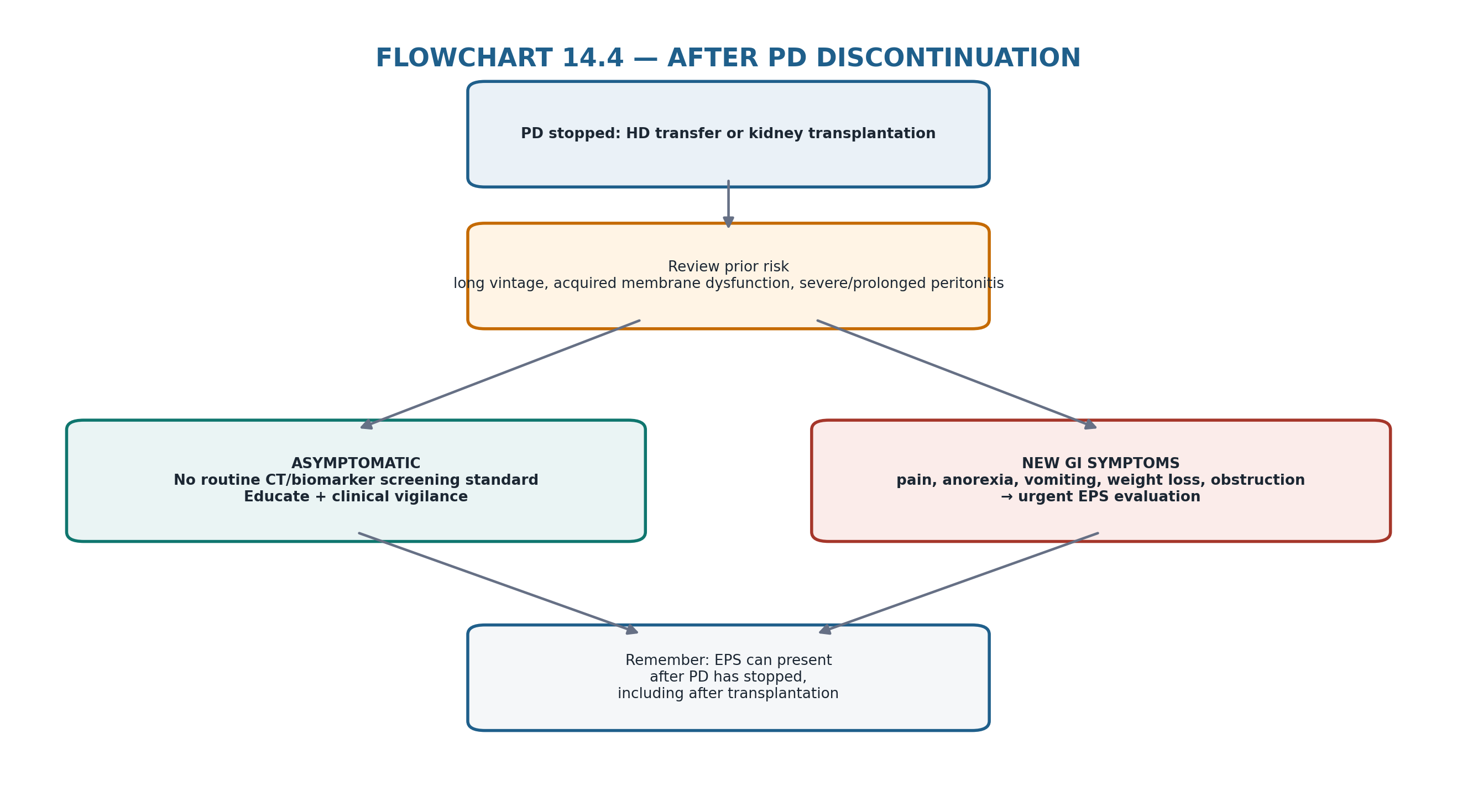
19. Retention tables: pattern recognition
Table 14.19 — If you see this, think this first.
| Finding | First hypothesis | Immediate action |
|---|---|---|
| Rising glucose requirement + falling UF | Acquired membrane dysfunction or non-membrane fluid problem | Chapter 8 differential + PET when indicated |
| 4-h high-glucose PET UF <400 mL | Low UF capacity | Identify fast PSTR vs intrinsic dysfunction vs non-membrane cause |
| 1-h sodium dip ≤5 mmol/L | Impaired free-water/osmotic conductance physiology | Discuss acquired intrinsic dysfunction if new/progressive |
| Long PD vintage but clinically well | Higher EPS risk, not EPS | Shared discussion; no automatic stop date |
| Recurrent vomiting + weight loss after PD stopped | Possible EPS | CT + urgent multidisciplinary assessment |
| Calcification on CT without symptoms | Chronic peritoneal injury; not diagnostic alone | Interpret in clinical context |
| Fixed recurrent bowel obstruction + cocooning CT | Advanced EPS phenotype | Nutrition + expert surgical referral |
| Asymptomatic high-risk patient asks for “screening CT” | No validated routine strategy | Explain limits; monitor clinically/functionally |
Table 14.20 — What not to confuse.
| Do not confuse | With | Correction |
|---|---|---|
| Long PD duration | A diagnosis of EPS | Duration is risk context only |
| Fast PSTR | EPS | Fast transport has many causes |
| Low UF capacity | Intrinsic membrane failure | Exclude leak/catheter/prescription/fluid absorption |
| Low sodium dip | Bowel encapsulation | It measures FWT physiology, not EPS anatomy |
| Peritoneal calcification | EPS by itself | Diagnosis requires syndrome + imaging constellation |
| Stopping PD | Eliminating EPS risk | Post-PD EPS is well recognized |
| Tamoxifen/corticosteroid use | Evidence-based cure | Evidence is observational |
| Parenteral nutrition | Failure of treatment | Often essential intestinal-failure support |
20. Clinical pearls
Membrane dysfunction is a functional diagnosis; EPS is a bowel-encapsulating clinical syndrome.
Follow the patient’s transport trajectory rather than a static “high/low transporter” label.
The 4-h high-glucose PET UF threshold and 1-h sodium dip are decision tools—not predictions of EPS.
Acquired intrinsic membrane dysfunction after years on PD should trigger a conversation about continued PD, alternatives and EPS risk.
There is no evidence-based universal maximum PD duration.
Discuss EPS before a crisis—ISPD considers the 3–4-year point reasonable for long-term risk discussion.
Stopping PD does not stop vigilance; EPS often presents after HD transfer or transplantation.
Recurrent subacute bowel obstruction plus weight loss in a former long-term PD patient is a high-value EPS clue.
CT diagnosis depends on a constellation: encapsulation/tethering, peritoneal change, loculation and obstruction—not calcification alone.
No blood or effluent biomarker is validated for routine EPS screening.
Nutrition is central treatment because intestinal failure and malnutrition drive outcomes.
Corticosteroids and tamoxifen are specialist, phenotype-guided options with low-certainty evidence.
Advanced fixed obstruction is an anatomical disease; expert enterolysis may be life-saving.
Early specialist referral is preferable to operating after profound malnutrition has developed.
21. Common pitfalls — and the correction
Table 14.21 — High-frequency errors in long-term membrane/EPS care.
| Pitfall | Why it fails | Correction |
|---|---|---|
| Stopping PD automatically at year 5 | No evidence supports a universal clock rule | Use individualized function, risk and alternatives |
| Calling fast transport “pre-EPS” | Poor specificity | Separate transport phenotype from EPS syndrome |
| Diagnosing EPS from calcification alone | Calcification is not specific | Require compatible symptoms + imaging pattern |
| Escalating dextrose indefinitely for falling UF | May worsen exposure while hiding membrane failure | Re-diagnose mechanism; discuss modality transition |
| Forgetting post-transplant EPS | Symptoms misattributed to drugs/infection | Keep EPS in differential after PD cessation |
| Routine biomarkers as screening tests | Not validated for clinical screening | Use research tests only in appropriate settings |
| Waiting for complete obstruction before referral | Allows malnutrition and frailty to progress | Refer early when recurrent obstruction develops |
| Treating with steroids before excluding infection | Can worsen occult infection | Exclude/treat infection first |
| Presenting tamoxifen as proven therapy | Observational evidence only | Shared specialist decision with adverse-effect review |
| Viewing parenteral nutrition as “support only” | Underestimates intestinal failure | Treat nutrition as core management |
22. Mini-cases: decisions, not trivia
Case 1 — The long-vintage patient who is still doing well
A 58-year-old has completed 6 years of PD. Volume control is good, PET pattern is stable, peritonitis burden is low and the patient strongly prefers PD.
| BEST NEXT STEP Do not transfer solely because of duration. Discuss EPS as a long-term risk, document shared decision-making, continue clinically indicated membrane surveillance and preserve alternative modality planning. |
|---|
Case 2 — Rising glucose requirement
A patient on PD for 7 years needs progressively stronger glucose to maintain the same UF. Mechanical causes and sodium intake have been addressed.
| BEST NEXT STEP Perform structured membrane reassessment. If acquired intrinsic UF insufficiency is confirmed, discuss the risks/benefits of continued PD versus transition rather than simply escalating glucose indefinitely. |
|---|
Case 3 — Blunted sodium dip
A high-glucose PET shows UF <400 mL at 4 h and a 1-h sodium dip of 3 mmol/L.
| BEST NEXT STEP This supports low UF capacity with intrinsic membrane dysfunction physiology. It does not diagnose EPS. Integrate the longitudinal trajectory, volume control, RKF and modality options, and discuss EPS risk in context. |
|---|
Case 4 — Vomiting after transplantation
Six months after kidney transplantation, a former long-term PD patient develops recurrent vomiting, early satiety and 8-kg weight loss.
| BEST NEXT STEP Post-PD/post-transplant EPS must be considered. Obtain urgent clinical and CT assessment rather than attributing the syndrome solely to mycophenolate or infection. |
|---|
Case 5 — Calcification without symptoms
CT performed for another reason shows peritoneal calcification in an asymptomatic former PD patient.
| BEST NEXT STEP Calcification alone is not EPS. Review PD history and symptoms, but do not diagnose the syndrome without compatible clinical/radiologic features. |
|---|
Case 6 — Inflammatory EPS phenotype
A patient with characteristic CT findings has abdominal pain, inflammatory features and progressive anorexia but no fixed complete obstruction. Infection has been excluded.
| BEST NEXT STEP Activate the multidisciplinary EPS pathway: stop ongoing PD exposure if still present, optimize nutrition and consider specialist phenotype-guided medical therapy; evidence for corticosteroids/tamoxifen is observational. |
|---|
Case 7 — Recurrent fixed obstruction
Despite nutritional and medical management, the patient has repeated admissions with obstructive symptoms and cannot sustain enteral intake.
| BEST NEXT STEP Refer to an experienced EPS surgical centre for enterolysis/peritonectomy assessment while optimizing nutrition and correcting electrolyte deficits. |
|---|
Case 8 — “Can you screen me every year?”
An asymptomatic patient with 8 years of PD asks for annual CT and effluent biomarkers to make sure EPS is not developing.
| BEST NEXT STEP Explain that no routine CT/biomarker screening strategy is validated. Use clinical vigilance, membrane trajectory and shared decision-making; investigate promptly if symptoms or function change. |
|---|
23. Active recall
MUST MEMORIZE
Table 14.22 — Core facts.
| Prompt | Answer |
|---|---|
| Long-term membrane dysfunction = EPS? | No. They are related but distinct. |
| ISPD maximum safe PD duration? | None established. |
| When is EPS discussion reasonable? | Around years 3–4 of long-term PD, then individualized thereafter. |
| 4-h high-glucose PET low-UF threshold? | <400 mL using 3.86% glucose/4.25% dextrose. |
| 4-h 2.27% glucose/2.5% dextrose low-UF threshold? | <100 mL. |
| 1-h sodium dip indicating intrinsic UF insufficiency? | ≤5 mmol/L. |
| 1-h sodium sieving ratio anchor? | ≤0.03. |
| Does low sodium dip diagnose EPS? | No. |
| Core EPS clinical phenotype? | Recurrent/progressive bowel obstruction with pain, vomiting, anorexia/weight loss ± inflammation. |
| Principal imaging test? | CT abdomen/pelvis. |
| Classic CT constellation? | Peritoneal thickening/enhancement/calcification + bowel tethering/encapsulation + loculation/obstruction. |
| Routine asymptomatic CT/biomarker screening? | Not established. |
| Can EPS present after PD stops? | Yes—after HD transfer or transplantation. |
| Most important supportive treatment? | Early nutrition/intestinal-failure support. |
| Corticosteroids/tamoxifen evidence level? | Observational/low certainty. |
| When is surgery central? | Persistent/recurrent fixed obstruction or advanced fibrotic disease in an expert centre. |
USE AS REFERENCE
Exact corticosteroid or tamoxifen drug choice, dose and duration.
Tamoxifen contraindications and thrombosis/endometrial-risk assessment.
Parenteral-nutrition formulation and refeeding protocol.
Timing of PD-catheter removal when infection, transplantation or unusual lavage protocols coexist.
Choice/timing of surgery and operative technique.
CT contrast protocol when kidney function, allergy or bowel ischemia concern modifies imaging.
Local pathways for intestinal failure, home parenteral nutrition and specialist EPS referral.
24. Flashcards: spaced repetition
1. Q: What is the key difference between membrane dysfunction and EPS? A: Membrane dysfunction is a transport/UF phenotype; EPS is a bowel-encapsulating obstructive clinical syndrome.
2. Q: Does ISPD recommend stopping PD after a fixed number of years? A: No.
3. Q: When should long-term EPS risk be discussed? A: Reasonably around 3–4 years of PD and revisited as circumstances change.
4. Q: Low UF on 4-h 3.86%/4.25% PET? A: <400 mL.
5. Q: Low UF on 4-h 2.27%/2.5% PET? A: <100 mL.
6. Q: Intrinsic UF dysfunction: sodium dip? A: ≤5 mmol/L at 1 h on high-glucose PET.
7. Q: Intrinsic UF dysfunction: sodium sieving ratio? A: ≤0.03 at 1 h.
8. Q: What does a blunted sodium dip measure? A: Reduced free-water/osmotic conductance physiology—not EPS anatomy.
9. Q: Core EPS symptom cluster? A: Pain, anorexia, nausea/vomiting, weight loss and recurrent/complete bowel obstruction.
10. Q: Best imaging test? A: CT abdomen/pelvis.
11. Q: Is peritoneal calcification diagnostic? A: No.
12. Q: Can EPS appear after kidney transplantation? A: Yes.
13. Q: Routine annual CT in asymptomatic long-term PD? A: Not established.
14. Q: Routine effluent biomarkers for screening? A: Not established.
15. Q: Why is nutrition central? A: Obstruction and inflammation cause intestinal failure and malnutrition, which drive morbidity/mortality.
16. Q: Corticosteroid evidence? A: Low-certainty observational; considered mainly for inflammatory phenotype after infection excluded.
17. Q: Tamoxifen evidence? A: Observational and conflicting; specialist selective use.
18. Q: When to refer for surgery? A: Persistent/recurrent obstruction, progressive nutritional failure or advanced fibrotic disease.
19. Q: Why expert surgery? A: Enterolysis/peritonectomy is complex with risks of enterotomy, fistula, sepsis and recurrence.
20. Q: Final mental model? A: Trajectory → membrane phenotype → individualized risk → GI symptoms → CT → nutrition/medical/surgery → reassess.
25. Rapid differential / troubleshooting
Table 14.23 — Long-term membrane/EPS troubleshooting from problem to action.
| Problem | Differential | First actions |
|---|---|---|
| Falling UF | Fast PSTR; low OCG; leak; catheter; increased absorption; sodium excess | Verify delivery/mechanics + PET/UF capacity when indicated |
| High glucose dependence | RKF loss; sodium excess; fast PSTR; intrinsic dysfunction | Do not just increase glucose; localize mechanism |
| Fast PSTR after peritonitis | Inflammatory change vs chronic remodeling | Repeat when clinically appropriate; optimize dwell architecture |
| Long PD duration, no symptoms | Risk context only | Shared discussion; no automatic modality transfer |
| Anorexia/weight loss after PD stopped | EPS; malignancy; drug toxicity; infection | CT + broad differential |
| Recurrent partial SBO | EPS; adhesions; malignancy | CT pattern + surgical review |
| Peritoneal calcification | Chronic injury vs EPS | Look for symptoms + encapsulation/tethering |
| Elevated inflammatory markers | EPS inflammation vs infection | Exclude infection and other inflammatory causes |
| Severe malnutrition | EPS intestinal failure; PEW; malignancy/infection | Dietitian + intestinal-failure team; consider parenteral support |
| No response to medical therapy | Fixed fibrosis/obstruction or wrong diagnosis | Reassess imaging/diagnosis; expert surgical referral |
26. Final revision sheet
| CORE CONCEPT Long-term PD should be managed as a changing membrane trajectory. Acquired intrinsic UF dysfunction raises concern and may justify modality transition, but EPS is a separate clinical–radiologic bowel-encapsulation syndrome. No fixed PD duration prevents EPS; diagnosis depends on symptoms + CT; treatment depends on nutrition, disease phenotype and expert surgery when obstruction is fixed. |
|---|
Table 14.24 — One-minute revision.
| Domain | Must remember |
|---|---|
| Long-term PD | Risk rises with time; no universal stop year |
| Low UF capacity | <400 mL on 4-h 3.86%/4.25% PET or <100 mL on 2.27%/2.5% PET |
| Intrinsic dysfunction | 1-h sodium dip ≤5 mmol/L and/or sodium sieving ratio ≤0.03 |
| EPS discussion | Reasonable around PD years 3–4; individualize thereafter |
| EPS syndrome | Bowel encapsulation/obstruction + pain/vomiting/anorexia/weight loss ± inflammation |
| Post-PD risk | EPS may present after HD transfer or kidney transplantation |
| Diagnosis | Clinical syndrome + characteristic CT; biomarkers not routine screening |
| CT pattern | Encapsulation/tethering + peritoneal change + loculation/obstruction |
| Nutrition | Core treatment; parenteral support when intestinal failure prevents adequate intake |
| Medical therapy | Corticosteroids/tamoxifen may be considered selectively; evidence observational |
| Surgery | Expert enterolysis/peritonectomy for persistent/recurrent obstruction |
| Prevention | Reduce avoidable membrane stress; prevent inflammation; monitor function; no guarantee of EPS prevention |
| TEN TAKE-HOME RULES 1) Membrane failure is not EPS. 2) Follow trajectory, not clock. 3) No fixed maximum PD duration is evidence based. 4) Acquired low OCG/falling FWT should trigger risk and modality discussion. 5) EPS can present after PD stops. 6) Recurrent bowel obstruction + weight loss is the key clinical signal. 7) CT diagnoses the pattern; calcification alone does not. 8) No validated routine CT/biomarker screening strategy exists. 9) Nutrition is central treatment. 10) Fixed obstruction belongs in an experienced EPS surgical pathway. |
|---|
Table 14.25 — One-minute bedside synthesis.
| If you see… | Think… | Do now… |
|---|---|---|
| Long PD + stable function | Higher risk but clinically viable PD | Discuss EPS risk; continue individualized PD |
| Falling UF + blunted sodium dip | Acquired intrinsic membrane dysfunction | Discuss continuation vs modality transition |
| Vomiting + weight loss after transplant | Post-PD EPS possible | CT + multidisciplinary assessment |
| Calcification only | Chronic injury, not necessarily EPS | Correlate clinically |
| Cocooning + obstruction | Established EPS phenotype | Nutrition + specialist medical/surgical plan |
| Severe malnutrition | Intestinal failure | Urgent nutrition support |
| Persistent obstruction despite conservative care | Fixed fibrotic disease | Expert enterolysis referral |
| FINAL MENTAL MODEL Long-term exposure → membrane trajectory → functional testing when indicated → acquired dysfunction? → individualized EPS-risk discussion → GI symptoms? → CT syndrome → nutrition + phenotype-guided medical therapy + expert surgery when needed → patient-important recovery. |
|---|
Rapid oral viva
Explain why long PD duration is a risk factor but not a reason for automatic transfer.
Give the ISPD low-UF and 1-hour sodium-dip anchors and explain what each does and does not diagnose.
Differentiate fast PSTR from acquired intrinsic low osmotic conductance.
Describe the clinical syndrome that should trigger urgent EPS evaluation.
List the characteristic CT findings and explain why calcification alone is insufficient.
Explain why EPS may first appear after kidney transplantation or HD transfer.
Build a management plan for inflammatory EPS without fixed obstruction.
Build a management plan for recurrent fixed obstruction with severe weight loss.
Explain why nutrition and surgical timing affect outcome.
Explain the evidence limitations for corticosteroids and tamoxifen.
| SAFETY BOUNDARY This chapter teaches longitudinal membrane reasoning and EPS diagnostic/management architecture. Exact drug doses, duration of corticosteroid/tamoxifen therapy, anticoagulation implications, parenteral-nutrition formulation, refeeding protocols, operative timing/technique and catheter-removal timing require specialist protocols and individualized multidisciplinary assessment. Acute obstruction, perforation, ischemia and sepsis override routine outpatient PD logic. |
|---|
27. Selected authoritative references
1. Brown EA, Bargman J, van Biesen W, et al. Length of Time on Peritoneal Dialysis and Encapsulating Peritoneal Sclerosis—Position Paper for ISPD: 2017 Update. Perit Dial Int. 2017;37(4):362–374. https://doi.org/10.3747/pdi.2017.00018. PMID: 28676507.
2. Morelle J, Stachowska-Pietka J, Öberg C, et al. ISPD recommendations for the evaluation of peritoneal membrane dysfunction in adults: classification, measurement, interpretation and rationale for intervention. Perit Dial Int. 2021;41(4):352–372. https://doi.org/10.1177/0896860820982218. PMID: 33563110.
3. Pepereke S, Shah AD, Brown EA. Encapsulating peritoneal sclerosis: Your questions answered. Perit Dial Int. 2023;43(2):119–127. https://doi.org/10.1177/08968608221125606. PMID: 36189954.
4. Nakayama M, Miyazaki M, Hamada C, Ito Y, Honda K; Peritoneal Biopsy Study Group of the Japanese Society for Peritoneal Dialysis. Pathophysiology of encapsulating peritoneal sclerosis: lessons from findings of the past three decades in Japan. Clin Exp Nephrol. 2023;27(9):717–727. https://doi.org/10.1007/s10157-023-02360-y. PMID: 37278945.
5. Li D, Li Y, Zeng H, Wu Y. Risk factors for Encapsulating Peritoneal Sclerosis in patients undergoing peritoneal dialysis: A meta-analysis. PLoS One. 2022;17(3):e0265584. https://doi.org/10.1371/journal.pone.0265584. PMID: 35312717.
6. Latus J, Habib SM, Kitterer D, et al. Histological and clinical findings in patients with post-transplantation and classical encapsulating peritoneal sclerosis: a European multicenter study. PLoS One. 2014;9(8):e106511. https://doi.org/10.1371/journal.pone.0106511. PMID: 25171219.
7. Jagirdar RM, Bozikas A, Zarogiannis SG, Bartosova M, Schmitt CP, Liakopoulos V. Encapsulating Peritoneal Sclerosis: Pathophysiology and Current Treatment Options. Int J Mol Sci. 2019;20(22):5765. https://doi.org/10.3390/ijms20225765. PMID: 31744097.
8. Srisajjakul S, Prapaisilp P, Bangchokdee S. Imaging pearls and differential diagnosis of encapsulating peritoneal sclerosis: Emphasis on computed tomography. Clin Imaging. 2023;94:116–124. https://doi.org/10.1016/j.clinimag.2022.12.001. PMID: 36527797.
9. Wang Y, Zhang J, Ma X, et al. Mechanisms underlying the involvement of peritoneal macrophages in the pathogenesis and novel therapeutic strategies for dialysis-induced peritoneal fibrosis. Front Immunol. 2024. PMID: 39749340.
10. Davenport A. Late presentation of encapsulating peritoneal sclerosis following renal transplantation and the potential under-reporting of the incidence and prevalence of encapsulating peritoneal sclerosis. Nephrology (Carlton). 2015;20(7):499–501. https://doi.org/10.1111/nep.12477. PMID: 26063486.
11. Lopes Barreto D, Sampimon DE, Struijk DG, Krediet RT. Early Detection of Imminent Encapsulating Peritoneal Sclerosis: Free Water Transport, Selected Effluent Proteins, or Both? Perit Dial Int. 2019;39(1):83–89. https://doi.org/10.3747/pdi.2017.00194. PMID: 30478138.
12. Vlijm A, Stoker J, Bipat S, et al. Computed tomographic findings characteristic for encapsulating peritoneal sclerosis: a case-control study. Perit Dial Int. 2009;29(5):517–522. https://doi.org/10.1177/089686080902900508. PMID: 19776044.
13. Danford CJ, Lin SC, Smith MP, Wolf JL. Encapsulating peritoneal sclerosis. World J Gastroenterol. 2018;24(28):3101–3111. https://doi.org/10.3748/wjg.v24.i28.3101. PMID: 30065556.
14. Korte MR, Fieren MWJA, Sampimon DE, Lingsma HF, Weimar W, Betjes MGH; Dutch Multicentre EPS Study. Tamoxifen is associated with lower mortality of encapsulating peritoneal sclerosis: results of the Dutch Multicentre EPS Study. Nephrol Dial Transplant. 2011;26(2):691–697. https://doi.org/10.1093/ndt/gfq362. PMID: 20584735.
15. Kawanishi H, Banshodani M, Yamashita M, Shintaku S, Dohi K. Surgical Treatment for Encapsulating Peritoneal Sclerosis: 24 Years’ Experience. Perit Dial Int. 2019;39(2):169–174. https://doi.org/10.3747/pdi.2018.00042. PMID: 29991561.
16. Brown EA, Van Biesen W, Finkelstein FO, et al. Length of time on peritoneal dialysis and encapsulating peritoneal sclerosis: position paper for ISPD. Perit Dial Int. 2009;29(6):595–600. PMID: 19910558.
17. Vlijm A, van Schuppen J, Lamers AB, Struijk DG, Krediet RT. Imaging in encapsulating peritoneal sclerosis. NDT Plus. 2011;4(4):281–284. https://doi.org/10.1093/ndtplus/sfr068.
18. Blake PG, Bargman JM. Peritoneal dialysis: long-term membrane complications and encapsulating peritoneal sclerosis. In: Brenner & Rector’s The Kidney. 12th ed. Elsevier; 2024.
| SOURCE NOTE ISPD guideline status and contemporary EPS/membrane evidence were checked 3 September 2026. The 2017 ISPD EPS position paper remains the dedicated EPS guidance listed by ISPD; the 2021 membrane-dysfunction recommendations provide the current functional testing framework. Because EPS treatment evidence remains largely observational, this chapter deliberately avoids presenting drug doses, treatment durations, CT-screening schedules or operative timing as universal standards. |
|---|