Applied Nephrology Master Series
Chapter 15
Nutrition, Protein-Energy Wasting and Metabolic Health in PD
Assessment | Protein | Energy | Dialysate Glucose | PEW | Sarcopenia | Supplements | Exercise | Metabolic Health
| CHAPTER MISSION Build a bedside nutrition system for peritoneal dialysis: detect nutritional decline before serum albumin becomes the only alarm; separate fluid, fat and muscle change; diagnose protein-energy wasting from a multidimensional assessment; account for peritoneal protein losses and dialysate-derived glucose calories; prescribe protein and energy using current KDOQI guidance; remove reversible barriers to eating; escalate from counselling to oral or enteral support when needed; and improve metabolic health without sacrificing lean mass, strength or life participation. |
|---|
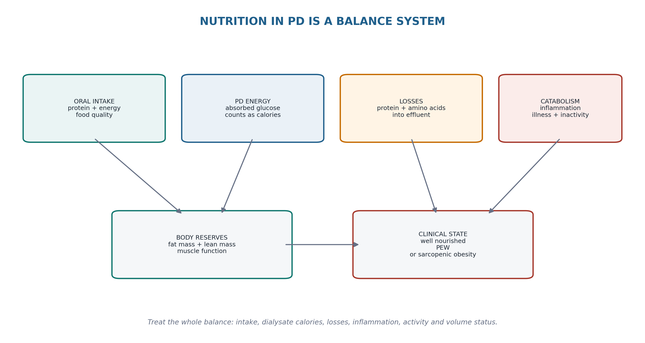
| MASTER PRINCIPLE Treat the TRAJECTORY, not the ALBUMIN. Nutrition in PD is not a single laboratory value and not a generic “renal diet.” The useful question is whether the person is maintaining adequate intake, lean tissue and function while avoiding excessive metabolic burden from dialysate glucose and unnecessary dietary restriction. |
|---|
0. One-page chapter map
Table 15.1 — The eight decisions that govern nutrition care in PD.
| Decision | Core question | Bedside output |
|---|---|---|
| 1. Screen | Is there a new risk signal for undernutrition, PEW, sarcopenia or unhealthy weight gain? | Risk status + trigger for full assessment |
| 2. Separate | Is weight change fluid, fat, lean tissue—or mixed? | Named body-composition phenotype |
| 3. Diagnose | Does the patient have PEW, isolated low intake, inflammation-driven hypoalbuminemia or sarcopenia? | Nutritional phenotype |
| 4. Quantify | Are protein and energy needs being met, including calories absorbed from dialysate? | Individual intake prescription |
| 5. Find cause | What reversible factor is suppressing intake or increasing catabolism/losses? | Cause-directed plan |
| 6. Intervene | Can counselling correct the gap, or are oral/enteral/specialized supports needed? | Nutrition support ladder |
| 7. Build muscle | Is resistance/aerobic activity feasible and safe? | Exercise-nutrition plan |
| 8. Verify | Did intake, SGA, muscle/function and patient goals improve without excess glucose burden? | Longitudinal reassessment |
Learning outcomes
Define protein-energy wasting (PEW) as a loss of body protein and energy stores rather than a synonym for low serum albumin.
Use the 7-point Subjective Global Assessment (SGA), appetite/intake history, weight trajectory, examination of fat and muscle, functional measures and biochemical context as a multidimensional nutritional assessment.
Apply current KDOQI 2020 recommendations for routine nutrition screening, comprehensive dietitian assessment, protein intake, energy intake and oral nutritional supplementation in adults on PD.
Explain why calories absorbed from glucose-containing dialysate must be included in total energy assessment and why increasing glucose exposure can coexist with loss of lean tissue.
Recognize that PD causes daily protein losses into effluent and that peritonitis/inflammation can amplify losses and catabolism.
Distinguish hypoalbuminemia caused by inflammation, overhydration or protein losses from true inadequate intake/PEW.
Recognize sarcopenia and sarcopenic obesity, where BMI can look reassuring despite low muscle mass or strength.
Use oral nutritional supplements when counselling alone cannot meet requirements and understand when enteral or specialized support should be considered.
Use amino-acid dialysate selectively, recognizing that evidence for patient-important outcomes is limited and product-specific safety rules apply.
Integrate exercise and physical activity with nutrition to preserve strength, function and life participation.
Avoid indiscriminate potassium, phosphorus or calorie restriction that worsens protein-energy intake; individualize according to serum values and food-source bioavailability.
Cross-reference Chapter 8 for sodium/volume balance, Chapter 13 for dysglycemia and hypokalemia, Chapter 14 for EPS-related intestinal failure, and Chapter 16 for frailty/obesity/special-population adaptations.
| EVIDENCE POSTURE KDOQI 2020 is the principal current guideline for adult CKD/dialysis nutrition. For metabolically stable adults on PD it recommends 1.0–1.2 g protein/kg ideal body weight/day (opinion) and 25–35 kcal/kg ideal body weight/day, individualized for age, activity, body composition, weight goals, illness and inflammation; PD glucose calories are included in total energy intake. It recommends routine nutrition screening at least biannually and comprehensive dietitian assessment within the first 90 days of dialysis, annually, or when screening/provider referral indicates. A minimum 3-month trial of oral nutritional supplements is suggested when counselling alone cannot meet requirements. ISPD goal-directed PD and exercise recommendations add the patient-centred, life-participation and physical-function framework. Evidence for amino-acid dialysate, specific anabolic therapies and hard-outcome benefits of nutritional supplements remains limited. [1–8] |
|---|
1. Core concept: nutrition is an outcome of the whole PD system
Nutritional status in PD emerges from the interaction of food intake, calories absorbed from dialysate, daily nutrient losses, inflammation, residual kidney function, acid-base status, comorbidity, gastrointestinal symptoms, physical activity and treatment burden. A patient may therefore gain fat while losing muscle, have a low albumin despite adequate intake, or have a “normal” BMI while becoming functionally sarcopenic. The chapter begins by refusing these false shortcuts. [1,3,9]
Table 15.2 — Four common nutrition phenotypes that look deceptively similar.
| Phenotype | Typical pattern | Main danger |
|---|---|---|
| True PEW | Low intake + weight/muscle loss ± inflammation | Loss of protein/energy stores and function |
| Inflammatory hypoalbuminemia | Low albumin with CRP/infection/illness; intake may be adequate | Treating a biomarker instead of the disease |
| Sarcopenic obesity | High/normal BMI with low muscle strength or lean tissue | Muscle loss hidden by adiposity |
| Fluid-associated weight change | Rapid “gain” or “loss” with congestion/decongestion | Misclassifying water as tissue |
| BEDSIDE TRANSLATION Ask “what changed in tissue and function?” before asking “what is the albumin?” |
|---|
2. Protein-energy wasting: define the syndrome precisely
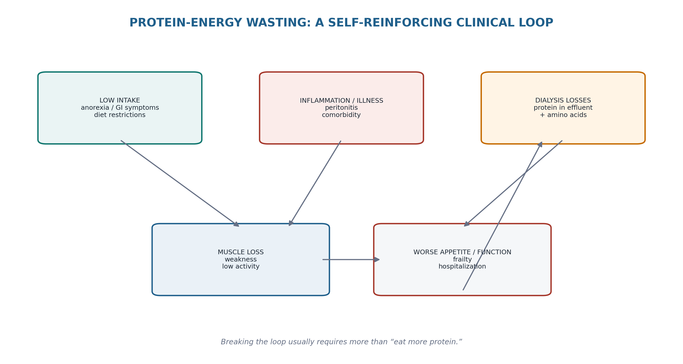
The International Society of Renal Nutrition and Metabolism defines PEW as a state of decreased body stores of protein and energy fuels. The proposed diagnostic framework uses four domains—serum chemistry, body mass, muscle mass and dietary intake—with evidence from multiple domains rather than one isolated measurement. In contemporary practice, SGA/MIS and longitudinal assessment often operationalize the syndrome more usefully than applying rigid research criteria at a single visit. [2,10]
Table 15.3 — PEW domains: what each contributes.
| Domain | Examples | Limitation |
|---|---|---|
| Intake | Reduced protein/energy intake, anorexia, meal skipping | Self-report error; short-term intake may not reflect chronic state |
| Body mass | Unintentional weight/fat loss | Fluid shifts can mimic change |
| Muscle | Visible wasting, reduced strength/function, low lean mass | Method dependent; obesity can hide loss |
| Biochemistry | Albumin/prealbumin/cholesterol in context | Inflammation and volume strongly confound |
| Inflammation/function | CRP, illness burden, frailty, reduced activity | Important mechanism/risk signal but not a stand-alone PEW definition |
| DEFINITION DISCIPLINE Low albumin is not PEW. Low BMI is not PEW. Low intake alone is not PEW. Diagnose a syndrome of declining protein/energy stores using several converging signals. |
|---|
3. Why PD creates a distinctive nutrition physiology
PD differs from non-dialysis CKD because dietary protein restriction is no longer the central strategy. Patients lose protein continuously into dialysate, absorb variable amounts of glucose from dextrose solutions, may experience early satiety from intraperitoneal fill, and can develop peritonitis-related catabolism. Contemporary literature reviews report daily PD protein losses commonly in the range of roughly 4–10 g/day, with marked interpatient variation; classic studies and clinical experience show greater loss during peritonitis. These losses are one reason PD protein requirements are higher than in non-dialysis CKD. [9,11,12]
Table 15.4 — PD-specific nutritional forces.
| PD feature | Nutrition consequence | Clinical implication |
|---|---|---|
| Daily protein loss into effluent | Raises protein replacement requirement | Do not import low-protein CKD diets into PD |
| Glucose absorption | Adds non-oral calories; may increase fat mass/glycemic burden | Count dialysate energy in total assessment |
| Intraperitoneal volume | Early satiety/reflux/abdominal fullness in some patients | Adjust meal timing/prescription when relevant |
| Peritonitis/inflammation | Higher protein losses + catabolic drive + anorexia | Nutrition reassessment after episodes |
| Continuous home therapy | Meal timing can be flexible but burden can impair eating | Design food plan around real treatment schedule |
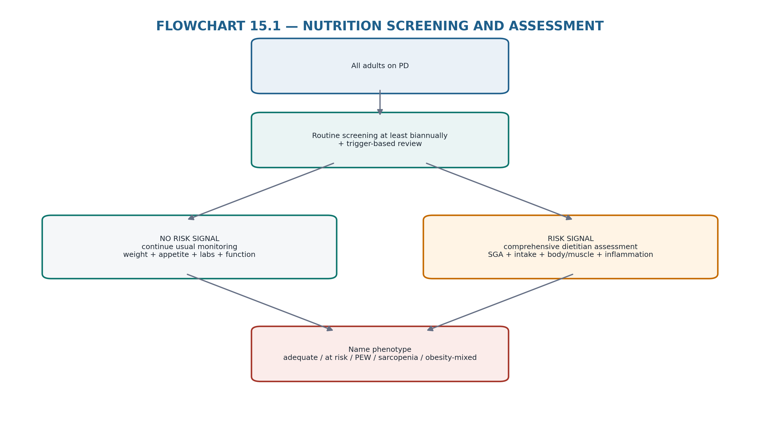
4. Screening and comprehensive assessment: build a repeatable system
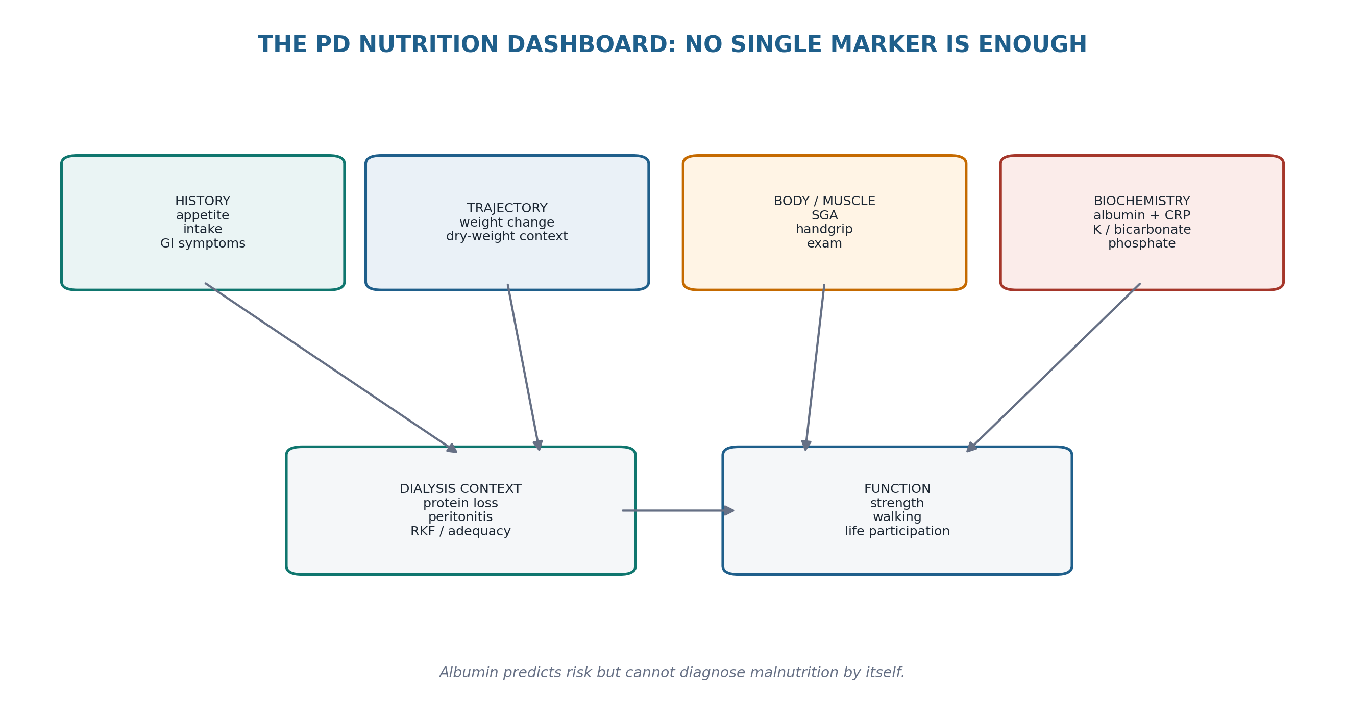
KDOQI 2020 states that adults with CKD 3–5D should undergo routine nutrition screening at least biannually. A registered dietitian nutritionist or equivalent should perform comprehensive assessment within the first 90 days of dialysis, annually, or sooner when screening or clinician concern indicates. In CKD 5D, the 7-point SGA is recommended as a valid and reliable nutritional assessment tool. [1]
Table 15.5 — Comprehensive PD nutrition assessment.
| Domain | What to document | What changes management |
|---|---|---|
| Appetite/intake | Meals, protein sources, dietary recall, ONS use, food insecurity | Identifies actual deficit and barriers |
| Weight trajectory | Usual weight, target/dry-weight context, unintentional change | Separates tissue loss from fluid |
| SGA / physical examination | Fat stores, muscle wasting, GI symptoms, intake change | Global PEW phenotype |
| Function | Handgrip, sit-to-stand/walking capacity, ADL/life participation | Detects clinically meaningful muscle decline |
| Biochemistry | Albumin, CRP/inflammation, bicarbonate, K, phosphate, glucose/HbA1c | Finds confounders and treatment targets |
| Dialysis context | Peritonitis, effluent protein loss when relevant, RKF, UF/glucose exposure | Explains losses and metabolic burden |
| Psychosocial | Depression, cognition, dentition, finances, caregiver burden | Often determines whether a plan can work |
| SCREENING RULE The scheduled screen is the minimum. New weight loss, poor appetite, hospitalization, peritonitis, functional decline or repeated low intake should trigger assessment immediately rather than waiting for the annual review. |
|---|
5. Weight and body composition: the scale does not tell you what changed
Body weight in PD is simultaneously affected by extracellular water, dialysate in the abdomen, fat mass and lean tissue. Serial trends are therefore interpreted against a consistent volume state and with attention to whether the abdomen is full or drained. KDOQI recommends considering body composition together with weight/BMI; DEXA is the reference method when feasible, while evidence is insufficient to recommend bioimpedance as a specific body-composition standard in PD. [1]
Table 15.6 — Weight change: four different diagnoses.
| Observed change | Possible explanation | How to separate it |
|---|---|---|
| Rapid gain + edema/BP rise | Fluid overload | Chapter 8 volume assessment |
| Slow gain + stable volume + rising waist/fat | Adipose gain | Diet + dialysate glucose + body composition |
| Weight stable + weaker / visible muscle loss | Sarcopenia masked by fat or fluid | Strength + SGA + lean-mass assessment |
| Weight loss after decongestion | Water loss, not necessarily malnutrition | Re-establish tissue baseline after euvolemia |
| NUMERICAL HUMILITY A BMI of 30 kg/m² does not prove good nutrition. A 2-kg fall after correcting edema does not prove PEW. Always identify the compartment that changed. |
|---|
6. Protein prescription: current KDOQI target for stable PD
For metabolically stable adults with CKD on maintenance PD, KDOQI 2020 recommends prescribing dietary protein intake of 1.0–1.2 g/kg ideal body weight/day to maintain stable nutritional status. The strength of evidence for PD is opinion-level, reflecting the limited direct trial base. The same range is considered reasonable for adults on dialysis who have diabetes, with individualized adjustment when glycemic instability or other clinical circumstances alter requirements. [1]
Table 15.7 — Protein prescription reasoning.
| Situation | Direction | Do not do |
|---|---|---|
| Stable PD, adequate nutrition | Aim for 1.0–1.2 g/kg IBW/day | Carry forward a pre-dialysis low-protein diet |
| Poor intake / PEW risk | Identify deficit and increase usable protein-energy intake | Focus on phosphate restriction at the expense of protein |
| Peritonitis / acute catabolic illness | Reassess intake and losses; individualize with dietitian/clinical team | Assume the stable outpatient target is automatically sufficient |
| Obesity | Use ideal/appropriate reference weight and body-composition goals | Prescribe from actual weight without considering adiposity |
| Diabetes | Maintain protein adequacy while coordinating carbohydrate/glucose exposure | Lower protein merely to manage glucose |
| PROTEIN RULE In PD, “renal diet” must not mean chronic protein restriction. The prescription has changed because the patient is now dialyzed and loses protein through the peritoneum. |
|---|
7. Energy prescription: include dialysate glucose calories
KDOQI 2020 recommends 25–35 kcal/kg ideal body weight/day for metabolically stable adults with CKD 1–5D, individualized according to age, sex, physical activity, body composition, weight goals, CKD stage, concurrent illness and inflammation. For PD, calories absorbed from glucose-containing dialysate are part of total energy intake; they should not be added on top of the desired energy budget as if metabolically invisible. [1]
Measured glucose absorption varies with membrane transport, dwell duration, glucose concentration and prescription. Observational PD data show that greater glucose absorption is associated with fat-mass gain and may coexist with declining fat-free mass. This is why “enough calories” and “healthy body composition” are different goals. [13]
Table 15.8 — Energy balance in PD.
| Problem | Mechanism | Correction direction |
|---|---|---|
| Low oral calories + low weight/muscle | Insufficient usable energy | Increase nutrient-dense oral intake/ONS |
| High dialysate glucose + fat gain | Non-oral caloric load | Reduce unnecessary hypertonic glucose; optimize sodium/volume prescription |
| High glucose calories + poor oral protein | Calories displace appetite without replacing protein | Protect protein intake; review meal timing and solution strategy |
| Obesity + low muscle | Energy excess with anabolic deficit/inactivity | Modest energy adjustment + protein adequacy + resistance activity |
8. Food quality: stop creating malnutrition with indiscriminate restriction
A PD diet is individualized, not a list of universal prohibitions. KDOQI recommends adjusting dietary phosphorus to maintain serum phosphate in the normal range and considering the bioavailability of phosphorus sources; phosphate additives are more readily absorbed than many plant sources. Potassium intake should be adjusted to maintain serum potassium within the normal range rather than restricted automatically. [1]
Table 15.9 — Restriction discipline.
| Issue | Better strategy | Common harmful shortcut |
|---|---|---|
| Hyperphosphatemia | Target additives, bioavailable sources, binder timing and total intake | Cut all high-protein foods |
| Hypokalemia | Increase dietary/supplemental potassium according to need; search causes | Continue a generic low-potassium diet |
| Sodium/volume | Reduce sodium to support thirst/volume control | Compensate high salt intake with more glucose dialysate |
| Diabetes/obesity | Reduce refined energy excess and unnecessary dialysate glucose exposure | Protein restriction to reduce calories |
| Poor appetite | Prioritize nutrient density and achievable meals | Add multiple simultaneous restrictions |
| DIET QUALITY RULE When nutrition is fragile, remove the least useful restriction first. A technically “renal” diet that the patient cannot eat is not therapeutic. |
|---|
9. Peritoneal protein losses: important, variable and amplified by inflammation
Peritoneal protein loss is a real component of PD physiology but is highly variable between patients. Recent literature review data report daily protein losses in PD broadly around 4–10 g/day; classic CAPD studies found similar order-of-magnitude losses and demonstrated substantially greater losses during peritonitis. Peritoneal protein clearance also reflects membrane biology and inflammation, so a high loss should not be interpreted only as dietary failure. [9,11,12]
Table 15.10 — When protein loss matters clinically.
| Context | Interpretation | Action |
|---|---|---|
| Stable patient, good intake/function | Expected PD loss may be compensated | Maintain target protein intake |
| Recurrent peritonitis | Loss + catabolism + anorexia | Early post-infection nutrition reassessment |
| Persistent hypoalbuminemia | Loss may contribute but is rarely the only cause | Assess inflammation, volume, liver/urine/GI losses and intake |
| High transport/inflammatory phenotype | Greater protein permeability may coexist | Treat underlying inflammation/volume and monitor tissue/function |
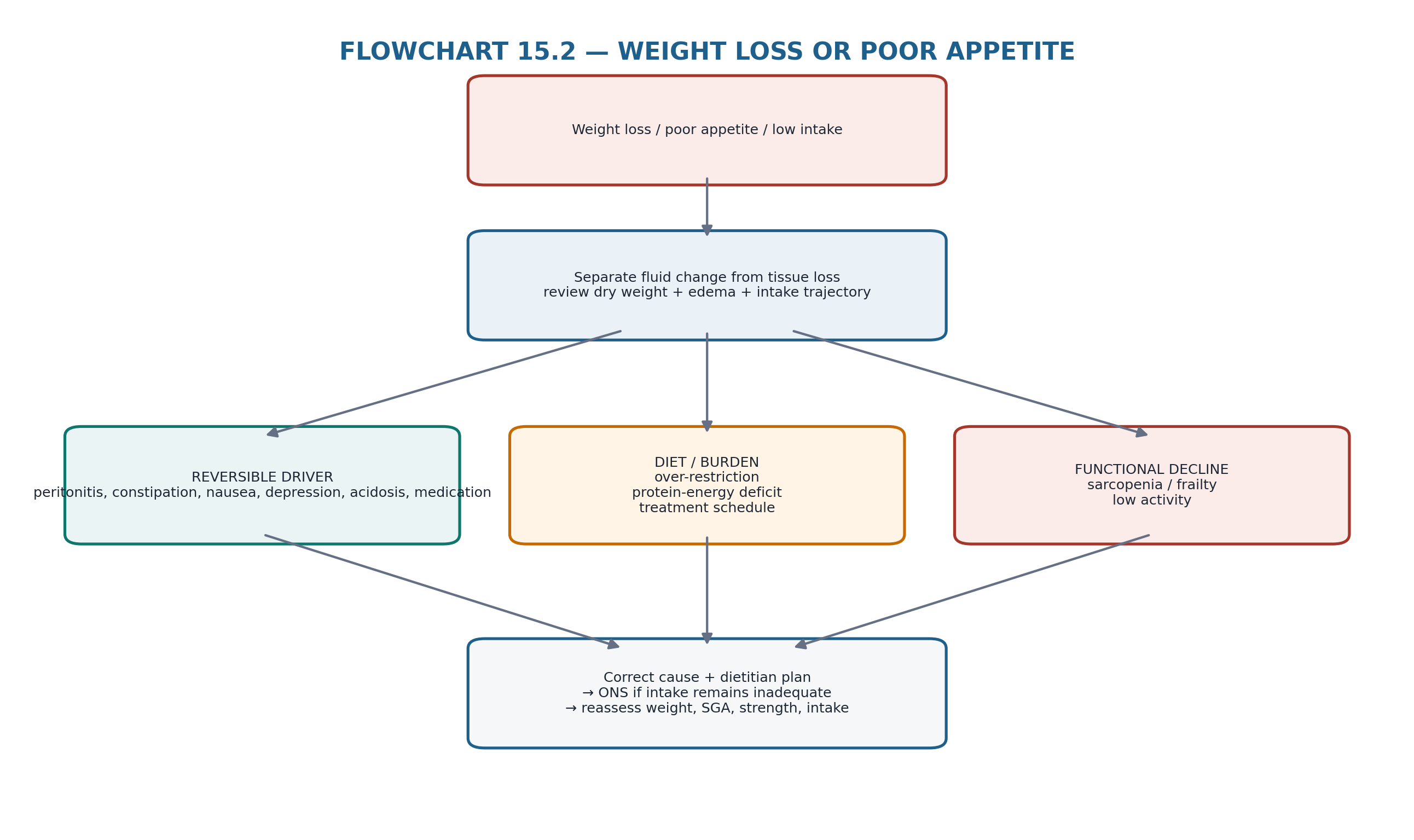
10. Appetite and gastrointestinal symptoms: treat the barrier before prescribing supplements
Poor appetite is often the proximal event that drives PEW. Causes include uremia or underdialysis, constipation, reflux/early satiety from intraperitoneal volume, nausea, gastroparesis, medications, depression, infection, oral/dental disease, food insecurity and overly restrictive diets. EPS-related obstructive symptoms belong to Chapter 14 and should not be managed as routine anorexia. [3,7]
Table 15.11 — Poor appetite: mechanism-based review.
| Clue | Likely contributor | First response |
|---|---|---|
| Fullness worse with daytime fill | Intraperitoneal volume / meal timing | Review fill/dwell timing and smaller frequent meals |
| Constipation | GI burden + catheter effects | Treat bowel dysfunction |
| After peritonitis | Inflammation/catabolism + aversion | Reassess intake, symptoms and losses |
| Depression/social isolation | Psychosocial anorexia | Screen/treat; engage caregiver/team |
| Multiple dietary bans | Iatrogenic restriction | Dietitian simplification |
| Persistent vomiting/obstruction | Not routine nutrition problem | Chapter 14 / urgent GI-surgical pathway |
11. Hypoalbuminemia: risk marker, not stand-alone nutrition diagnosis
Serum albumin is strongly associated with outcomes in dialysis populations, but its concentration is influenced by inflammation, hydration/dilution, peritoneal and urinary losses, liver synthesis and acute illness. Studies including PD patients show that inflammation explains a substantial part of the albumin–mortality association. Nutritional assessment should therefore use albumin as a prognostic/context marker rather than as proof that a patient simply needs more protein. [14,15]
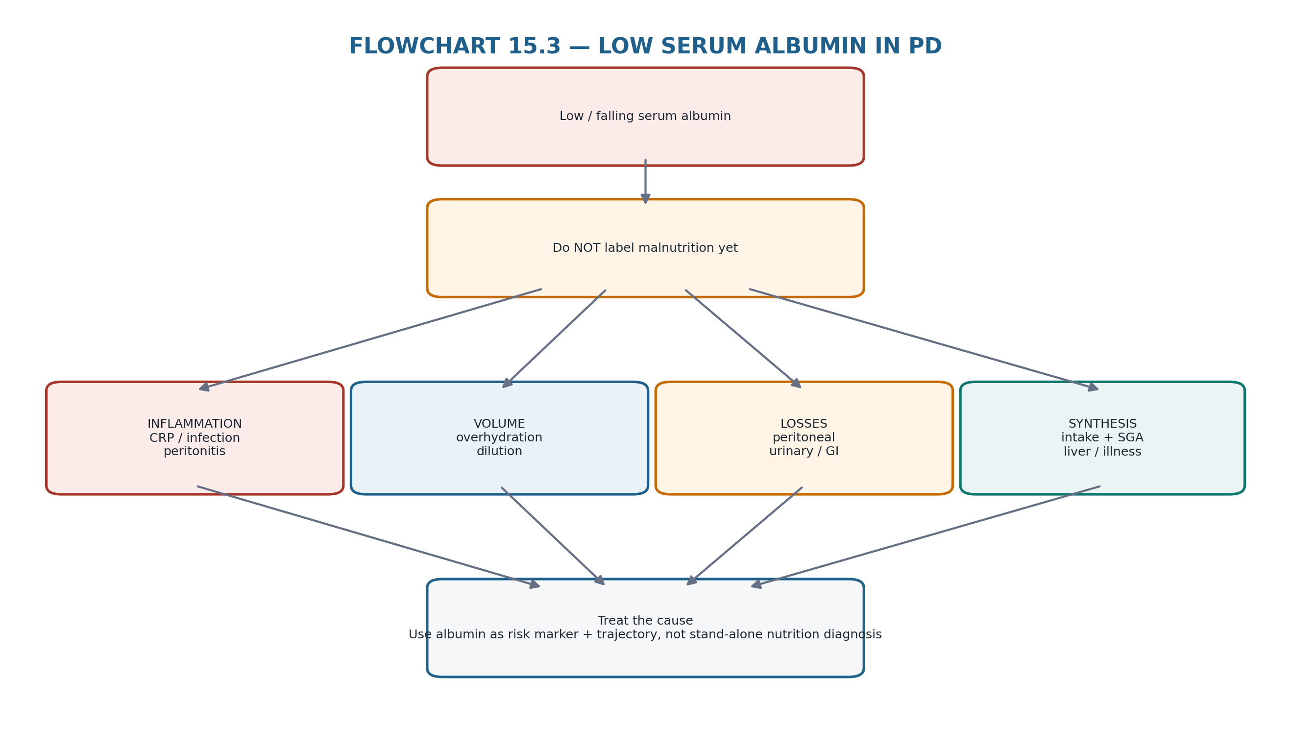
| ALBUMIN RULE Do not “treat the albumin.” Treat infection/inflammation, volume overload, inadequate intake, protein losses or liver disease—and then follow the albumin trajectory as one part of recovery. |
|---|
12. Muscle and sarcopenia: function is a nutritional outcome
Muscle loss can occur despite stable weight and can be accelerated by inflammation, inactivity, hospitalization, inadequate protein-energy intake and comorbidity. Sarcopenia and low strength are associated with adverse outcomes across dialysis cohorts. In PD, observational data show low physical activity and declining fitness/strength over time, making muscle function an essential longitudinal outcome rather than an optional research measure. [16,17]
Table 15.12 — Practical muscle surveillance.
| Measure | What it adds | Limitation |
|---|---|---|
| SGA muscle examination | Low-cost bedside estimate of wasting | Examiner dependent |
| Handgrip strength | Functional muscle signal; easy serial trend | Affected by arthritis/neurologic disease |
| Sit-to-stand / gait tests | Life-relevant lower-limb function | Need safe testing environment |
| DEXA / body composition | Quantifies lean/fat compartments | Volume and access/cost limitations |
| Activity history | Detects deconditioning and participation loss | Self-report bias |
13. Obesity and sarcopenic obesity: metabolic improvement must preserve lean tissue
PD can produce a mixed phenotype in which dialysate glucose and dietary energy promote adipose gain while inactivity and catabolic illness reduce lean tissue. Observational PD data demonstrate that greater glucose absorption can be associated with increasing fat mass and decreasing fat-free mass. Therefore weight-loss strategies that ignore muscle preservation can worsen the clinically important compartment even while BMI improves. [13]
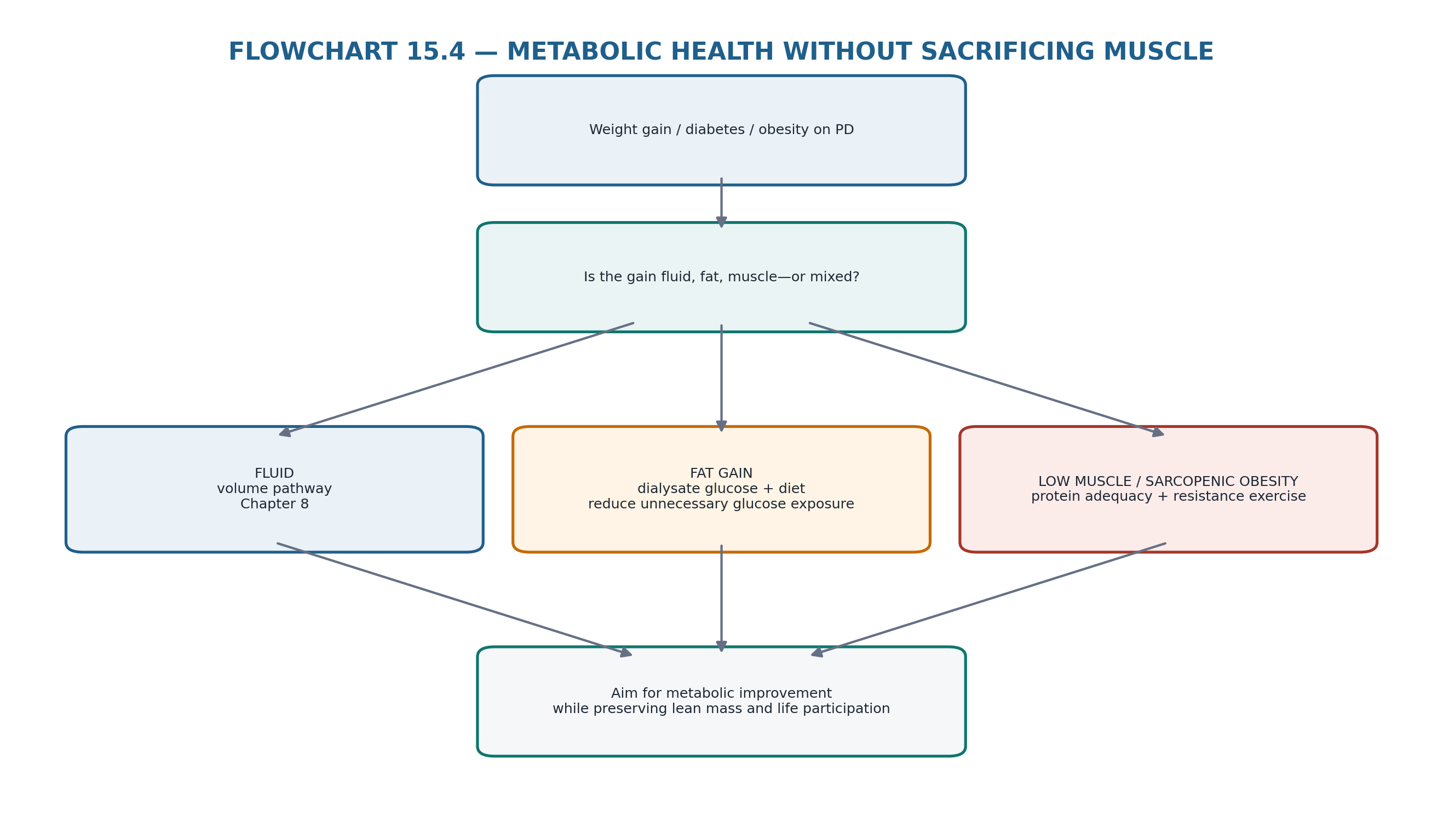
Table 15.13 — Obesity phenotype → management focus.
| Phenotype | Priority | Avoid |
|---|---|---|
| Obesity with preserved strength | Metabolic risk reduction + activity | Crash diets / protein inadequacy |
| Sarcopenic obesity | Resistance exercise + adequate protein + careful energy deficit | Judging success only by scale weight |
| Fluid + obesity | Treat congestion and metabolic risk separately | Calling all gain adipose |
| Diabetes + high glucose exposure | Optimize PD osmotic strategy + diabetes treatment + diet quality | Solving volume solely with stronger glucose bags |
14. Intervention ladder: fix causes first, then escalate support
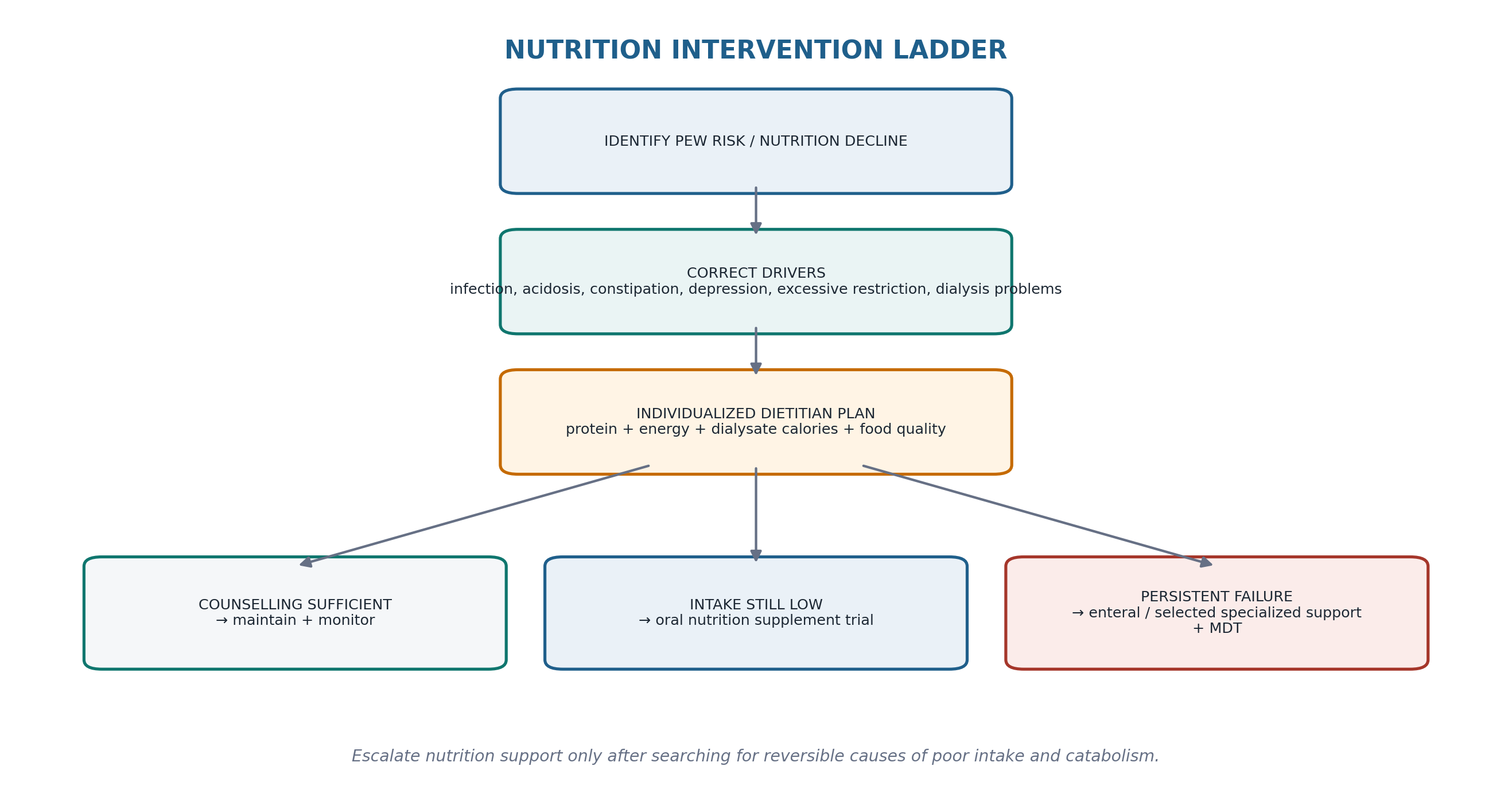
KDOQI places medical nutrition therapy and dietary counselling before nutritional supplementation. For adults with CKD 3–5D at risk of or with PEW, a minimum 3-month trial of oral nutritional supplements is suggested when counselling alone does not achieve sufficient protein and energy intake. If chronic intake remains inadequate despite counselling and ONS, enteral tube feeding is a reasonable next consideration when medically appropriate. [1]
Table 15.14 — Nutrition support ladder.
| Level | When | What to reassess |
|---|---|---|
| 1. Cause correction | Any decline | Infection, acidosis, constipation, depression, medications, dialysis delivery, restrictions |
| 2. Dietitian-led food plan | Intake gap or risk | Protein/energy intake, meal pattern, glucose calories, acceptability |
| 3. Oral nutrition supplements | Counselling insufficient; PEW risk/presence | Adherence, GI tolerance, intake, SGA, weight/muscle |
| 4. Enteral feeding | Chronically inadequate intake despite oral strategy and appropriate GI tract | Goals, tolerance, aspiration/volume/electrolytes |
| 5. Specialized support | Selected severe/refractory cases | MDT risk–benefit; underlying disease and route-specific complications |
| ONS EVIDENCE Meta-analyses of dialysis trials show modest improvements in albumin, BMI, protein intake indices and sometimes handgrip/MIS, but hard-outcome evidence remains limited and most trials are hemodialysis-dominant. PD-specific pilot and small randomized studies support feasibility and nutritional improvement, not a guaranteed survival benefit. [18–20] |
|---|
15. Amino-acid dialysate and specialized nutrition support: selective tools, not routine escalation
Amino-acid-containing PD solutions can replace part of the dialysate nutrient loss and improve short-term protein kinetics when adequate non-protein calories are available. Small randomized and crossover studies suggest biochemical or nitrogen-balance benefit, but long-term patient-important outcome evidence is weak. Amino-acid dialysate should therefore be viewed as a selective specialist option for patients who cannot meet protein needs orally/enterally, not as a routine substitute for food or treatment of unexplained hypoalbuminemia. [21–23]
Table 15.15 — Amino-acid dialysate: evidence-calibrated use.
| Potential role | Important condition | Caution |
|---|---|---|
| Supplement protein delivery | Needs adequate concurrent energy intake for anabolism | Does not correct inflammation or obstruction |
| Glucose-sparing exchange | May reduce one glucose exposure | Not equivalent to eliminating metabolic burden |
| PEW with poor oral tolerance | Consider only in specialist individualized plan | Product-specific acid-base/urea and formulation issues |
| Long-term outcome improvement | Not established | Do not promise mortality benefit |
16. Exercise–nutrition synergy: feeding muscle without using it is incomplete therapy
The 2021 ISPD/Global Renal Exercise Network practice recommendations support physical activity and individualized aerobic/resistance exercise in people receiving PD, while acknowledging a sparse trial base. A home-based randomized trial showed improvement in exercise capacity and several quality-of-life domains, and observational data show declining fitness and activity during PD. Nutrition and resistance exercise are therefore complementary strategies for preserving muscle, function and life participation. [7,17,24]
Table 15.16 — Exercise–nutrition pairing.
| Problem | Nutrition arm | Activity arm |
|---|---|---|
| Low muscle strength | Adequate protein/energy intake | Progressive resistance activity when safe |
| Frailty/deconditioning | Prevent energy/protein deficit | Graded aerobic + strength + balance plan |
| Obesity with low muscle | Moderate energy strategy preserving protein | Resistance training to protect lean mass |
| Post-hospitalization decline | Early nutrition reassessment | Rehabilitation/physio-led reconditioning |
| FUNCTION RULE If the patient is eating more but getting weaker, the nutrition plan is not yet successful. Strength, mobility and life participation belong on the follow-up sheet. |
|---|
17. Longitudinal monitoring: success is a stable or improving phenotype
The aim is not to normalize every biomarker. A useful nutrition follow-up asks whether appetite and intake are adequate, tissue weight and muscle function are stable or improving, fluid status is controlled, inflammatory drivers have resolved, metabolic burden is acceptable and the plan is sustainable for the person. Serial change in SGA is prognostically meaningful in dialysis cohorts. [15]
Table 15.17 — Nutrition follow-up dashboard.
| Domain | Success looks like | Escalation trigger |
|---|---|---|
| Intake/appetite | Requirements met with tolerable meal plan | Persistent deficit or anorexia |
| Weight/body composition | Stable desired tissue trajectory | Unintentional loss or rapid fat gain |
| Muscle/function | Stable/improving strength and activity | New weakness/falls/functional decline |
| SGA | Stable or improving score/phenotype | Deteriorating SGA |
| Inflammation/albumin | Cause addressed; trajectory consistent with recovery | Persistent CRP/infection or unexplained albumin decline |
| Metabolic health | Acceptable glycemia/lipids/weight goals without high glucose burden | Escalating hypertonic glucose + worsening metabolic phenotype |
| Patient burden | Plan affordable, culturally acceptable and feasible | Nonadherence caused by complexity or restrictions |
18. Major clinical algorithms
19. Retention tables: pattern recognition
Table 15.18 — If you see this, think this first.
| Finding | First hypothesis | Immediate action |
|---|---|---|
| Falling weight + edema improving | Could be decongestion, not tissue loss | Re-establish euvolemic baseline |
| Stable BMI + weaker grip/legs | Sarcopenia may be hidden | SGA + strength/body composition assessment |
| Low albumin + high CRP | Inflammation-driven hypoalbuminemia likely contributes | Find/treat inflammatory source |
| Low albumin + edema | Dilution/volume contributes | Volume pathway + nutrition assessment |
| Poor appetite + many food bans | Iatrogenic dietary restriction | Dietitian simplification |
| Hyperphosphatemia + PEW risk | Protein cannot simply be cut | Target additives/binders/bioavailability |
| Obesity + high dialysate glucose | Metabolic burden may be prescription-linked | Optimize sodium/UF and glucose-sparing strategy |
| After peritonitis + weight/muscle loss | Catabolism + increased losses | Early nutrition recovery plan |
Table 15.19 — What not to confuse.
| Do not confuse | With | Correction |
|---|---|---|
| Serum albumin | Direct measurement of protein intake | Inflammation/volume/losses strongly affect it |
| Body weight | Lean mass | Fluid and fat can move in opposite directions |
| Obesity | Good nutrition | Sarcopenic obesity exists |
| Dialysate glucose calories | Nutritionally balanced energy | Adds calories without protein/micronutrients |
| Protein restriction | Phosphate treatment | Use source/bioavailability/binders while preserving protein |
| ONS prescription | Treatment success | Verify intake, SGA, muscle/function and cause correction |
| Amino-acid dialysate | Evidence-based routine PEW cure | Selective low-certainty adjunct |
| Higher Kt/V alone | Nutrition treatment | Adequacy may help appetite but PEW is multifactorial |
20. Clinical pearls
1. Nutrition screening is scheduled; nutrition assessment is triggered by change.
2. In PD, always ask what proportion of apparent weight change is water, fat and muscle.
3. The 7-point SGA is more useful than albumin alone for nutritional phenotype.
4. KDOQI 2020 protein target for metabolically stable PD is 1.0–1.2 g/kg ideal body weight/day.
5. KDOQI energy target is 25–35 kcal/kg ideal body weight/day—and PD glucose calories count.
6. Peritoneal protein loss is real but does not explain every low albumin.
7. Peritonitis is both an infection event and a nutrition event.
8. A high BMI can conceal clinically important muscle wasting.
9. Hyperphosphatemia should not be solved by starving a patient of protein.
10. Potassium restriction should be individualized; PD patients frequently develop hypokalemia.
11. ONS is an escalation step after individualized food counselling—not a replacement for cause-finding.
12. Muscle strength and mobility are nutrition outcomes.
13. Reducing unnecessary glucose exposure can improve metabolic health, but never at the expense of safe volume control.
14. Food plans must be culturally and financially feasible or they will not be delivered.
15. Persistent vomiting or obstructive symptoms in long-vintage PD belongs to the EPS pathway, not routine nutrition counselling.
21. Common pitfalls — and the correction
Table 15.20 — High-frequency errors in PD nutrition care.
| Pitfall | Why it fails | Correction |
|---|---|---|
| Calling low albumin “malnutrition” | Inflammation/volume/losses confound | Use multidimensional assessment |
| Using actual obese body weight blindly for protein/energy targets | Can overprescribe substantially | Use guideline reference/ideal weight and individual goals |
| Ignoring dialysate glucose calories | Underestimates energy exposure | Include absorbed PD calories |
| Keeping a pre-dialysis low-protein diet | Worsens protein deficit in PD | Use dialysis protein targets |
| Restricting all potassium routinely | PD often has low potassium | Individualize to serum level and causes |
| Cutting protein to control phosphate | May worsen PEW | Target phosphate additives/source + binders |
| ONS without investigating poor appetite | Does not fix constipation, depression, infection or obstruction | Treat cause + supplement if still needed |
| Judging obesity treatment by kilograms alone | May lose muscle while weight falls | Track strength/body composition |
| Assuming exercise is unsafe in all PD patients | Promotes deconditioning | Use ISPD/GREX individualized activity guidance |
| Treating PEW with higher dialysis dose alone | PEW is not simply underdialysis | Review adequacy but address intake/inflammation/function |
22. Mini-cases: decisions, not trivia
Case 1 — Low albumin after peritonitis
A stable PD patient had albumin 36 g/L before peritonitis. Two weeks after treatment it is 27 g/L; CRP remains elevated, appetite is only mildly reduced and weight is unchanged.
| BEST NEXT STEP Do not diagnose dietary protein failure from albumin alone. Reassess inflammation, volume, intake and recovery trajectory; post-peritonitis protein loss/catabolism likely contribute. |
|---|
Case 2 — “Healthy BMI” but weaker
A patient with BMI 28 kg/m² has stable weight but reports difficulty rising from a chair. Examination shows reduced quadriceps bulk.
| BEST NEXT STEP Suspect sarcopenia/sarcopenic obesity. Add SGA, strength/function assessment and dietary review rather than reassuring from BMI. |
|---|
Case 3 — Hyperphosphatemia and poor appetite
A frail patient eats little but phosphate remains high. The family has removed meat, dairy, legumes and nuts.
| BEST NEXT STEP Do not intensify global protein restriction. Review phosphate additives, binder timing, source bioavailability and preserve adequate protein-energy intake. |
|---|
Case 4 — Weight gain after starting PD
Over 12 months a patient gains 7 kg, edema is absent, waist increases and glucose-containing dialysate exposure is high.
| BEST NEXT STEP Separate metabolic gain from volume. Review total energy including PD glucose, dietary quality, sodium/UF strategy and physical activity while preserving muscle. |
|---|
Case 5 — Intake remains low after counselling
A patient with SGA deterioration continues to reach only a fraction of prescribed protein/energy despite tailored food advice.
| BEST NEXT STEP KDOQI supports a minimum 3-month ONS trial when counselling alone is insufficient. Define reassessment endpoints. |
|---|
Case 6 — Persistent vomiting in long-vintage PD
A 9-year PD patient is losing weight and repeatedly vomits after meals.
| BEST NEXT STEP Do not manage as simple PEW. Enter Chapter 14 EPS/obstruction pathway urgently while providing nutrition/intestinal-failure support. |
|---|
Case 7 — Hypokalemia and “renal diet”
A PD patient with poor appetite has K 3.2 mmol/L and avoids nearly all fruits and vegetables because of prior CKD advice.
| BEST NEXT STEP Individualize potassium intake; do not maintain reflex restriction. Search GI losses, medications and intake causes and manage Chapter 13 hypokalemia pathway. |
|---|
Case 8 — Obesity with falling function
An obese diabetic patient wants rapid calorie restriction. Grip strength and walking capacity are already low.
| BEST NEXT STEP Prioritize lean-mass preservation: adequate protein, modest individualized energy strategy, glucose-exposure review and resistance/aerobic activity rather than aggressive caloric restriction. |
|---|
23. Active recall
MUST MEMORIZE
Table 15.21 — Core facts.
| Prompt | Answer |
|---|---|
| Routine nutrition screening in CKD 3–5D? | At least biannually (KDOQI opinion). |
| Comprehensive dietitian assessment? | Within first 90 days of dialysis, annually, or when screening/referral indicates. |
| Validated CKD 5D assessment tool? | 7-point SGA (KDOQI 1B). |
| Stable PD protein target? | 1.0–1.2 g/kg ideal body weight/day. |
| Stable CKD 1–5D energy target? | 25–35 kcal/kg ideal body weight/day, individualized. |
| Do PD glucose calories count? | Yes—include absorbed dialysate calories in total energy assessment. |
| PEW definition? | Loss of body protein and energy stores; multidimensional syndrome. |
| Does albumin diagnose PEW? | No. |
| What can lower albumin besides low intake? | Inflammation, overhydration/dilution, peritoneal/urinary/GI losses, liver disease. |
| ONS when counselling fails? | KDOQI suggests minimum 3-month trial in PEW risk/presence. |
| If oral strategy remains chronically inadequate? | Consider enteral feeding when medically appropriate. |
| Phosphate restriction principle? | Preserve protein; consider source bioavailability/additives and binders. |
| Potassium restriction? | Individualize to serum potassium and clinical need. |
| Obesity protects from sarcopenia? | No. |
| Peritonitis nutrition effect? | Anorexia/catabolism and increased peritoneal protein loss. |
| Amino-acid dialysate outcome evidence? | Limited; selective adjunct, no proven hard-outcome benefit. |
| Exercise role? | Individualized aerobic/resistance activity supports function and muscle. |
| Final mental model? | Screen → separate fluid/fat/muscle → diagnose phenotype → quantify intake → remove drivers → support → exercise → verify. |
USE AS REFERENCE
Exact oral supplement formulation and electrolyte content.
Enteral formula choice and route-specific feeding protocols.
Amino-acid dialysate product labelling, dose/exchange schedule and contraindications.
Diabetes medication/insulin adjustment for PD glucose exposure.
Phosphate binder selection and timing.
Potassium replacement dose and monitoring.
Individual calorie deficit strategies in severe obesity.
Disease-specific nutrition for cirrhosis, cancer, critical illness or pregnancy.
24. Flashcards: spaced repetition
1. Q: What is PEW? A: Loss of body protein and energy stores in kidney disease.
2. Q: Best single lab test for PEW? A: There is none.
3. Q: KDOQI PD protein range? A: 1.0–1.2 g/kg ideal body weight/day if metabolically stable.
4. Q: KDOQI energy range? A: 25–35 kcal/kg ideal body weight/day, individualized.
5. Q: Dialysate glucose calories? A: Count them in total energy intake.
6. Q: Routine screening frequency? A: At least biannually.
7. Q: Comprehensive assessment timing? A: Within 90 days, annually, or earlier if indicated.
8. Q: Recommended assessment tool in CKD 5D? A: 7-point SGA.
9. Q: Low albumin + high CRP means? A: Inflammation likely contributes; do not diagnose malnutrition from albumin alone.
10. Q: Stable BMI + weak muscles? A: Think sarcopenia or sarcopenic obesity.
11. Q: Peritoneal protein loss magnitude? A: Variable; modern literature commonly reports roughly 4–10 g/day.
12. Q: Peritonitis effect on protein loss? A: Increases losses and catabolism.
13. Q: ONS indication? A: PEW risk/presence when counselling cannot meet protein/energy needs.
14. Q: ONS trial duration? A: Minimum 3 months per KDOQI.
15. Q: Persistent inadequate oral intake? A: Consider enteral feeding if medically appropriate.
16. Q: Amino-acid dialysate? A: Selective adjunct; limited outcome evidence.
17. Q: High phosphate + PEW? A: Do not cut protein indiscriminately; target additives/bioavailability/binders.
18. Q: Low potassium + low intake? A: Individualize potassium upward as appropriate; no automatic restriction.
19. Q: Exercise type for muscle? A: Progressive resistance activity, with aerobic activity as tolerated.
20. Q: Success endpoint? A: Intake, SGA, tissue/muscle/function and patient goals improve—not just albumin.
25. Rapid differential / troubleshooting
Table 15.22 — PD nutrition troubleshooting from problem to action.
| Problem | Differential | First actions |
|---|---|---|
| Unintentional weight loss | Decongestion vs PEW, depression, GI disease, infection, malignancy, EPS | Confirm volume state; SGA/intake; symptom-directed evaluation |
| Low albumin | Inflammation, volume, losses, liver disease, low intake | CRP/infection + volume + losses + SGA/intake |
| Poor appetite | Constipation, nausea, intraperitoneal fullness, drugs, depression, underdialysis, EPS | Treat reversible cause; simplify diet; dietitian plan |
| Weight gain | Fluid vs fat vs mixed; dialysate glucose | Volume assessment + waist/body composition + glucose exposure |
| Weakness with normal BMI | Sarcopenia, inactivity, neuropathy, anemia | Strength/function + nutrition + exercise review |
| High phosphate + low intake | Additives/binder timing vs protein source | Protect protein; target high-bioavailability phosphate |
| Hypokalemia | Low intake, GI loss, insulin, residual diuretics, dialysate losses | Individualized dietary/supplemental replacement + cause search |
| ONS not working | Poor adherence/tolerance, unresolved inflammation, wrong diagnosis | Reassess cause and route; consider enteral/specialist support |
| Persistent vomiting/obstruction | EPS or surgical disease | Chapter 14 urgent pathway |
26. Final revision sheet
| CORE CONCEPT PD nutrition is a longitudinal balance of oral intake, dialysate-derived glucose calories, protein losses, inflammation, body composition and physical function. PEW is a multidimensional loss of protein and energy reserves—not a low albumin result. |
|---|
Table 15.23 — One-minute revision.
| Domain | Must remember |
|---|---|
| Screen | At least biannually; assess sooner with any risk signal. |
| Assess | 7-point SGA + intake + weight/body composition + function + biochemical/inflammatory context. |
| Protein | 1.0–1.2 g/kg IBW/day in metabolically stable PD. |
| Energy | 25–35 kcal/kg IBW/day; include absorbed PD glucose calories. |
| Albumin | Risk marker; inflammation, volume and losses confound. |
| PEW | Loss of protein/energy stores across multiple domains. |
| Muscle | Track strength/function; BMI can hide sarcopenia. |
| Food quality | Individualize K/P restrictions; preserve protein and nutrient density. |
| ONS | Minimum 3-month trial when counselling alone is insufficient. |
| Enteral | Consider if chronic needs cannot be met orally. |
| AA dialysate | Selective adjunct; limited hard-outcome evidence. |
| Exercise | Pair nutrition with individualized resistance/aerobic activity. |
| Cross-references | Chapter 8 volume; 13 metabolic/K; 14 EPS; 16 special populations. |
Table 15.24 — One-minute bedside synthesis.
| If you see… | Think… | Do now… |
|---|---|---|
| Low albumin + CRP | Inflammation | Find/treat cause; assess nutrition separately |
| Stable weight + low strength | Hidden sarcopenia | SGA + functional assessment |
| Weight gain + no edema | Fat/metabolic gain | Count PD glucose + diet/activity review |
| PEW + high phosphate | Competing priorities | Protect protein; target phosphate source/binders |
| Poor intake after counselling | Need support escalation | 3-month ONS trial |
| Persistent oral failure | Enteral/specialized support | MDT route decision |
| After peritonitis | Catabolic recovery need | Early nutrition reassessment |
| Long-vintage PD + vomiting | Possible EPS | Chapter 14 urgent evaluation |
| FINAL MENTAL MODEL SCREEN → separate WATER / FAT / MUSCLE → identify PEW or metabolic phenotype → quantify PROTEIN + ENERGY including PD glucose → remove reversible DRIVERS → escalate FOOD → ONS → ENTERAL/specialized support → pair with EXERCISE → verify INTAKE + SGA + FUNCTION + metabolic burden. |
|---|
Rapid oral viva
Explain why serum albumin cannot diagnose malnutrition in PD.
Give the KDOQI 2020 protein and energy targets for metabolically stable PD and explain the weight basis.
Explain how dialysate glucose changes energy assessment.
Walk through the nutritional assessment of a patient losing weight after peritonitis.
Differentiate PEW, sarcopenia and sarcopenic obesity.
Explain why hyperphosphatemia should not automatically trigger protein restriction.
Describe the escalation from dietitian counselling to ONS and enteral feeding.
Explain the evidence limitations of amino-acid dialysate.
Build a combined nutrition–exercise plan for a frail PD patient.
| SAFETY BOUNDARY This chapter teaches assessment and management architecture. Exact supplement composition, tube-feeding formulas/rates, amino-acid dialysate prescriptions, insulin adjustment, potassium replacement, phosphate binder dosing and nutrition in pregnancy, cirrhosis, cancer, critical illness or severe intestinal failure require current product information, renal dietitian/pharmacy input and disease-specific protocols. |
|---|
27. Selected authoritative references
1. Ikizler TA, Burrowes JD, Byham-Gray LD, et al. KDOQI Clinical Practice Guideline for Nutrition in CKD: 2020 Update. Am J Kidney Dis. 2020;76(3 Suppl 1):S1–S107. https://doi.org/10.1053/j.ajkd.2020.05.006. PMID: 32829751.
2. Fouque D, Kalantar-Zadeh K, Kopple J, et al. A proposed nomenclature and diagnostic criteria for protein-energy wasting in acute and chronic kidney disease. Kidney Int. 2008;73(4):391–398. https://doi.org/10.1038/sj.ki.5002585. PMID: 18094682.
3. Brown EA, Blake PG, Boudville N, et al. International Society for Peritoneal Dialysis practice recommendations: Prescribing high-quality goal-directed peritoneal dialysis. Perit Dial Int. 2020;40(3):244–253. https://doi.org/10.1177/0896860819895364. PMID: 32063219.
4. KDOQI US Commentary on the 2020 ISPD Practice Recommendations for Prescribing High-Quality Goal-Directed Peritoneal Dialysis. Am J Kidney Dis. 2021. PMID: 33341315.
5. National Kidney Foundation. Pocket Guide to Nutrition Assessment of the Patient with Kidney Disease. Updated to align with KDOQI 2020 Nutrition Guideline.
6. Guedes AM. Peritoneal Protein Loss, Leakage or Clearance In Peritoneal Dialysis, Where Do We Stand? Perit Dial Int. 2019;39(3):201–209. https://doi.org/10.3747/pdi.2018.00138. PMID: 31088933.
7. Bennett PN, Bohm C, Harasemiw O, et al. Physical activity and exercise in peritoneal dialysis: ISPD and Global Renal Exercise Network practice recommendations. Perit Dial Int. 2022;42(1):8–24. https://doi.org/10.1177/08968608211055290. PMID: 34743628.
8. de Mutsert R, Grootendorst DC, Boeschoten EW, et al. Subjective global assessment of nutritional status is strongly associated with mortality in chronic dialysis patients. Am J Clin Nutr. 2009;89(3):787–793. https://doi.org/10.3945/ajcn.2008.26970. PMID: 19144733.
9. Mafrici B, Viramontes-Hörner D, Gardner DS, Smith K, Taal MW, Selby NM, et al. Amino acid and protein losses in adult patients receiving maintenance dialysis: a literature review. J Ren Nutr. 2026;36(1):30–48. https://doi.org/10.1053/j.jrn.2025.07.012. PMID: 40759352.
10. Law S, Davenport A. Glucose absorption from peritoneal dialysate is associated with a gain in fat mass and a reduction in lean body mass in prevalent peritoneal dialysis patients. Br J Nutr. 2020;123(11):1269–1276. https://doi.org/10.1017/S0007114520000306. PMID: 31992383.
11. Blumenkrantz MJ, Gahl GM, Kopple JD, et al. Protein losses during peritoneal dialysis. Kidney Int. 1981;19(4):593–602. https://doi.org/10.1038/ki.1981.57. PMID: 7241892.
12. Rubin J, McFarland S, Hellems EW, Bower JD. Peritoneal dialysis during peritonitis. Kidney Int. 1981;19(3):460–464. https://doi.org/10.1038/ki.1981.40. PMID: 7241884.
13. de Mutsert R, Grootendorst DC, Indemans F, et al. Association between serum albumin and mortality in dialysis patients is partly explained by inflammation, and not by malnutrition. J Ren Nutr. 2009. PMID: 19218039.
14. CANUSA Peritoneal Dialysis Study Group. Adequacy of dialysis and nutrition in continuous peritoneal dialysis: association with clinical outcomes. J Am Soc Nephrol. 1996. PMID: 8785388.
15. Kwon YE, Kee YK, Yoon CY, et al. Change of Nutritional Status Assessed Using Subjective Global Assessment Is Associated With All-Cause Mortality in Incident Dialysis Patients. Medicine (Baltimore). 2016;95(7):e2714. https://doi.org/10.1097/MD.0000000000002714. PMID: 26886609.
16. Modifiable Physical Factors That Influence Physical Function for People Receiving Peritoneal Dialysis. Kidney Int Rep. 2024. PMID: 38707811.
17. Uchiyama K, Washida N, Morimoto K, et al. Home-based Aerobic Exercise and Resistance Training in Peritoneal Dialysis Patients: A Randomized Controlled Trial. Sci Rep. 2019. PMID: 30796338.
18. Liu et al. Oral nutritional supplement helps to improve nutritional status of dialysis dependent patients: a systematic review and meta-analysis of randomized controlled trials. 2023. PMID: 38075233.
19. Salamon KM, Lambert K. Oral nutritional supplementation in patients undergoing peritoneal dialysis: a randomised, crossover pilot study. J Ren Care. 2018;44(2):73–81. https://doi.org/10.1111/jorc.12224. PMID: 29151280.
20. González-Espinoza L, Gutiérrez-Chávez J, del Campo FM, Martínez-Ramírez HR, Cortés-Sanabria L, Rojas-Campos E, et al. Randomized, open label, controlled clinical trial of oral administration of an egg albumin-based protein supplement to patients on continuous ambulatory peritoneal dialysis. Perit Dial Int. 2005;25(2):173–80. PMID: 15796146.
21. Li FK, Chan LYY, Woo J, et al. A 3-year, prospective, randomized, controlled study on amino acid dialysate in patients on CAPD. Am J Kidney Dis. 2003. PMID: 12830470.
22. Tjiong HL, van den Berg JWB, Wattimena JLD, et al. Dialysate as food: combined amino acid and glucose dialysate improves protein anabolism in renal failure patients on automated peritoneal dialysis. J Am Soc Nephrol. 2005. PMID: 15800130.
23. Tjiong HL, Rietveld T, Wattimena JLD, et al. Peritoneal dialysis with solutions containing amino acids plus glucose promotes protein synthesis during oral feeding. Clin J Am Soc Nephrol. 2007. PMID: 17699390.
24. Cheng et al. Sarcopenia and Its Individual Traits Independently Predict Mortality in Patients on Dialysis: A Systematic Review and Meta-Analysis. 2025. PMID: 41069070.
SOURCE NOTE Guideline status and contemporary evidence were checked 3 September 2026. KDOQI 2020 remains the principal comprehensive adult CKD/dialysis nutrition guideline, while the ISPD repository continues to list 2020 goal-directed PD and 2021/2022 exercise guidance as relevant current standards. Exact food plans, supplements, amino-acid solutions, glucose exposure, body-weight reference method and electrolyte replacement must be individualized by the renal dietitian and PD team.