Chapter Preamble
Signals declared
Sig-D — Diagnostic (primary). Exclude pseudohyperkalaemia, read the ECG (and its limits), and find the mechanism — shift, impaired excretion, or intake.
Sig-T — Therapeutic (strong). The three-pronged emergency — stabilise, shift, remove — the agents, and chronic management including binders.
Sig-M — Mechanistic (strong). Potassium balance, impaired excretion as the main chronic cause, and the membrane effect that threatens the heart.
Sig-V — Evidence-dense (strong). The evidence on calcium, insulin, bicarbonate, the older and modern binders, and the unreliability of the ECG — graded and reflected on.
Levels populated and omitted
Populated (20): L1–L14, L17–L22. As a four-signal flagship it fires nearly everything — concept maps and triads (Sig-M), the absolute-risk table and templates (Sig-T), and the reflective prompts (Sig-V).
L15 / L16 preference-sensitive map and SDM scripts — omitted. No Sig-E; treating hyperkalaemia is effective, protocol-driven care.
| 01 | PHASE A · LEVEL 1 · ORIENTATION & KNOWLEDGE Learning Objectives |
By the end of this chapter you should be able to:
Recognise pseudohyperkalaemia and confirm a true elevation without delaying treatment.
Classify hyperkalaemia as shift, impaired excretion, or excess intake, with impaired excretion the main chronic cause.
Interpret the ECG progression and understand why the ECG is an unreliable sole guide.
Explain how a high extracellular potassium threatens cardiac conduction.
Apply the three-pronged emergency: stabilise the myocardium, shift potassium into cells, and remove it.
Use calcium, insulin-glucose, beta-agonists, bicarbonate, binders, and dialysis appropriately.
Manage chronic hyperkalaemia, including using binders to continue RAAS blockade.
Summarise the evidence behind each intervention.
| 02 | PHASE A · LEVEL 2 · ORIENTATION & KNOWLEDGE Executive Summary |
Hyperkalaemia is the mirror of hypokalaemia — a shift out of cells, impaired excretion, or excess intake — but its danger makes the emergency management the centrepiece.
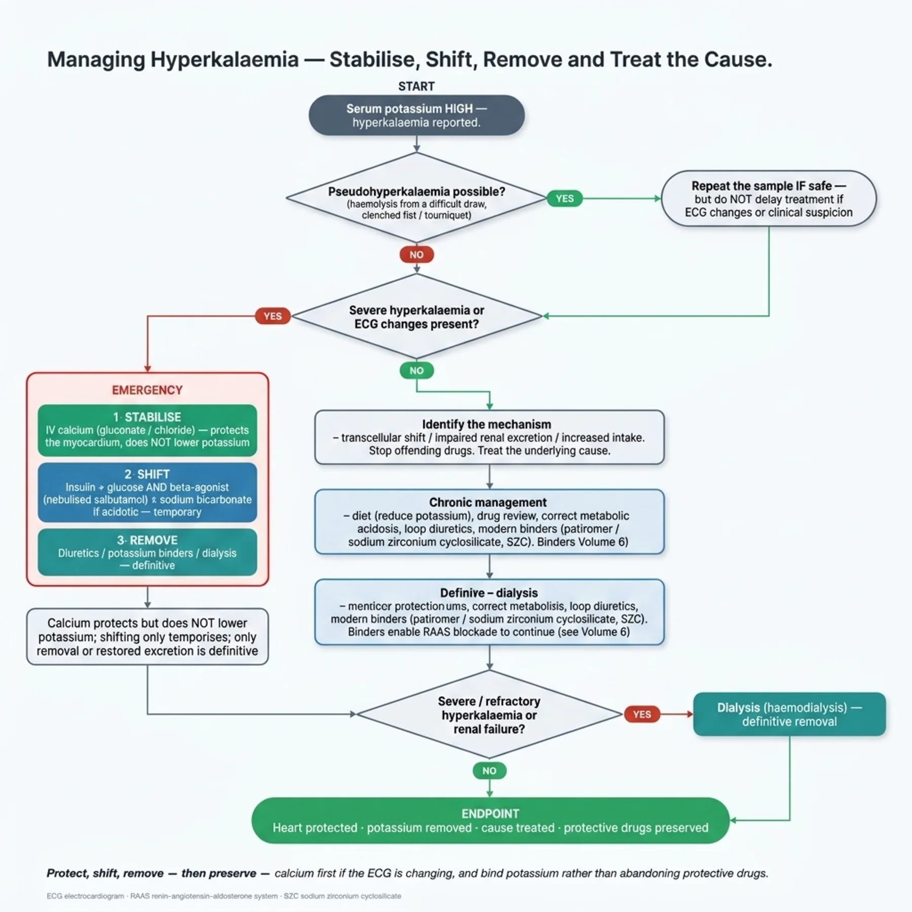
Pseudohyperkalaemia, from in-vitro potassium release (haemolysis, fist-clenching, high cell counts, delayed processing), must be considered — but treatment is not delayed when ECG changes or clinical suspicion are present.
True hyperkalaemia arises from a shift out of cells (acidosis, insulin deficiency, cell lysis, certain drugs), from impaired excretion (the main chronic cause), or from excess intake in the setting of impaired excretion.
Impaired excretion reflects reduced GFR, reduced aldosterone, and a long list of drugs — RAAS blockers, potassium-sparing diuretics, NSAIDs, trimethoprim, calcineurin inhibitors.
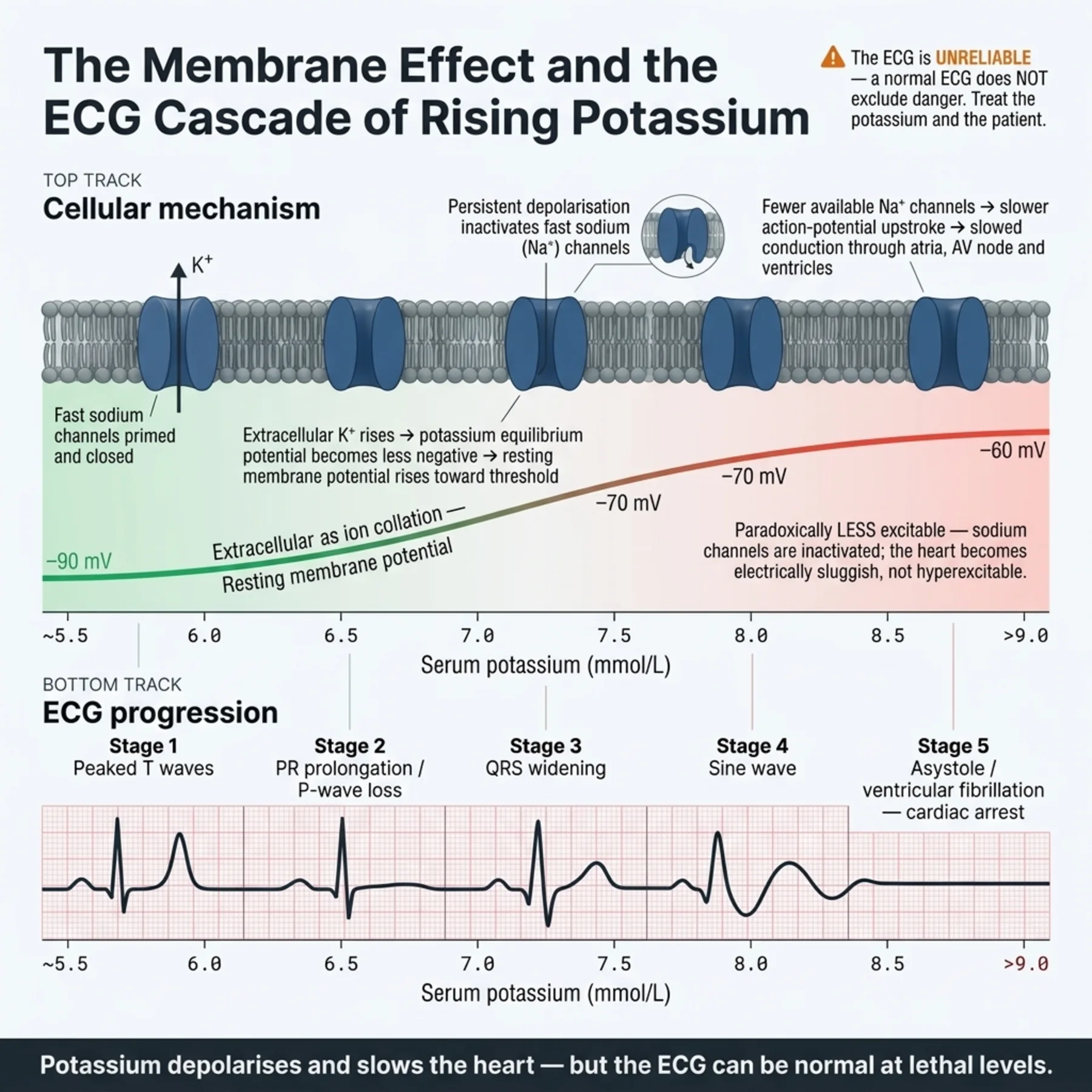
A high extracellular potassium depolarises the resting membrane and slows cardiac conduction, threatening ventricular fibrillation and asystole.
The ECG progresses from peaked T waves through PR prolongation and P-wave loss to QRS widening, a sine wave, and arrest — but it is insensitive and unreliable, so its absence does not exclude danger.
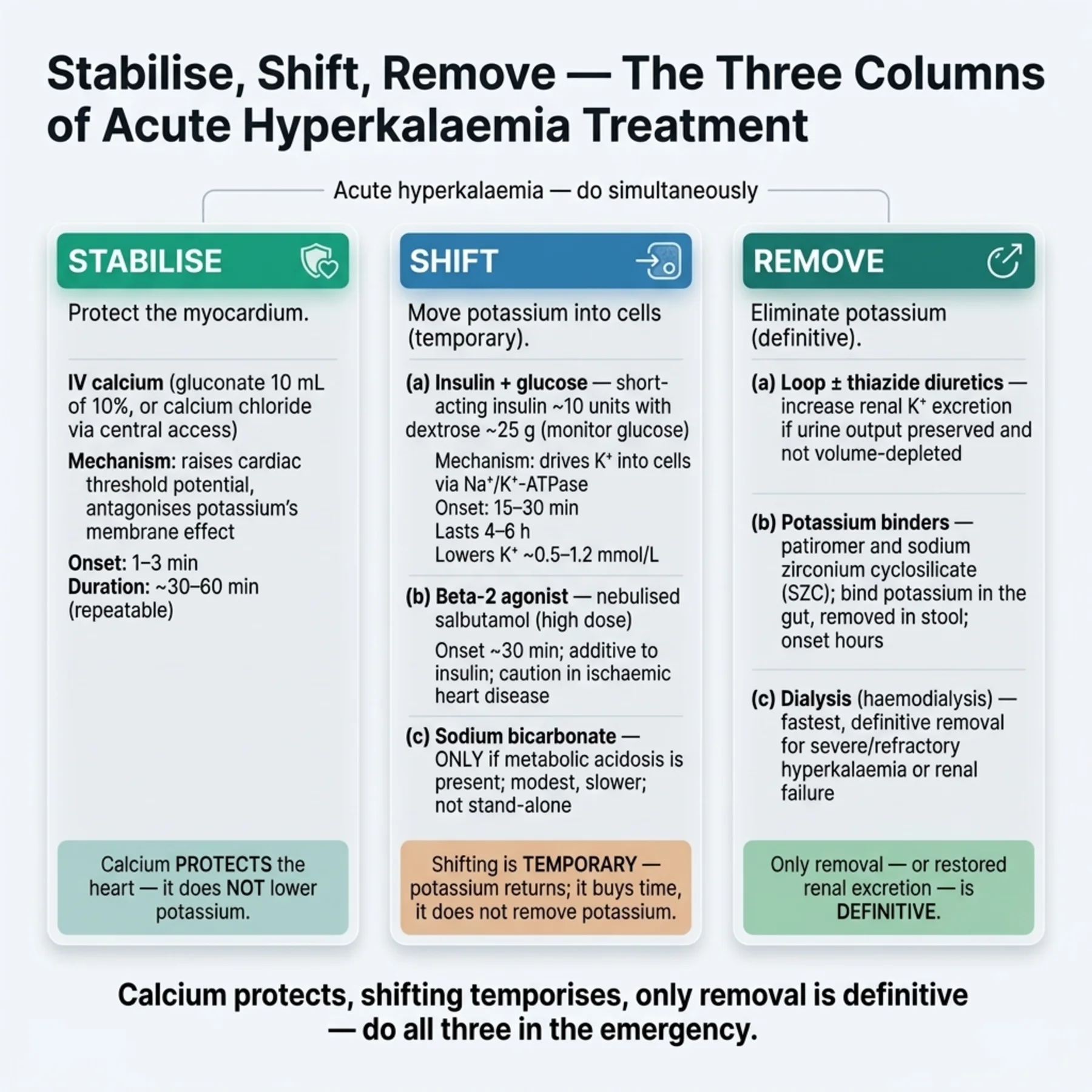
Severe or ECG-positive hyperkalaemia is managed in three prongs: stabilise, shift, and remove.
Calcium stabilises the myocardium immediately but does not lower the potassium, and is repeated if the ECG persists.
Insulin with glucose is the mainstay shift, with a beta-agonist additive and bicarbonate reserved for acidosis; these temporise but do not remove potassium.
Potassium is removed by diuretics (if urine is being made), gut binders, and dialysis — the definitive treatment in severe or refractory cases or renal failure.
The modern binders, sodium zirconium cyclosilicate and patiromer, are effective and faster than the older sodium polystyrene sulfonate, which carries a gastrointestinal-injury risk.
Insulin-glucose causes hypoglycaemia and bicarbonate is a weak monotherapy, so each agent's limits matter.
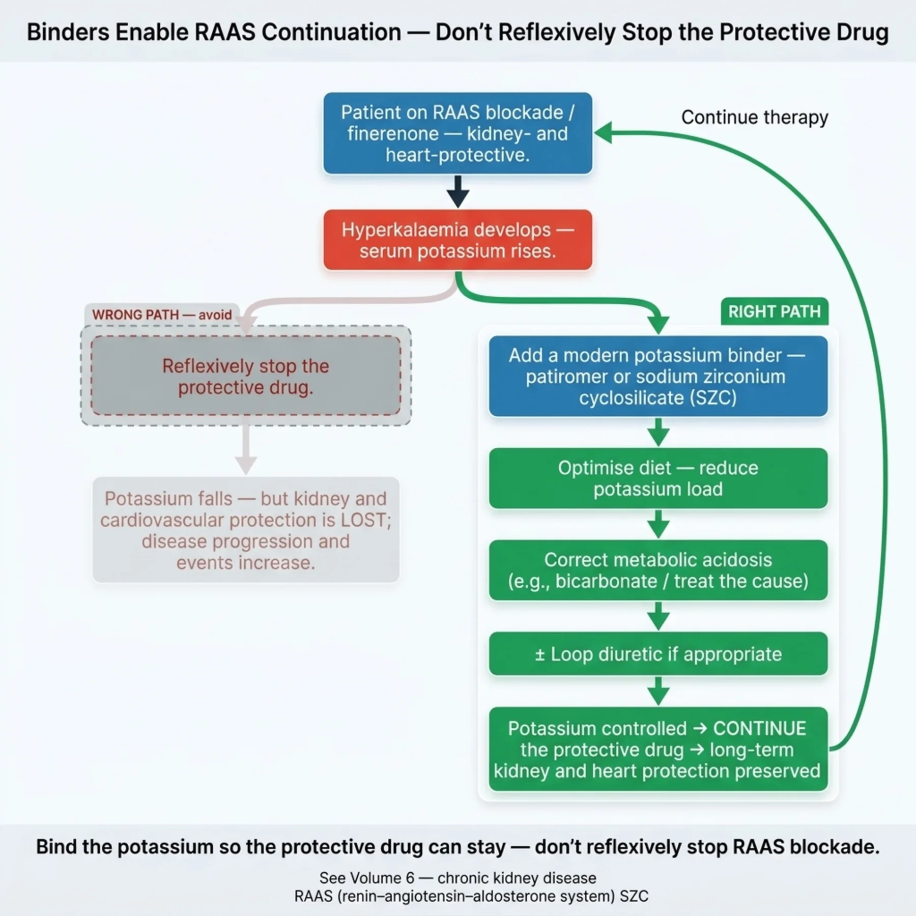
Chronic hyperkalaemia is managed with diet, drug review, acidosis correction, loop diuretics, and binders — which crucially allow RAAS blockade to be continued for its kidney and cardiovascular benefit.
Throughout, the offending drugs are reviewed and the underlying cause is treated.
| 03 | PHASE A · LEVEL 3 · ORIENTATION & KNOWLEDGE Main Narrative |
Hyperkalaemia is the electrolyte emergency. A high potassium can stop the heart within minutes, and the management — stabilise, shift, remove — is among the most important sequences in acute medicine. But it is also a chronic problem, the commonest reason the protective drugs of the CKD volume are stopped, so the chapter has two faces: the emergency that must be acted on at once, and the chronic disorder that must be managed without sacrificing renal protection. Both rest on the same potassium physiology, read in the opposite direction from the last chapter.
— Confirm it — but don't be paralysed
The first question is whether the hyperkalaemia is real. Pseudohyperkalaemia — potassium released in the test tube rather than in the patient — is common: haemolysis of the sample, fist-clenching or a prolonged tourniquet during venepuncture, very high platelet or white-cell counts, and delayed or cold processing all liberate potassium in vitro. A haemolysed sample or a clenched-fist draw should prompt a repeat, ideally a plasma sample handled promptly. But this caution has a strict limit: when there are ECG changes or a clinical context that makes severe hyperkalaemia likely (renal failure, a missed dialysis session, crush injury), treatment is not delayed to chase a confirmatory sample. The rule is to confirm where it is safe to do so and to treat where waiting would be dangerous — the lethal potential of true hyperkalaemia outweighs the inconvenience of treating an occasional pseudo-elevation.
— The mechanisms
True hyperkalaemia, like hypokalaemia, divides by mechanism. A shift out of cells occurs in acidosis (mineral acids shift potassium more than organic ones), in insulin deficiency and hyperglycaemia (where hypertonicity also drags potassium out), in cell lysis (rhabdomyolysis, tumour lysis, massive haemolysis), and with certain drugs (beta-blockers, digoxin toxicity poisoning the sodium-potassium pump, succinylcholine). Impaired excretion is the dominant chronic cause and the one most relevant to the kidney patient: a reduced GFR (advanced or oliguric kidney disease), reduced aldosterone effect (type 4 renal tubular acidosis, hyporeninaemic hypoaldosteronism of diabetes, adrenal insufficiency), reduced distal sodium delivery, and a long list of drugs — RAAS blockers, potassium-sparing diuretics and mineralocorticoid antagonists, NSAIDs, trimethoprim, calcineurin inhibitors, heparin. Excess intake — supplements, salt substitutes (which are potassium chloride), transfusion — rarely causes hyperkalaemia alone and almost always requires impaired excretion to coexist. As with hypokalaemia, identifying the mechanism directs both the urgency and the definitive treatment.
— Why it kills, and why the ECG misleads
The danger is cardiac. A rising extracellular potassium raises (makes less negative) the resting membrane potential, which initially increases excitability but soon impairs the recovery of sodium channels and slows conduction throughout the heart, culminating in ventricular fibrillation or asystole. The electrocardiogram reflects this in a classic progression: peaked T waves first, then PR prolongation and flattening or loss of the P wave, then widening of the QRS, merging finally into a sine-wave pattern and arrest. It is tempting to use the ECG to gauge severity, but here is the crucial caveat: the ECG is insensitive and unreliable in hyperkalaemia. The changes correlate poorly with the potassium level, and a dangerously high potassium can be present with a normal or near-normal ECG — so the absence of ECG changes does not exclude lethal hyperkalaemia. Treatment is therefore guided by the potassium level, the rate of rise, and the clinical context together with the ECG, not by the ECG alone; relying on a 'normal' ECG to defer treatment is a recognised and dangerous error.
— The emergency: stabilise, shift, remove
Severe hyperkalaemia, or any hyperkalaemia with ECG changes, is managed in three prongs that do different jobs and must not be confused. First, stabilise the myocardium: intravenous calcium (gluconate or chloride) acts within minutes to antagonise the membrane effects of potassium and protect the heart — but it does not lower the potassium at all, and its effect is short-lived, so it is repeated if the ECG changes persist, buying time for the other measures (and given with caution in digoxin toxicity). Second, shift potassium into cells to lower the plasma level temporarily: insulin with glucose is the mainstay (monitoring for the hypoglycaemia it commonly causes), a nebulised beta-agonist such as salbutamol adds to it, and sodium bicarbonate has a role only in the acidotic patient and is a weak agent alone. These shifts buy time but do not remove a single milligram of potassium from the body. Third, remove potassium: loop or thiazide diuretics if the patient makes urine, gut binders, and — definitively — dialysis for severe, refractory, or renal-failure cases. The conceptual key is that calcium protects, shifting temporises, and only removal (or restored excretion) is definitive; all three are deployed together in the emergency, and the patient is not 'treated' until potassium has actually left the body.
— The agents, and the binders
Each agent has a defined role and limits. Calcium is the protector — immediate, repeatable, but not potassium-lowering. Insulin-glucose is the workhorse shift, effective but reliably causing hypoglycaemia that must be anticipated with glucose and monitoring. Beta-agonists shift additively. Bicarbonate's role is narrow: trials of bicarbonate as a stand-alone potassium-lowering agent have been disappointing, so it is reserved for the genuinely acidotic patient rather than used reflexively. For removal, the binder story has evolved. The older agent, sodium polystyrene sulfonate, is slow, of debated efficacy, and carries a risk of gastrointestinal injury including colonic necrosis, so it is being displaced. The modern binders — sodium zirconium cyclosilicate (with an onset of around an hour) and patiromer (slower) — are supported by randomised trials, are better tolerated, and have transformed both acute and chronic management. Dialysis remains the definitive removal route when the potassium is dangerous and the kidney cannot excrete it.
— Chronic hyperkalaemia and the RAAS dilemma
Beyond the emergency lies the chronic problem, and its central tension was met in the CKD volume: hyperkalaemia is the commonest reason RAAS blockade and mineralocorticoid antagonists — drugs that protect the kidney and heart — are stopped. Chronic management aims to control the potassium without that sacrifice. The toolkit is dietary potassium restriction (thoughtfully, given the value of plant-rich diets), review of the contributing drugs (stopping the dispensable, such as NSAIDs and trimethoprim), correction of metabolic acidosis (which itself lowers potassium), loop diuretics to enhance excretion, and — the key enabler — the modern potassium binders, which lower potassium reliably enough to let RAAS blockade and finerenone continue at effective doses rather than be withdrawn. Treating hypoaldosteronism (fludrocortisone in selected cases) addresses a specific cause. The governing principle, carried directly from the CKD volume, is to manage the potassium so the protective drug can stay, reserving discontinuation for hyperkalaemia that genuinely cannot be controlled.
— Where the evidence is firm, and where it is qualified
The firm parts: calcium stabilises the membrane without lowering potassium; insulin-glucose effectively shifts it (and causes hypoglycaemia); the modern binders are effective and enable RAAS continuation; and dialysis is definitive. The qualified parts are the ones that overturn old habits — bicarbonate is a weak potassium-lowering agent and is reserved for acidosis rather than used routinely, sodium polystyrene sulfonate's efficacy and safety are questioned, and, most importantly, the ECG is an unreliable guide whose normality must not reassure. The disciplined position is to confirm pseudohyperkalaemia only where safe, treat by level and context rather than by the ECG alone, deploy the three prongs in the emergency knowing what each does, prefer the modern binders, use dialysis when removal is needed and the kidney cannot, and — chronically — use binders to keep patients on their protective drugs. The mirror with hypokalaemia is exact: same physiology, opposite direction, but here the stakes and the speed are higher.
| 04 | PHASE A · LEVEL 4 · ORIENTATION & KNOWLEDGE Reference Tables |
Table 8.1 — Causes of hyperkalaemia
| Mechanism | Examples |
| Pseudohyperkalaemia | In-vitro release: haemolysis, fist-clenching, thrombocytosis/leucocytosis, delayed processing |
| Shift out of cells | Acidosis, insulin deficiency/hyperglycaemia, cell lysis, beta-blockers, digoxin toxicity, succinylcholine |
| Impaired excretion (main chronic) | Low GFR; low aldosterone (type 4 RTA, hypoaldosteronism); drugs (RAAS blockers, K-sparing, NSAIDs, trimethoprim, calcineurin inhibitors) |
| Excess intake | Supplements, salt substitutes, transfusion — usually needs impaired excretion too |
Table 8.2 — The ECG (and its unreliability)
| Stage | Detail |
| Peaked T waves | Earliest change |
| PR prolongation / P-wave loss | Conduction slowing |
| QRS widening → sine wave | Pre-arrest |
| Arrhythmia | Ventricular fibrillation / asystole |
| Caveat | ECG is insensitive — a normal ECG does NOT exclude danger; treat by level/context |
Table 8.3 — The three-pronged emergency
| Prong | Action | What it does |
| Stabilise | IV calcium (gluconate/chloride) | Protects the myocardium — does NOT lower potassium |
| Shift | Insulin + glucose; beta-agonist; bicarbonate (if acidotic) | Lowers plasma K temporarily — does NOT remove it |
| Remove | Diuretics, binders, dialysis | Definitive — removes potassium from the body |
Table 8.4 — The agents
| Agent | Onset / role | Caveat |
| Calcium | Minutes — stabilise | Doesn't lower K; repeat; caution in digoxin |
| Insulin + glucose | Shift (mainstay) | Hypoglycaemia — give glucose, monitor |
| Beta-agonist (salbutamol) | Shift (additive) | Tachycardia |
| Bicarbonate | Shift — only if acidotic | Weak monotherapy |
| Binders / dialysis | Remove (definitive) | Modern binders preferred; dialysis for severe/renal failure |
Table 8.5 — Chronic hyperkalaemia management
| Measure | Detail |
| Diet | Thoughtful potassium restriction |
| Drug review | Stop dispensable culprits (NSAIDs, trimethoprim) |
| Correct acidosis | Bicarbonate lowers potassium |
| Loop diuretics | Enhance excretion |
| Binders | Patiromer/SZC — enable RAAS blockade to continue (Volume 6) |
Table 8.6 — The evidence
| Statement | Note |
| Calcium stabilises without lowering potassium | Physiology / consensus |
| Insulin-glucose effectively shifts; hypoglycaemia common | Clinical data |
| Bicarbonate is weak monotherapy | Disappointing trials — reserve for acidosis |
| SPS efficacy/safety questioned (GI injury) | Modern binders preferred |
| ECG unreliable as a sole guide | Poor correlation with level |
| 05 | PHASE B · LEVEL 5 · VISUALISE & MAP Imaging & Flowchart Specifications |
| 06 | PHASE B · LEVEL 6 · VISUALISE & MAP Concept Maps |
Each chain runs from physiology to a named bedside action; read the arrows as “leads to.”
Balance and excretion. Shift out of cells / impaired excretion (main chronic) / excess intake → high plasma potassium → ACTION: identify the mechanism, with impaired excretion (low GFR, low aldosterone, drugs) the usual chronic culprit.
The membrane effect. High extracellular potassium → raised resting membrane potential → slowed cardiac conduction → VF/asystole → ACTION: treat by level and context, since the ECG is an unreliable sole guide.
Pseudohyperkalaemia. In-vitro potassium release (haemolysis, clenched fist, high cell counts) → falsely high reading → ACTION: repeat where safe, but never delay treatment when ECG changes or clinical suspicion are present.
Stabilise / shift / remove. Calcium protects the membrane (no K lowering); insulin/beta shift K into cells (temporary); diuretics/binders/dialysis remove it (definitive) → ACTION: deploy all three in the emergency, knowing what each does.
Binders and RAAS. RAAS blockade raises potassium → commonest reason the protective drug is stopped → modern binders lower potassium → ACTION: bind the potassium to keep RAAS blockade going rather than withdrawing it.
| 07 | PHASE B · LEVEL 7 · VISUALISE & MAP Decision Pathways |
| R1 | IF hyperkalaemia is reported, THEN consider pseudohyperkalaemia and repeat where safe — but do NOT delay treatment if there are ECG changes or clinical suspicion of true severe hyperkalaemia. |
| R2 | IF assessing severity, THEN treat by the potassium level, the rate of rise, and the context — not by the ECG alone, which is unreliable. |
| R3 | IF hyperkalaemia is severe or there are ECG changes, THEN give intravenous calcium first to stabilise the myocardium — knowing it does not lower the potassium. |
| R4 | IF lowering the potassium, THEN shift it with insulin-glucose (± beta-agonist) to temporise, monitoring for hypoglycaemia — and recognise this does not remove it. |
| R5 | IF potassium must be removed, THEN use diuretics (if urine is made), a modern binder, or dialysis — the definitive options. |
| R6 | IF using bicarbonate, THEN reserve it for the acidotic patient — it is a weak potassium-lowering agent alone. |
| R7 | IF managing chronic hyperkalaemia, THEN use diet, drug review, acidosis correction, loop diuretics, and binders — to keep RAAS blockade going rather than stopping it. |
| R8 | IF the hyperkalaemia is drug-related, THEN stop the dispensable culprits (NSAIDs, trimethoprim) and treat the underlying cause. |
| 08 | PHASE C · LEVEL 8 · CLINICAL REASONING Clinical Cases |
| CASE 1 | THE EMERGENCY Stabilise, shift, remove Severe hyperkalaemia with ECG changes |
Presentation
A dialysis patient who missed a session has a potassium of 7.4 with peaked T waves and a widening QRS. The team gives insulin and glucose and waits for the level to fall.
❖ Pause and reflect Is insulin-glucose alone the right response to ECG changes? |
Analysis
No — the ECG changes signal an immediate cardiac threat, and insulin-glucose, though appropriate as a shift, does not protect the heart and only temporises. The missing first step is intravenous calcium to stabilise the myocardium within minutes (it does not lower the potassium, but it buys time), and the missing last step is definitive removal — in a dialysis patient with renal failure and a potassium of 7.4, dialysis. The three prongs — stabilise, shift, remove — are deployed together, not the shift alone.
Plan
Give intravenous calcium immediately to stabilise the myocardium (repeat if ECG changes persist), continue insulin-glucose (with a beta-agonist) to shift, and arrange urgent dialysis to remove the potassium. Treat the missed-session cause.
Teaching point
In severe hyperkalaemia with ECG changes, give calcium first to protect the heart, shift to temporise, and remove definitively — all three prongs.
Cross-reference
Exercises rules R3, R4, R5; the stabilise-shift-remove concept map; Figures 8.1–8.2; Tables 8.3, 8.4.
| CASE 2 | TOO HIGH TO BE TRUE? Confirm — but don't be paralysed Pseudohyperkalaemia |
Presentation
A well outpatient with normal renal function has a potassium of 6.8 on a sample noted to be haemolysed, with no ECG changes and no clinical features of hyperkalaemia.
❖ Pause and reflect Should this be treated as a hyperkalaemic emergency? |
Analysis
Probably not as an emergency, but it must be handled carefully. The haemolysed sample, the well patient, normal renal function, and the absence of ECG changes all suggest pseudohyperkalaemia — potassium released in the tube, not in the patient. The right step is to repeat the sample promptly (a clean, well-handled plasma sample). The contrast with case 1 is the point: there, ECG changes and renal failure mandated immediate treatment; here, the safe context permits confirmation first. The judgement is to confirm where safe and treat where waiting is dangerous.
Plan
Repeat the potassium on a properly drawn, promptly processed sample, with an ECG; if the repeat is normal, no emergency treatment is needed. Had there been ECG changes or a high-risk context, treatment would not have waited.
Teaching point
Consider pseudohyperkalaemia (haemolysis, clenched fist) and repeat where safe — but never delay treatment when ECG changes or clinical risk are present.
Cross-reference
Exercises rule R1; the pseudohyperkalaemia concept map; Table 8.1.
| CASE 3 | KEEP THE PILLAR Bind, don't stop Chronic hyperkalaemia on RAAS blockade |
Presentation
A patient with proteinuric CKD on an ACE inhibitor has a chronic potassium of 5.6. The plan is to stop the ACE inhibitor to bring the potassium down.
❖ Pause and reflect Is stopping the ACE inhibitor the best way to manage this chronic hyperkalaemia? |
Analysis
Stopping it would lower the potassium but forfeit the kidney and cardiovascular protection the ACE inhibitor provides — and chronic hyperkalaemia is usually manageable without that sacrifice. Dietary potassium restriction, reviewing other contributing drugs, correcting any acidosis, a loop diuretic, and a modern potassium binder can control the potassium while the ACE inhibitor continues. This is the chronic-management lesson carried directly from the CKD volume: manage the potassium to keep the protective pillar on board.
Plan
Keep the ACE inhibitor, restrict dietary potassium thoughtfully, correct acidosis, optimise a loop diuretic, review other culprit drugs, and start a modern binder (patiromer or sodium zirconium cyclosilicate) to control the potassium. Reserve discontinuation for uncontrollable hyperkalaemia.
Teaching point
Chronic hyperkalaemia on RAAS blockade is managed by binding the potassium to keep the protective drug — not by reflexively stopping it.
Cross-reference
Exercises rules R7 and R8; the binders-and-RAAS concept map; Figure 8.3; Table 8.5; the same lesson in Volume 6 Chapter 9.
| CASE 4 | THE NORMAL ECG TRAP Don't be reassured The unreliable ECG |
Presentation
A patient with acute kidney injury has a potassium of 7.0 but a near-normal ECG. A trainee proposes to defer treatment because 'the ECG is fine.'
❖ Pause and reflect Does a normal ECG make a potassium of 7.0 safe to defer? |
Analysis
No — this is the normal-ECG trap. The ECG is insensitive in hyperkalaemia and correlates poorly with the level, so a dangerously high potassium can coexist with a near-normal ECG, and arrhythmia can occur without a warning ECG progression. Deferring treatment because the ECG looks fine is a recognised and dangerous error. A potassium of 7.0 in acute kidney injury is treated on the level and context, regardless of the reassuring ECG.
Plan
Treat the hyperkalaemia on the basis of the level and context — stabilise, shift, and arrange removal — rather than being reassured by the near-normal ECG. Use the ECG as one input, not the gatekeeper to treatment.
Teaching point
A normal ECG does not exclude dangerous hyperkalaemia — treat by the level and context, not by the ECG alone.
Cross-reference
Exercises rules R2 and R3; the membrane/ECG concept map; Figure 8.1; Table 8.2.
| 09 | PHASE C · LEVEL 9 · CLINICAL REASONING Clinical Implications |
One triad per mechanism the narrative exposed: the physiology, why it matters, and the bedside move.
MECHANISM Hyperkalaemia arises from a shift out of cells, impaired excretion, or excess intake. |
WHY IT MATTERS Impaired excretion is the main chronic cause and the one most relevant to the kidney patient. |
ACTION Identify the mechanism — low GFR, low aldosterone, or drugs — to direct treatment. |
MECHANISM A rising extracellular potassium depolarises the resting membrane and slows cardiac conduction. |
WHY IT MATTERS This threatens ventricular fibrillation and asystole, but the ECG correlates poorly with the level. |
ACTION Treat by the potassium level and context, not by the ECG alone. |
MECHANISM Potassium is released in vitro by haemolysis, fist-clenching, and high cell counts. |
WHY IT MATTERS Pseudohyperkalaemia can mislead, but true hyperkalaemia can kill while a sample is repeated. |
ACTION Repeat where safe, but never delay treatment when ECG changes or clinical risk are present. |
MECHANISM Calcium antagonises the membrane effect, shifting moves potassium into cells, and only removal clears it. |
WHY IT MATTERS Confusing what each prong does leaves a 'treated' patient with potassium still in the body. |
ACTION Deploy all three prongs, knowing calcium protects, shifting temporises, and removal is definitive. |
MECHANISM RAAS blockade raises potassium and is the commonest reason it is stopped. |
WHY IT MATTERS Stopping it forfeits the kidney and cardiovascular protection it provides. |
ACTION Use modern binders to control the potassium and keep the protective drug going. |
| 10 | PHASE C · LEVEL 10 · CLINICAL REASONING Clinical Pearls |
| Hyperkalaemia: shift out / impaired excretion (main chronic) / excess intake. | Pseudohyperkalaemia (haemolysis, clenched fist, high cell counts) — repeat where safe. |
| Don't delay treatment for confirmation if ECG changes/clinical risk. | Impaired excretion: low GFR, low aldosterone, drugs (RAAS, K-sparing, NSAIDs, trimethoprim). |
| High K → raised resting membrane potential → slowed conduction → VF/asystole. | ECG: peaked T → PR prolongation/P loss → wide QRS → sine wave → arrest. |
| ECG is UNRELIABLE — normal ECG doesn't exclude danger; treat by level/context. | Emergency = three prongs: STABILISE, SHIFT, REMOVE. |
| Calcium stabilises the myocardium — does NOT lower potassium; repeat; caution digoxin. | Insulin + glucose = mainstay shift; monitor for hypoglycaemia. |
| Beta-agonist additive; bicarbonate only if acidotic (weak alone). | Shifting temporises — only removal/restored excretion is definitive. |
| Remove: diuretics (if urine), binders, dialysis. | Modern binders (SZC, patiromer) preferred; SPS — GI necrosis risk. |
| Dialysis = definitive for severe/refractory/renal failure. | Chronic: diet, drug review, correct acidosis, loop diuretics, binders. |
| Binders enable RAAS blockade to CONTINUE — don't reflexively stop it. | Stop dispensable culprits (NSAIDs, trimethoprim); treat the cause. |
| 11 | PHASE D · LEVEL 11 · SAFETY & EVIDENCE Red Flags & Never-Do |
Panel A — Red flags
| ▲ | Peaked T waves or a widening QRS — give intravenous calcium immediately to stabilise the myocardium. |
| ▲ | A high potassium with a near-normal ECG — the ECG is unreliable; treat on the level and context. |
| ▲ | Severe hyperkalaemia in renal failure — shifting only temporises; arrange dialysis for definitive removal. |
| ▲ | RAAS blockade being stopped for a manageable chronic hyperkalaemia — bind the potassium instead. |
| ▲ | Hyperkalaemia on trimethoprim or an NSAID — dispensable culprits; stop them. |
Panel B — Never do
| ✖ NEVER — defer treatment of dangerous hyperkalaemia because the ECG looks normal. |
| ✖ NEVER — rely on calcium or shifting to remove potassium — only removal/restored excretion does. |
| ✖ NEVER — use bicarbonate as a stand-alone potassium-lowering agent in a non-acidotic patient. |
| ✖ NEVER — stop RAAS blockade for a manageable chronic hyperkalaemia when a binder would let it continue. |
| 12 | PHASE D · LEVEL 12 · SAFETY & EVIDENCE Common Pitfalls |
Pitfall 1 — Trusting the ECG
| ✖ | WRONG Deferring treatment of a potassium of 7.0 because the ECG is normal. |
| ✓ | RIGHT Treating on the level and context. |
| ✉ | WHY The ECG is insensitive — dangerous hyperkalaemia can have a normal ECG. |
Pitfall 2 — Shift without protect/remove
| ✖ | WRONG Giving insulin-glucose alone for ECG-positive hyperkalaemia. |
| ✓ | RIGHT Giving calcium first and arranging definitive removal too. |
| ✉ | WHY Shifting protects neither the heart (calcium does) nor removes potassium (dialysis/binders do). |
Pitfall 3 — Reflex bicarbonate
| ✖ | WRONG Giving bicarbonate routinely to lower potassium. |
| ✓ | RIGHT Reserving bicarbonate for the acidotic patient. |
| ✉ | WHY Bicarbonate is a weak potassium-lowering agent alone. |
Pitfall 4 — Stopping the pillar
| ✖ | WRONG Stopping RAAS blockade for a chronic potassium of 5.6. |
| ✓ | RIGHT Binding the potassium to keep the protective drug going. |
| ✉ | WHY Withdrawal forfeits kidney and cardiovascular protection for a manageable problem. |
Pitfall 5 — Treating the tube
| ✖ | WRONG Treating a haemolysed-sample 'hyperkalaemia' in a well patient as an emergency. |
| ✓ | RIGHT Repeating the sample where the context is safe. |
| ✉ | WHY Pseudohyperkalaemia is potassium released in vitro, not in the patient. |
| 13 | PHASE D · LEVEL 13 · SAFETY & EVIDENCE Evidence Grading |
GRADE A HIGH CONFIDENCE The effect is real and the estimate is stable. RCTs at low risk of bias; multiple concordant prospective cohorts; meta-analyses. |
GRADE B MODERATE CONFIDENCE The effect is likely real but may shift with new data. Observational studies, registries, mechanistic human studies. |
GRADE C LOW CONFIDENCE Rests on physiology, reasoning, or consensus rather than outcomes. Pathophysiological reasoning; extrapolation; consensus without outcomes. |
Graded statements (by evidence type)
| Statement | Grade | Basis (evidence type) |
| Calcium stabilises the myocardium without lowering potassium. | A | Established physiology and consensus |
| Insulin-glucose effectively shifts potassium and commonly causes hypoglycaemia. | A | Clinical and pharmacological data |
| The ECG is insensitive and unreliable for grading hyperkalaemia severity. | B | Diagnostic-accuracy data |
| Bicarbonate is a weak potassium-lowering agent as monotherapy. | B | Disappointing trial data |
| Modern binders (SZC, patiromer) lower potassium and enable RAAS continuation. | A | RCTs |
| Sodium polystyrene sulfonate has questioned efficacy and GI-injury risk. | B | Observational and case data |
| Dialysis is definitive for severe/refractory hyperkalaemia in renal failure. | A | Physiology and clinical consensus |
| 14 | PHASE E · LEVEL 14 · PATIENT DECISIONS Absolute Risk in Natural Frequency |
Natural-frequency estimates for orientation, from hyperkalaemia management; they vary with severity and renal function. They convey the size of the decisions, expressed per 100 comparable patients.
| Per 100 patients… | Outcome | Roughly how many | See |
| Given insulin-glucose for hyperkalaemia | Develop hypoglycaemia | A notable share — hence monitor and give glucose | L13 row 2 |
| With dangerous hyperkalaemia and a normal ECG | Have a normal ECG despite the risk | A meaningful share — hence don't trust the ECG | L13 row 3 |
| On RAAS blockade given a binder | Continue the protective drug rather than stopping | Many more than without a binder | L13 row 5 |
| With severe hyperkalaemia in renal failure given dialysis | Achieve definitive potassium removal | Most | L13 row 7 |
★ How to read these Read these as orientation, not promises; outcomes vary with severity and renal function. The stable signals: insulin-glucose causes hypoglycaemia, the ECG can be normal at lethal levels, binders keep patients on protective drugs, and dialysis is definitive. Communicate them as people out of 100, not as a hazard ratio. |
| 17 | PHASE F · LEVEL 17 · APPLY & TEST Documentation Templates |
Paste-ready notes. Tick the boxes that apply and delete the rest; make the three prongs and the don't-trust-the-ECG point explicit.
Template 1 — Acute hyperkalaemia protocol
Template 2 — Chronic hyperkalaemia management
| 18 | PHASE F · LEVEL 18 · APPLY & TEST Cheat Sheet |
| Causes: shift out / impaired excretion (main chronic) / excess intake. | Pseudohyperkalaemia — repeat where safe; don't delay if ECG/clinical risk. |
| Impaired excretion: low GFR, low aldosterone, drugs. | High K → raised resting potential → slowed conduction → arrest. |
| ECG: peaked T → wide QRS → sine wave; UNRELIABLE — treat by level/context. | EMERGENCY = STABILISE, SHIFT, REMOVE. |
| Calcium = stabilise (NO K lowering); repeat; caution digoxin. | Insulin + glucose = mainstay shift (watch hypoglycaemia). |
| Beta-agonist additive; bicarbonate only if acidotic (weak alone). | Shifting temporises; only removal is definitive. |
| Remove: diuretics (if urine), binders, dialysis. | Modern binders (SZC/patiromer) > SPS (GI necrosis risk). |
| Dialysis = definitive (severe/refractory/renal failure). | Chronic: diet, drug review, correct acidosis, loop diuretics, binders. |
| Binders enable RAAS blockade to CONTINUE. | Stop dispensable culprits (NSAIDs, trimethoprim). |
| 19 | PHASE F · LEVEL 19 · APPLY & TEST Flashcards |
| CARD 1 | Q. What are the mechanisms of hyperkalaemia? A. A shift out of cells (acidosis, insulin deficiency, cell lysis, certain drugs), impaired excretion (low GFR, low aldosterone, drugs — the main chronic cause), and excess intake (usually only with impaired excretion). DETAILED. Pseudohyperkalaemia from in-vitro release must also be considered. CLINICAL. Identify the mechanism to direct treatment. |
| CARD 2 | Q. What is pseudohyperkalaemia, and how is it handled? A. Potassium released in the test tube — by haemolysis, fist-clenching, high platelet or white-cell counts, or delayed processing — giving a falsely high reading; repeat on a clean sample where safe. DETAILED. Treatment is not delayed if ECG changes or clinical suspicion are present. CLINICAL. Confirm where safe; treat where waiting is dangerous. |
| CARD 3 | Q. Why is the ECG an unreliable guide in hyperkalaemia? A. Although it progresses from peaked T waves through QRS widening to a sine wave, the changes correlate poorly with the potassium level, so a dangerously high potassium can coexist with a normal or near-normal ECG. DETAILED. A normal ECG does not exclude lethal hyperkalaemia. CLINICAL. Treat by the level, the rate of rise, and the context. |
| CARD 4 | Q. What are the three prongs of emergency management? A. Stabilise the myocardium with intravenous calcium, shift potassium into cells with insulin-glucose (and a beta-agonist), and remove potassium with diuretics, binders, or dialysis. DETAILED. Calcium protects but does not lower potassium; shifting temporises; only removal is definitive. CLINICAL. Deploy all three together in the emergency. |
| CARD 5 | Q. What does calcium do in hyperkalaemia? A. It antagonises the membrane effects of potassium within minutes, stabilising the myocardium, but it does not lower the potassium and is short-acting, so it is repeated if ECG changes persist. DETAILED. It buys time for the shifting and removal measures. CLINICAL. Give it first for ECG changes — but follow with shift and removal. |
| CARD 6 | Q. What is the role and limit of bicarbonate? A. It can shift potassium into cells but is a weak potassium-lowering agent as monotherapy, with disappointing trial evidence, so it is reserved for the genuinely acidotic patient. DETAILED. It is not a reflexive treatment for hyperkalaemia. CLINICAL. Use it only in acidosis, not routinely. |
| CARD 7 | Q. Which binders are preferred, and why? A. The modern binders sodium zirconium cyclosilicate (onset ~1 hour) and patiromer are effective, better tolerated, and supported by trials; sodium polystyrene sulfonate is slow, of debated efficacy, and risks gastrointestinal injury. DETAILED. Modern binders also enable RAAS blockade to continue. CLINICAL. Prefer the modern binders for removal. |
| CARD 8 | Q. How is chronic hyperkalaemia managed on RAAS blockade? A. With dietary potassium restriction, drug review, acidosis correction, loop diuretics, and modern binders — which lower potassium enough to continue RAAS blockade and finerenone rather than stopping them. DETAILED. Hyperkalaemia is the commonest reason these protective drugs are stopped. CLINICAL. Bind the potassium to keep the protective drug going. |
| CARD 9 | Q. What is definitive removal in severe hyperkalaemia with renal failure? A. Dialysis — it removes potassium from the body when the kidney cannot, and is the definitive treatment for severe or refractory hyperkalaemia in renal failure. DETAILED. Shifting and binders are not definitive in this setting. CLINICAL. Arrange dialysis for definitive removal. |
| 20 | PHASE F · LEVEL 20 · APPLY & TEST One-Minute Preceptor |
| SCENE 1 | The intern who only shifted |
GET A COMMITMENT. “You gave insulin-glucose for this potassium of 7.4 with a widening QRS — is that enough?”
PROBE FOR EVIDENCE. “It'll bring the potassium down” — ask: “What protects the heart right now, and what actually removes the potassium?”
TEACH A GENERAL RULE. Insulin-glucose only shifts; calcium protects the heart and dialysis removes the potassium — the emergency needs all three prongs, stabilise, shift, remove.
REINFORCE WHAT WAS RIGHT. Starting the shift was a correct component.
CORRECT A MISTAKE. Give calcium now and arrange urgent dialysis.
| SCENE 2 | The resident reassured by the ECG |
GET A COMMITMENT. “You're deferring treatment of this potassium of 7.0 because the ECG is normal — why?”
PROBE FOR EVIDENCE. “No ECG changes, so it's safe” — ask: “How well does the ECG correlate with the potassium level?”
TEACH A GENERAL RULE. The ECG is insensitive and unreliable — a dangerous potassium can have a normal ECG, so treat by the level and context, not the ECG alone.
REINFORCE WHAT WAS RIGHT. Checking the ECG was appropriate.
CORRECT A MISTAKE. Treat the hyperkalaemia on the level — don't be reassured by a normal ECG.
| 21 | PHASE F · LEVEL 21 · APPLY & TEST Reflective Prompts |
Genuine tensions this evidence leaves open; sit with them rather than resolving them too quickly.
The ECG is taught as the window onto hyperkalaemic danger, yet it is unreliable. How do you unlearn a heuristic that feels authoritative but can fatally mislead?
Pseudohyperkalaemia and true hyperkalaemia demand opposite responses — wait versus act — and the context decides. How do you make that judgement quickly and safely under pressure?
Bicarbonate and the old binder persisted for decades on physiological plausibility before the evidence questioned them. What does that say about treatments that 'make sense'?
Hyperkalaemia is the commonest reason the kidney's best drugs are stopped. How much avoidable harm comes from treating the potassium by withdrawing protection rather than binding it?
The three prongs do different jobs, yet a 'normalised' potassium after shifting can create false security. How do you keep a team focused on removal, not just the number?
| 22 | PHASE F · LEVEL 22 · APPLY & TEST Board-Style Questions |
| Q 01 | The first treatment for severe hyperkalaemia with ECG changes is: |
| A | Insulin and glucose |
| B | Intravenous calcium to stabilise the myocardium |
| C | Sodium bicarbonate |
| D | A potassium binder |
Rationale Calcium protects the heart within minutes (though it doesn't lower potassium) and is given first for ECG changes (case 1, rule R3, Table 8.3). A shifts; C is weak; D is slow removal. |
| Q 02 | What does intravenous calcium do in hyperkalaemia? |
| A | Lowers the potassium |
| B | Stabilises the myocardial membrane without lowering potassium |
| C | Shifts potassium into cells |
| D | Removes potassium from the body |
Rationale Calcium antagonises the membrane effect and protects the heart but does not lower the potassium (Figure 8.2, Table 8.4). A, C, and D misattribute its action. |
| Q 03 | Why is the ECG an unreliable guide to hyperkalaemia severity? |
| A | It always shows peaked T waves |
| B | Its changes correlate poorly with the level — a normal ECG doesn't exclude danger |
| C | It overestimates the potassium |
| D | It is only abnormal in pseudohyperkalaemia |
Rationale The ECG is insensitive, so dangerous hyperkalaemia can have a normal ECG; treat by level and context (case 4, rule R2, Table 8.2). A, C, and D are incorrect. |
| Q 04 | Insulin-glucose and beta-agonists treat hyperkalaemia by: |
| A | Removing potassium from the body |
| B | Shifting potassium into cells (temporary) |
| C | Stabilising the membrane |
| D | Increasing renal excretion |
Rationale These shift potassium intracellularly, temporising without removing it (Table 8.3, rule R4). A is removal; C is calcium; D is diuretics/dialysis. |
| Q 05 | A haemolysed sample in a well patient with normal renal function and a normal ECG shows potassium 6.8. The best step is to: |
| A | Treat as an emergency |
| B | Repeat the sample on a clean specimen |
| C | Start dialysis |
| D | Give calcium |
Rationale The context suggests pseudohyperkalaemia, so a repeat is appropriate — though treatment would not wait if ECG changes or risk were present (case 2, rule R1). A, C, and D over-treat a likely artefact. |
| Q 06 | When is sodium bicarbonate appropriate in hyperkalaemia? |
| A | Routinely as a first-line shift |
| B | Only in the acidotic patient — it is weak monotherapy |
| C | To stabilise the membrane |
| D | To remove potassium |
Rationale Bicarbonate is a weak potassium-lowering agent and is reserved for acidosis (rule R6, Table 8.6). A overuses it; C is calcium; D is removal. |
| Q 07 | Which binder is preferred for hyperkalaemia, and why? |
| A | Sodium polystyrene sulfonate — it is fastest |
| B | A modern binder (SZC or patiromer) — effective, better tolerated, and enables RAAS continuation |
| C | No binder is effective |
| D | Calcium resonium only |
Rationale Modern binders are effective, safer, and allow RAAS blockade to continue, unlike SPS with its GI-injury risk (Table 8.5, rule R7). A, C, and D are incorrect. |
| Q 08 | A patient on an ACE inhibitor has a chronic potassium of 5.6. The best approach is to: |
| A | Stop the ACE inhibitor |
| B | Manage the potassium (diet, acidosis, diuretic, binder) and continue the ACE inhibitor |
| C | Ignore it |
| D | Add a potassium supplement |
Rationale Chronic hyperkalaemia is usually manageable, and the ACE inhibitor protects the kidney and heart, so bind the potassium and continue it (case 3, rule R7). A forfeits benefit; C is unsafe; D is harmful. |
| Q 09 | Across 100 patients given insulin-glucose for hyperkalaemia, a notable number will: |
| A | Have no side effects |
| B | Develop hypoglycaemia |
| C | Develop hyperglycaemia |
| D | Need dialysis |
Rationale Insulin-glucose commonly causes hypoglycaemia, which must be anticipated and monitored (L14, L13 row 2). A, C, and D are incorrect. |