Chapter Preamble
Signals declared
Sig-D — Diagnostic (primary). Recognise volume depletion and the types of oedema, and use the urine sodium and the clinical picture to find the cause.
Sig-T — Therapeutic (strong). Replacing a deficit appropriately, and treating oedema by restricting and removing sodium while protecting effective arterial blood volume.
Sig-M — Mechanistic (strong). How effective arterial blood volume is sensed and defended by RAAS, the sympathetic system, ADH, and the natriuretic peptides — and the underfill paradox of oedema.
Levels populated and omitted
Populated (19): L1–L14, L17–L20, L22. The mechanistic signal fires the concept maps (L6) and triads (L9); the therapeutic signal fires the absolute-risk table (L14) and templates (L17); the diagnostic signal drives the tables, rules, cases, pitfalls, and board items.
L15 / L16 preference-sensitive map and SDM scripts — omitted. No Sig-E; correcting a volume disorder is effective care.
L21 reflective prompts — omitted. No Sig-E/V; the chapter's tension (the underfill paradox) is worked through the cases and pitfalls.
| 01 | PHASE A · LEVEL 1 · ORIENTATION & KNOWLEDGE Learning Objectives |
By the end of this chapter you should be able to:
Explain how effective arterial blood volume is sensed and why it differs from total extracellular volume.
Describe the effectors — RAAS, the sympathetic system, ADH, and the natriuretic peptides — that defend volume.
Recognise volume depletion and identify its cause and the type of loss.
Use the urine sodium and laboratory clues to confirm avid sodium retention.
Distinguish underfill from overfill oedema and explain the underfill paradox.
Replace a volume deficit with the appropriate fluid and treat the cause.
Treat oedema by restricting and removing sodium, using diuretics effectively.
Recognise and manage diuretic resistance and the risk of over-diuresis in underfill states.
| 02 | PHASE A · LEVEL 2 · ORIENTATION & KNOWLEDGE Executive Summary |
Disorders of volume are disorders of sodium content — the extracellular-volume axis from the opening chapter.
What the body actually senses is not total extracellular volume but the effective arterial blood volume — the fullness of the arterial circulation perfusing tissues.
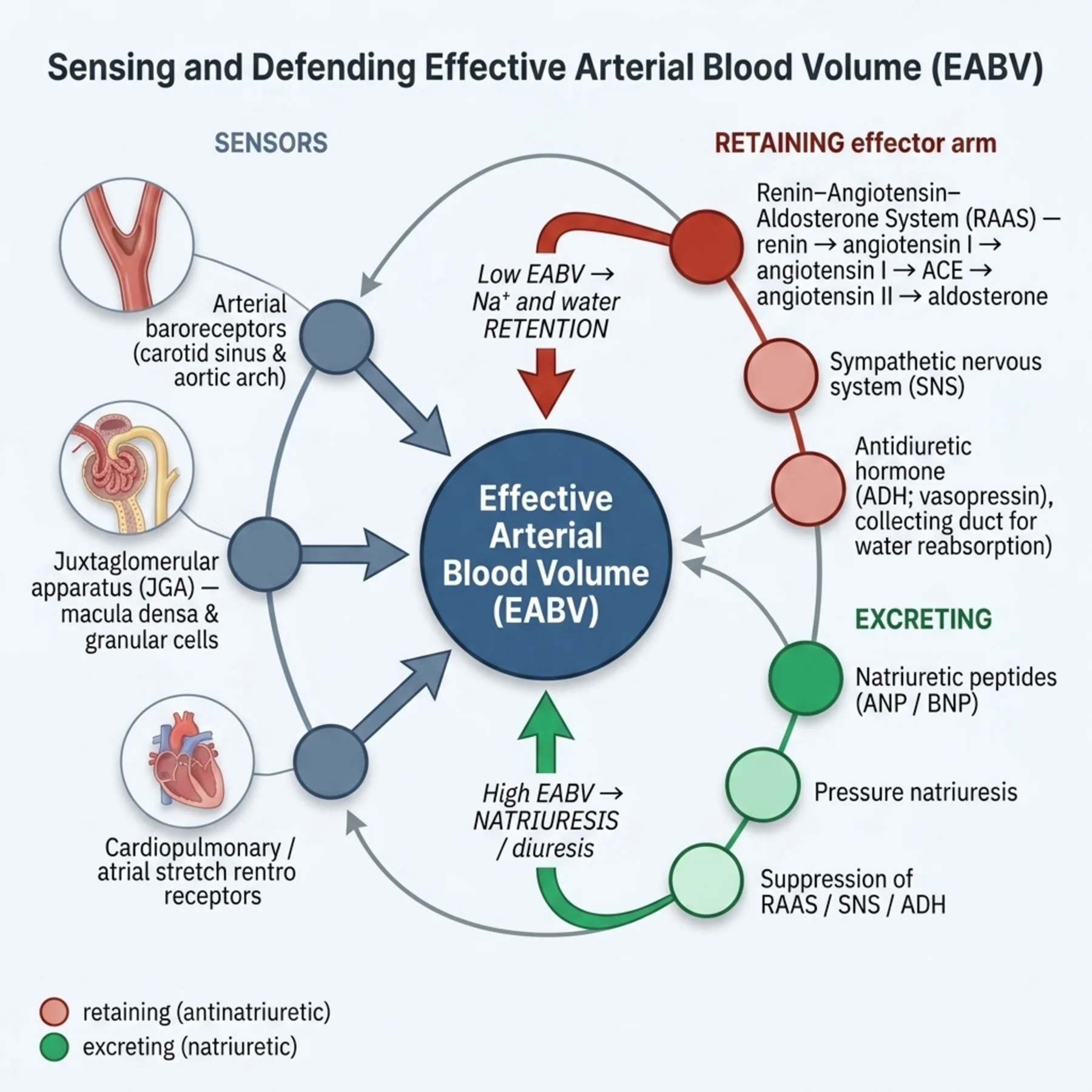
Effective arterial blood volume is sensed by arterial baroreceptors, the juxtaglomerular apparatus, and atrial stretch receptors.
A low effective arterial blood volume activates RAAS, the sympathetic system, and ADH, producing avid sodium and water retention; the natriuretic peptides oppose this when volume is high.
Volume depletion lowers the effective arterial blood volume, so the kidney retains sodium avidly — a low urine sodium, a low fractional excretion, concentrated oliguric urine, and prerenal azotaemia.
Depletion has gastrointestinal, renal, skin, third-space, and haemorrhagic causes, each replaced with appropriate fluid.
Oedema is sodium-content excess, and it comes in two physiologies.
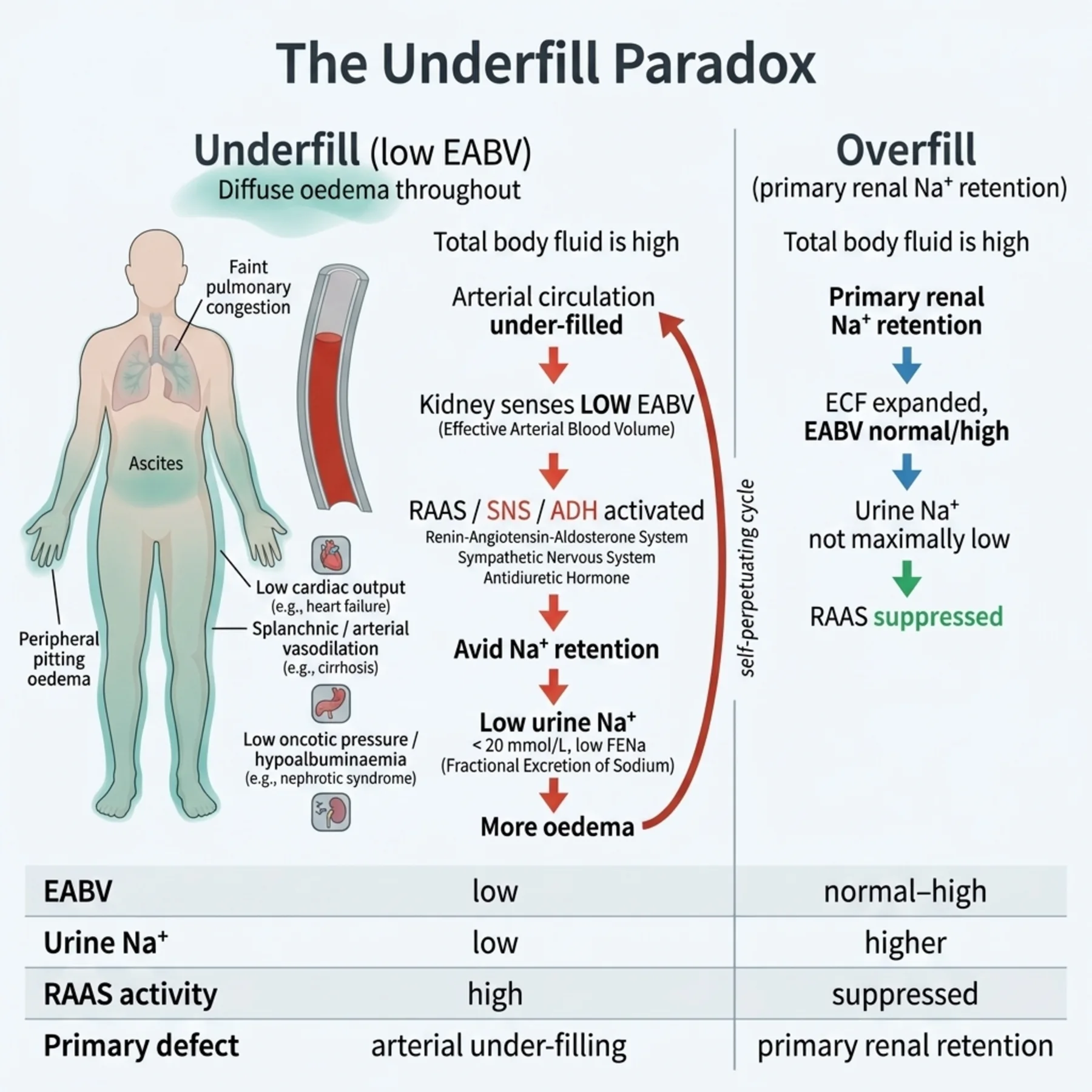
In underfill oedema — heart failure, cirrhosis, nephrotic syndrome — the effective arterial blood volume is low despite total overload, so the kidney retains sodium and the patient is paradoxically both overloaded and acting as if depleted, with a low urine sodium.
In overfill states — primary renal sodium retention in kidney disease — the kidney simply cannot excrete sodium, and the volume expands.
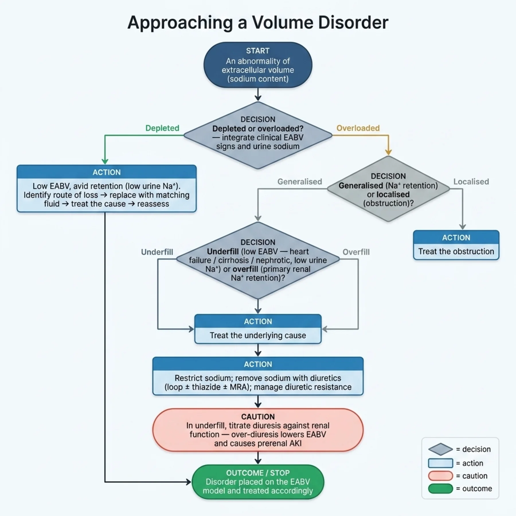
Generalised oedema reflects sodium retention; localised oedema reflects venous or lymphatic obstruction and is not a sodium problem.
Depletion is treated by replacing the deficit with the appropriate fluid and treating the cause.
Oedema is treated by addressing the cause, restricting sodium, and removing it with diuretics — loop as the mainstay, with thiazide for sequential blockade and a mineralocorticoid antagonist in aldosterone-driven states.
Diuretic resistance is anticipated and managed, and over-diuresis in underfill states can worsen the effective arterial blood volume and cause acute kidney injury, so diuresis is titrated.
| 03 | PHASE A · LEVEL 3 · ORIENTATION & KNOWLEDGE Main Narrative |
Volume disorders turn on a quantity the body never measures directly. It does not sense total extracellular volume; it senses the effective arterial blood volume — how well-filled the arterial circulation is, how adequately tissues are perfused — and it defends that. Usually the two move together, but in the great oedema-forming diseases they come apart, and a patient drowning in total body fluid can have a kidney behaving as though the tank were empty. Understanding what is sensed, and what defends it, makes both depletion and oedema intelligible.
— What is sensed, and what defends it
The effective arterial blood volume is monitored at several sites: the high-pressure arterial baroreceptors in the carotid sinus and aortic arch, the renal juxtaglomerular apparatus sensing afferent arteriolar stretch and distal sodium delivery, and the low-pressure atrial stretch receptors. When these sense a fall, they trigger the volume-retaining effectors. The renin-angiotensin-aldosterone system is central: renin generates angiotensin II, which constricts vessels and drives proximal sodium reabsorption, and stimulates aldosterone, which reabsorbs sodium distally. The sympathetic nervous system adds vasoconstriction, sodium retention, and more renin. ADH, released for volume as well as osmotic reasons, retains water. Opposing all of these are the natriuretic peptides — ANP and BNP — released when atrial and ventricular stretch signal a high volume, promoting natriuresis and vasodilation. Volume regulation is the balance of these retaining and excreting forces around the sensed effective arterial blood volume, and every volume disorder is read through them.
— Volume depletion
When fluid is lost, the effective arterial blood volume falls, the retaining effectors switch on, and the kidney becomes avid for sodium and water — the diagnostic signature of depletion. The urine sodium falls low (typically under 20), the fractional excretion of sodium or urea is low, the urine is concentrated and scant, and prerenal azotaemia appears with a urea rising out of proportion to creatinine. The clinical signs of the opening chapter — low jugular venous pressure, postural change, reduced turgor, weight loss — support the picture, integrated rather than read singly. The task is then to identify the route of loss: gastrointestinal (vomiting, diarrhoea, drainage), renal (diuretics, osmotic diuresis, salt-wasting, adrenal insufficiency), through the skin (sweating, burns), into a third space (pancreatitis, obstruction), or haemorrhage. The route matters because it dictates what is replaced — the composition of the loss guides the composition of the replacement.
— Oedema, and the underfill paradox
Oedema is the visible sign of sodium-content excess, but its physiology splits in two, and the split is the conceptual heart of the chapter. In the underfill states — heart failure, cirrhosis, and nephrotic syndrome — something lowers the effective arterial blood volume even though total body sodium and water are high: a failing heart pumps too little (low cardiac output), a cirrhotic splanchnic circulation vasodilates and pools blood, and in nephrotic syndrome low oncotic pressure and other factors reduce arterial filling. The kidney, sensing this low effective arterial blood volume, does exactly what it does in true depletion — it retains sodium avidly, with a low urine sodium — and that retention expands the already-overloaded extracellular space further, generating oedema. This is the paradox: the patient is grossly overloaded yet the kidney behaves as if depleted, because the thing the kidney senses, the effective arterial blood volume, is low. The other physiology is overfill: in primary renal sodium retention — acute or chronic kidney disease, nephritic states, primary hyperaldosteronism — the kidney simply cannot excrete the sodium presented to it, and volume expands directly. The urine sodium tends to be low in underfill (RAAS-driven avidity) and more variable in overfill. And not all swelling is sodium: localised oedema from venous or lymphatic obstruction is a Starling-force problem in one region, not a whole-body sodium problem, and is approached entirely differently.
— Treating depletion
Treating volume depletion is, in principle, simple: replace the deficit with a fluid that matches what was lost, and treat the cause of the loss. Extracellular and most gastrointestinal losses are replaced with isotonic crystalloid — balanced by preference, as the fluids chapter argued; haemorrhage is replaced with blood; and ongoing losses are matched in volume and electrolyte composition (a vomiting patient losing chloride and potassium needs those replaced, not just water). The replacement is titrated against the clinical response and, where relevant, volume responsiveness, stopping when the effective arterial blood volume is restored rather than overshooting into overload. And the cause is addressed — stopping the offending diuretic, treating the diarrhoea, controlling the haemorrhage — because replacing fluid into ongoing losses without stemming them is a losing game.
— Treating oedema
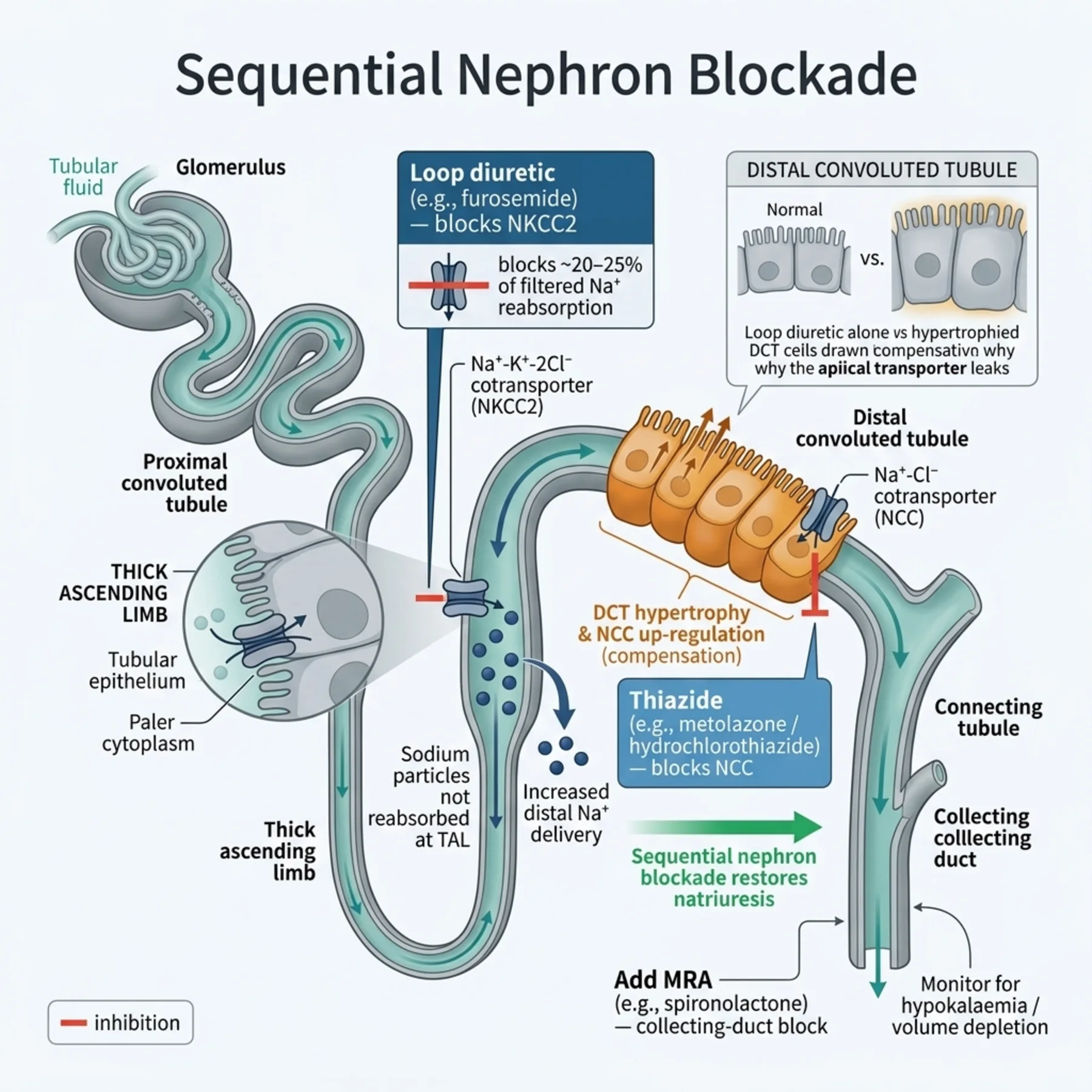
Oedema, being sodium-content excess, is treated by reducing sodium input and increasing sodium output, on top of treating the underlying disease. Sodium restriction reduces the input. Diuretics increase the output: the loop diuretics are the mainstay, blocking sodium reabsorption in the thick ascending limb; a thiazide added to a loop produces sequential nephron blockade that overcomes the distal compensation limiting loop efficacy; and a mineralocorticoid antagonist is particularly valuable in the aldosterone-driven underfill states of heart failure and cirrhosis. Diuretic resistance — a blunting of response — is common and has identifiable mechanisms (the braking phenomenon, distal tubular hypertrophy, hypoalbuminaemia reducing delivery, poor gut absorption, and reduced renal function), and it is managed by adequate dosing, the intravenous route, combination (sequential blockade), and addressing the contributing factors. For oedema refractory to all of this, mechanical fluid removal by ultrafiltration or dialysis is the backstop. The treatment is, in essence, restrict the sodium and remove the sodium.
— The over-diuresis trap in underfill states
The underfill paradox has a treatment hazard that the opening chapter foreshadowed. In a patient who is totally overloaded but has a low effective arterial blood volume — and often an intravascularly depleted compartment behind the oedema — aggressive diuresis can drop the effective arterial blood volume further, precipitating prerenal acute kidney injury and worsening the very perfusion the body was trying to defend. So in heart failure, cirrhosis, and nephrotic syndrome, diuresis is titrated carefully against renal function and volume status, removing fluid at a rate the intravascular space can refill from the oedema, and slowing or pausing when the kidney function deteriorates. The art is to remove the excess sodium and water without collapsing the effective arterial blood volume — which is why oedema in these states is treated thoughtfully rather than with maximal diuretics, and why the same urine that signals avid retention also warns against over-diuresis.
— Where the mechanisms meet the bedside
The payoff of the effective-arterial-blood-volume model is that it unifies what otherwise looks contradictory. The depleted patient and the underfill-oedema patient share a kidney phenotype — avid sodium retention, low urine sodium — because both have a low effective arterial blood volume, yet one needs fluid and the other needs fluid removed, and only the volume model explains why. The overfill patient retains sodium for a different reason — the kidney cannot excrete it — and is treated by forcing excretion. Localised oedema is not a sodium problem at all. Reading every volume disorder through what the body senses (effective arterial blood volume) and how it responds (the retaining and excreting effectors) turns a confusing array of presentations into a single, coherent framework, and prevents the cardinal error of treating the underfill-oedema kidney's avid retention as though it signalled a need for salt.
| 04 | PHASE A · LEVEL 4 · ORIENTATION & KNOWLEDGE Reference Tables |
Table 3.1 — Sensing and defending effective arterial blood volume
| Component | Role |
| Sensors | Arterial baroreceptors; juxtaglomerular apparatus; atrial stretch receptors |
| RAAS | Renin → angiotensin II (vasoconstriction, Na reabsorption) → aldosterone (distal Na) |
| Sympathetic system | Vasoconstriction, sodium retention, renin release |
| ADH | Water retention (volume and osmotic stimuli) |
| Natriuretic peptides | ANP/BNP — natriuresis and vasodilation when volume is high (oppose RAAS) |
Table 3.2 — Causes of volume depletion
| Route | Examples |
| Gastrointestinal | Vomiting, diarrhoea, drainage, fistulae |
| Renal | Diuretics, osmotic diuresis, salt-wasting, adrenal insufficiency |
| Skin | Sweating, burns |
| Third-space | Pancreatitis, bowel obstruction |
| Haemorrhage | Replace with blood |
Table 3.3 — Confirming avid sodium retention (depletion)
| Finding | Detail |
| Urine sodium | Low (typically < 20) with intact kidneys |
| FENa / FEUrea | Low |
| Urine | Concentrated, oliguric |
| Prerenal azotaemia | Urea rises out of proportion to creatinine |
| Clinical signs | Low JVP, postural change, weight loss — integrated (Chapter 1) |
Table 3.4 — Types of oedema
| Type | Mechanism | Urine sodium / clue |
| Underfill | Low EABV (heart failure, cirrhosis, nephrotic) → RAAS Na retention | Low — the overload-with-avidity paradox |
| Overfill | Primary renal Na retention (AKI/CKD, nephritic) | Variable / higher |
| Localised | Venous / lymphatic obstruction | Not a sodium problem |
Table 3.5 — Diuretics and resistance
| Agent / issue | Detail |
| Loop diuretic | Mainstay — blocks the thick ascending limb |
| Thiazide | Add for sequential nephron blockade (resistance) |
| Mineralocorticoid antagonist | Aldosterone-driven states (heart failure, cirrhosis) |
| Resistance mechanisms | Braking, distal hypertrophy, hypoalbuminaemia, poor absorption, CKD |
| Refractory | High-dose/IV loop, combination; ultrafiltration as backstop |
Table 3.6 — The treatment balance
| Disorder | Approach |
| Depletion | Replace deficit with matching fluid; treat the cause; reassess |
| Oedema | Treat cause; restrict sodium; remove sodium (diuretics) |
| Underfill caution | Titrate diuresis — over-diuresis lowers EABV → prerenal AKI |
| Localised oedema | Treat the obstruction — not a sodium problem |
| 05 | PHASE B · LEVEL 5 · VISUALISE & MAP Imaging & Flowchart Specifications |
| 06 | PHASE B · LEVEL 6 · VISUALISE & MAP Concept Maps |
Each chain runs from physiology to a named bedside action; read the arrows as “leads to.”
Sensing EABV. Baroreceptors/JGA/atrial stretch sense effective arterial blood volume (not total volume) → low EABV activates RAAS/sympathetic/ADH, high EABV releases natriuretic peptides → ACTION: read every volume disorder through what the body senses and how it responds.
Depletion. Fluid loss → ↓ EABV → RAAS/sympathetic/ADH → avid sodium and water retention (low urine Na, prerenal azotaemia) → ACTION: confirm avidity, replace the deficit with matching fluid, treat the cause.
Underfill oedema. Heart failure/cirrhosis/nephrotic → low EABV despite total overload → kidney retains sodium (low urine Na) → oedema worsens → ACTION: treat the cause, restrict and remove sodium, but titrate diuresis to protect EABV.
Overfill. Primary renal sodium retention (AKI/CKD, nephritic) → kidney cannot excrete sodium → volume expands → ACTION: force excretion (diuretics) and treat the renal cause.
Diuretic resistance. Loop blockade → distal compensation (hypertrophy/braking) + hypoalbuminaemia/poor absorption/CKD → blunted response → ACTION: adequate/IV dosing, add a thiazide (sequential blockade), address contributors; ultrafiltration if refractory.
| 07 | PHASE B · LEVEL 7 · VISUALISE & MAP Decision Pathways |
| R1 | IF assessing a volume disorder, THEN think in terms of effective arterial blood volume — what the body senses — not total extracellular volume alone. |
| R2 | IF a patient is volume-depleted, THEN expect avid sodium retention (low urine sodium, prerenal azotaemia), replace the deficit with matching fluid, and treat the cause. |
| R3 | IF replacing losses, THEN match the composition (blood for haemorrhage; replace chloride/potassium for vomiting) — not water alone. |
| R4 | IF a patient has generalised oedema, THEN classify it as underfill or overfill and treat the underlying cause, restricting and removing sodium. |
| R5 | IF oedema is localised, THEN treat the venous or lymphatic obstruction — it is not a whole-body sodium problem. |
| R6 | IF a diuretic response is inadequate, THEN manage resistance — adequate/IV dosing, sequential nephron blockade, and address hypoalbuminaemia, absorption, and renal function. |
| R7 | IF diuresing an underfill state, THEN titrate against renal function — over-diuresis lowers effective arterial blood volume and causes prerenal acute kidney injury. |
| R8 | IF a low urine sodium is found, THEN interpret it as avid retention — from depletion OR underfill oedema — not automatically as a need for salt. |
| 08 | PHASE C · LEVEL 8 · CLINICAL REASONING Clinical Cases |
| CASE 1 | AVID FOR SALT Reading depletion Volume depletion from losses |
Presentation
A patient with several days of vomiting and diarrhoea is hypotensive and oliguric, with a urine sodium of 8, a low fractional excretion of sodium, and a urea raised out of proportion to creatinine.
❖ Pause and reflect What do these findings tell you, and what does the patient need? |
Analysis
This is volume depletion with avid sodium retention. The losses have lowered the effective arterial blood volume, activating the retaining effectors, so the kidney conserves sodium — hence the urine sodium of 8, the low fractional excretion, and the prerenal azotaemia. The patient needs the deficit replaced and the losses stemmed. Because the vomiting and diarrhoea lose chloride, potassium, and bicarbonate as well as water, the replacement must match the composition, not be water alone.
Plan
Replace the deficit with isotonic (balanced) crystalloid, matching ongoing losses in composition and replacing potassium, titrate against the response, and treat the cause of the gastrointestinal losses. Reassess as volume is restored.
Teaching point
A low urine sodium with prerenal azotaemia signals avid retention from depletion — replace the deficit with matching fluid and treat the cause.
Cross-reference
Exercises rules R2 and R3; the depletion concept map; Tables 3.2 and 3.3; fluids in Chapter 2.
| CASE 2 | OVERLOADED, YET RETAINING The underfill paradox Oedema in heart failure |
Presentation
A patient with heart failure is grossly oedematous with a raised jugular venous pressure, yet the urine sodium is low at 10. A colleague is confused: 'how can the kidney be retaining sodium when the patient is so overloaded?'
❖ Pause and reflect Why does an overloaded kidney retain sodium so avidly? |
Analysis
This is the underfill paradox. The patient's total body sodium and water are high — hence the oedema — but the failing heart's low cardiac output reduces the effective arterial blood volume, the quantity the kidney actually senses. Reading a low effective arterial blood volume, the kidney activates RAAS and retains sodium avidly, giving the low urine sodium, and that retention worsens the oedema. The patient is overloaded and 'sensing depletion' at once, which only the effective-arterial-blood-volume model explains.
Plan
Treat the heart failure (improving cardiac output raises the effective arterial blood volume), restrict sodium, and remove sodium with diuretics — but titrate against renal function. Do not misread the low urine sodium as a need for salt.
Teaching point
In underfill oedema the kidney retains sodium despite overload because the effective arterial blood volume is low — treat the cause and remove sodium, don't give it.
Cross-reference
Exercises rules R4 and R8; the underfill concept map; Figure 3.2; Table 3.4.
| CASE 3 | THE DIURETIC THAT STOPPED WORKING Sequential blockade Diuretic resistance |
Presentation
A patient with refractory oedema on a high oral dose of a loop diuretic has a poor diuretic response and persistent fluid overload. The team keeps increasing the oral loop dose without effect.
❖ Pause and reflect Why has the loop diuretic stopped working, and what should change? |
Analysis
This is diuretic resistance. Chronic loop-diuretic use drives distal tubular hypertrophy and compensatory sodium reabsorption (the braking phenomenon) that blunts the response, and oral absorption and hypoalbuminaemia may further limit delivery. Simply escalating the oral loop dose does little. The response is to ensure adequate dosing by the intravenous route, add a thiazide for sequential nephron blockade of the distal compensation, and address contributors such as hypoalbuminaemia and renal function — with ultrafiltration as the backstop if truly refractory.
Plan
Give the loop diuretic intravenously at an adequate dose, add a thiazide for sequential blockade, correct contributing factors, and monitor electrolytes and renal function; consider ultrafiltration if refractory. Stop merely escalating the oral loop.
Teaching point
Diuretic resistance is overcome by adequate/IV dosing and sequential nephron blockade (add a thiazide), not by escalating the oral loop alone.
Cross-reference
Exercises rule R6; the diuretic-resistance concept map; Figure 3.3; Table 3.5.
| CASE 4 | DIURESED INTO AKI Protecting EABV Over-diuresis in underfill |
Presentation
A cirrhotic patient with tense ascites and oedema is diuresed aggressively to clear the fluid quickly. The creatinine rises sharply and the patient develops acute kidney injury.
❖ Pause and reflect Why did aggressive diuresis cause acute kidney injury here? |
Analysis
Aggressive diuresis outpaced the rate at which the intravascular space could refill from the oedema and ascites, dropping the already-low effective arterial blood volume further and precipitating prerenal acute kidney injury — a particular hazard in the underfill states, where the effective arterial blood volume is the limiting factor. The fluid needed to come off more slowly, at a rate the intravascular compartment could tolerate, with renal function watched.
Plan
Slow or pause the diuresis, allow the effective arterial blood volume to recover, and resume at a gentler rate titrated against renal function. In underfill states, remove fluid at a pace the intravascular space can refill, not as fast as possible.
Teaching point
Over-diuresis in underfill states lowers effective arterial blood volume and causes prerenal AKI — titrate diuresis against renal function.
Cross-reference
Exercises rule R7; the underfill concept map; Table 3.6; intravascular-versus-interstitial divergence in Chapter 1.
| 09 | PHASE C · LEVEL 9 · CLINICAL REASONING Clinical Implications |
One triad per mechanism the narrative exposed: the physiology, why it matters, and the bedside move.
MECHANISM The body senses effective arterial blood volume — arterial fullness — not total extracellular volume. |
WHY IT MATTERS This is why an overloaded patient can have a kidney behaving as if depleted. |
ACTION Read every volume disorder through the effective arterial blood volume. |
MECHANISM A low effective arterial blood volume activates RAAS, the sympathetic system, and ADH. |
WHY IT MATTERS Depletion therefore produces avid sodium retention with a low urine sodium and prerenal azotaemia. |
ACTION Confirm avidity, replace the deficit with matching fluid, and treat the cause. |
MECHANISM In heart failure, cirrhosis, and nephrotic syndrome the effective arterial blood volume is low despite total overload. |
WHY IT MATTERS The kidney retains sodium avidly and worsens the oedema — the underfill paradox. |
ACTION Treat the cause and remove sodium; do not misread the low urine sodium as a need for salt. |
MECHANISM Chronic loop-diuretic use drives distal compensation that blunts the response. |
WHY IT MATTERS Escalating the oral loop alone fails to overcome this resistance. |
ACTION Dose adequately/intravenously and add a thiazide for sequential nephron blockade. |
MECHANISM In underfill states the intravascular space refills from the oedema only so fast. |
WHY IT MATTERS Diuresing faster than it can refill drops the effective arterial blood volume and causes prerenal AKI. |
ACTION Titrate diuresis against renal function; remove fluid at a tolerable pace. |
| 10 | PHASE C · LEVEL 10 · CLINICAL REASONING Clinical Pearls |
| Volume disorders are sodium-content disorders (the ECF-volume axis). | The body senses EFFECTIVE arterial blood volume, not total ECF. |
| Sensors: arterial baroreceptors, JGA, atrial stretch. | Low EABV → RAAS + sympathetic + ADH (retain Na/water). |
| Natriuretic peptides (ANP/BNP) oppose — natriuresis when volume is high. | Depletion → avid retention: low urine Na (<20), low FENa, prerenal azotaemia. |
| Match replacement to losses (blood for haemorrhage; K/Cl for vomiting). | Oedema = sodium-content excess. |
| Underfill (HF/cirrhosis/nephrotic): low EABV despite overload → RAAS retention (low urine Na). | Overfill: primary renal Na retention (AKI/CKD, nephritic). |
| Generalised oedema = Na retention; localised = obstruction (not Na). | Treat oedema: cause + restrict Na + remove Na (diuretics). |
| Loop = mainstay; add thiazide for sequential blockade; MRA in aldosterone-driven states. | Diuretic resistance: braking, distal hypertrophy, hypoalbuminaemia, CKD — IV/combine/address. |
| Over-diuresis in underfill → ↓EABV → prerenal AKI — titrate. | A low urine Na means avid retention (depletion OR underfill) — not a need for salt. |
| 11 | PHASE D · LEVEL 11 · SAFETY & EVIDENCE Red Flags & Never-Do |
Panel A — Red flags
| ▲ | A low urine sodium in an oedematous patient — underfill avidity, not a need for salt; treat the cause and remove sodium. |
| ▲ | A rising creatinine during diuresis of heart failure or cirrhosis — over-diuresis lowering EABV; slow down. |
| ▲ | Escalating oral loop diuretic with no response — diuretic resistance; switch to IV and add sequential blockade. |
| ▲ | Prerenal azotaemia with a low urine sodium — depletion; replace the deficit and treat the cause. |
| ▲ | Localised limb oedema treated with systemic diuretics — it's an obstruction problem, not sodium overload. |
Panel B — Never do
| ✖ NEVER — read a low urine sodium as a need for salt without considering underfill oedema. |
| ✖ NEVER — diurese an underfill state without titrating against renal function. |
| ✖ NEVER — replace losses with water alone when the loss contains electrolytes. |
| ✖ NEVER — treat localised obstructive oedema as a whole-body sodium problem. |
| 12 | PHASE D · LEVEL 12 · SAFETY & EVIDENCE Common Pitfalls |
Pitfall 1 — Misreading the low urine sodium
| ✖ | WRONG Giving salt to an oedematous patient because the urine sodium is low. |
| ✓ | RIGHT Recognising underfill avidity and removing sodium instead. |
| ✉ | WHY A low urine sodium signals avid retention, not a need for salt. |
Pitfall 2 — Over-diuresing underfill
| ✖ | WRONG Aggressively diuresing heart failure or cirrhosis to clear fluid fast. |
| ✓ | RIGHT Titrating diuresis against renal function and intravascular refill. |
| ✉ | WHY Over-diuresis lowers effective arterial blood volume and causes prerenal AKI. |
Pitfall 3 — Escalating the oral loop
| ✖ | WRONG Increasing the oral loop diuretic dose repeatedly with no response. |
| ✓ | RIGHT Switching to IV and adding a thiazide for sequential blockade. |
| ✉ | WHY Distal compensation and poor absorption blunt the oral loop. |
Pitfall 4 — Replacing water alone
| ✖ | WRONG Replacing vomiting or diarrhoeal losses with water or dextrose alone. |
| ✓ | RIGHT Matching the composition — replacing chloride and potassium too. |
| ✉ | WHY The losses contain electrolytes, not just water. |
Pitfall 5 — Treating localised oedema systemically
| ✖ | WRONG Giving systemic diuretics for a swollen obstructed limb. |
| ✓ | RIGHT Treating the venous or lymphatic obstruction. |
| ✉ | WHY Localised oedema is a regional Starling problem, not sodium overload. |
| 13 | PHASE D · LEVEL 13 · SAFETY & EVIDENCE Evidence Grading |
GRADE A HIGH CONFIDENCE The effect is real and the estimate is stable. RCTs at low risk of bias; multiple concordant prospective cohorts; meta-analyses. |
GRADE B MODERATE CONFIDENCE The effect is likely real but may shift with new data. Observational studies, registries, mechanistic human studies. |
GRADE C LOW CONFIDENCE Rests on physiology, reasoning, or consensus rather than outcomes. Pathophysiological reasoning; extrapolation; consensus without outcomes. |
Graded statements (by evidence type)
| Statement | Grade | Basis (evidence type) |
| Effective arterial blood volume, not total volume, is the sensed quantity. | A | Established physiology |
| Low EABV activates RAAS, sympathetic, and ADH with avid sodium retention. | A | Established physiology |
| Underfill states cause oedema with avid renal sodium retention. | A | Physiology and clinical observation |
| A low urine sodium indicates avid retention (depletion or underfill). | A | Established physiology |
| Sequential nephron blockade overcomes loop-diuretic resistance. | B | Mechanistic and clinical data |
| Over-diuresis in underfill states causes prerenal AKI. | B | Clinical observation |
| Ultrafiltration removes fluid in diuretic-refractory overload. | B | Clinical and trial data |
| 14 | PHASE E · LEVEL 14 · PATIENT DECISIONS Absolute Risk in Natural Frequency |
Natural-frequency estimates for orientation, from volume-disorder management; they vary with the underlying disease. They convey the size of the decisions, expressed per 100 comparable patients.
| Per 100 patients… | Outcome | Roughly how many | See |
| Depleted, given matching fluid and cause treated | Recover volume and renal function | Most | L13 row 2 |
| With loop resistance given sequential blockade | Achieve effective diuresis | More than with loop escalation alone | L13 row 5 |
| With underfill oedema over-diuresed | Develop prerenal AKI | More than the carefully titrated | L13 row 6 |
| With diuretic-refractory overload given ultrafiltration | Achieve fluid removal | Most | L13 row 7 |
★ How to read these Read these as orientation, not promises; outcomes depend heavily on the underlying disease. The stable signals: matched replacement restores depletion, sequential blockade overcomes resistance, and over-diuresing underfill harms the kidney. Communicate them as people out of 100, not as a hazard ratio. |
| 17 | PHASE F · LEVEL 17 · APPLY & TEST Documentation Templates |
Paste-ready notes. Tick the boxes that apply and delete the rest; make the EABV reasoning and the diuresis caution explicit.
Template 1 — Volume depletion assessment and replacement
Template 2 — Oedema and diuretic management
| 18 | PHASE F · LEVEL 18 · APPLY & TEST Cheat Sheet |
| Volume disorders = sodium-content (ECF-volume) disorders. | Body senses EABV, not total ECF. |
| Sensors: baroreceptors, JGA, atrial stretch. | Low EABV → RAAS + SNS + ADH (retain). |
| Natriuretic peptides oppose (natriuresis when high). | Depletion: low urine Na (<20), low FENa, prerenal azotaemia. |
| Match replacement to losses (blood/K/Cl). | Oedema = sodium excess. |
| Underfill (HF/cirrhosis/nephrotic): ↓EABV despite overload → retention (low urine Na). | Overfill: primary renal Na retention. |
| Generalised = Na; localised = obstruction. | Treat oedema: cause + restrict Na + remove Na. |
| Loop mainstay; +thiazide (sequential); MRA in aldosterone states. | Resistance: IV/adequate dose, combine, address contributors. |
| Over-diuresis in underfill → prerenal AKI — titrate. | Low urine Na = avid retention, not a need for salt. |
| 19 | PHASE F · LEVEL 19 · APPLY & TEST Flashcards |
| CARD 1 | Q. What quantity does the body sense in volume regulation? A. The effective arterial blood volume — the fullness of the arterial circulation perfusing tissues — not the total extracellular volume. DETAILED. This is why an overloaded patient can have a kidney behaving as if depleted. CLINICAL. Read every volume disorder through the effective arterial blood volume. |
| CARD 2 | Q. What defends a low effective arterial blood volume? A. RAAS (renin–angiotensin–aldosterone), the sympathetic nervous system, and ADH retain sodium and water; the natriuretic peptides oppose this when volume is high. DETAILED. These effectors are activated in depletion and in underfill oedema. CLINICAL. Interpret retention through these effectors. |
| CARD 3 | Q. How does volume depletion present biochemically? A. Avid sodium retention — a low urine sodium (under 20), a low fractional excretion of sodium or urea, concentrated oliguric urine, and prerenal azotaemia. DETAILED. The low effective arterial blood volume drives the retention. CLINICAL. Replace the deficit with matching fluid and treat the cause. |
| CARD 4 | Q. What is the underfill paradox in oedema? A. In heart failure, cirrhosis, and nephrotic syndrome the effective arterial blood volume is low despite total overload, so the kidney retains sodium avidly (low urine sodium), worsening the oedema. DETAILED. The patient is overloaded yet the kidney senses depletion. CLINICAL. Treat the cause and remove sodium; don't give salt for the low urine sodium. |
| CARD 5 | Q. How do underfill and overfill oedema differ? A. Underfill: low effective arterial blood volume drives renal sodium retention (low urine sodium). Overfill: the kidney cannot excrete sodium (primary renal retention), and volume expands directly. DETAILED. Localised oedema is obstruction, not a sodium problem. CLINICAL. Classify the oedema and treat the underlying cause. |
| CARD 6 | Q. How is oedema treated? A. Treat the underlying cause, restrict sodium input, and remove sodium with diuretics — loop as the mainstay, a thiazide for sequential nephron blockade, and a mineralocorticoid antagonist in aldosterone-driven states. DETAILED. It is fundamentally restrict-and-remove sodium. CLINICAL. Address cause, restrict, and diurese. |
| CARD 7 | Q. How is diuretic resistance overcome? A. By ensuring adequate dosing (often intravenous), adding a thiazide for sequential nephron blockade of the distal compensation, and addressing hypoalbuminaemia, poor absorption, and renal function — with ultrafiltration as a backstop. DETAILED. Escalating the oral loop alone usually fails. CLINICAL. Dose adequately, combine, and address contributors. |
| CARD 8 | Q. Why can over-diuresis in underfill states cause AKI? A. The intravascular space refills from the oedema only so fast; diuresing faster drops the already-low effective arterial blood volume and precipitates prerenal acute kidney injury. DETAILED. Underfill states are limited by the effective arterial blood volume. CLINICAL. Titrate diuresis against renal function. |
| 20 | PHASE F · LEVEL 20 · APPLY & TEST One-Minute Preceptor |
| SCENE 1 | The intern who wants to give salt |
GET A COMMITMENT. “The urine sodium is low in this oedematous patient, and you want to give saline — why?”
PROBE FOR EVIDENCE. “Low urine sodium means salt depletion” — ask: “Can an overloaded kidney retain sodium avidly, and why?”
TEACH A GENERAL RULE. In underfill oedema the effective arterial blood volume is low despite overload, so the kidney retains sodium — the low urine sodium signals avidity, not a need for salt.
REINFORCE WHAT WAS RIGHT. Noticing the low urine sodium was a good observation.
CORRECT A MISTAKE. Treat the cause and remove sodium — don't give it.
| SCENE 2 | The resident escalating the oral loop |
GET A COMMITMENT. “You've pushed the oral furosemide dose up three times with no response — what now?”
PROBE FOR EVIDENCE. “Higher doses should work” — ask: “What distal compensation limits a loop diuretic, and how is it overcome?”
TEACH A GENERAL RULE. Chronic loop use drives distal hypertrophy that blunts the response; sequential nephron blockade with a thiazide, and adequate IV dosing, overcome it — not endless oral escalation.
REINFORCE WHAT WAS RIGHT. Recognising the inadequate response was correct.
CORRECT A MISTAKE. Switch to IV, add a thiazide, and address contributors.
| 22 | PHASE F · LEVEL 22 · APPLY & TEST Board-Style Questions |
| Q 01 | What does the body actually sense in regulating volume? |
| A | Total extracellular volume |
| B | Effective arterial blood volume |
| C | Intracellular volume |
| D | Plasma osmolality |
Rationale Volume regulation responds to the effective arterial blood volume — arterial fullness — not total ECF, which is why underfill oedema occurs (Figure 3.1, rule R1). A, C, and D are not the sensed quantity. |
| Q 02 | Which findings indicate avid sodium retention from volume depletion? |
| A | High urine sodium, high FENa |
| B | Low urine sodium, low FENa, prerenal azotaemia |
| C | Dilute urine |
| D | High natriuretic peptides |
Rationale Depletion lowers EABV, driving avid retention: low urine sodium, low fractional excretion, prerenal azotaemia (Table 3.3, rule R2). A and C are the opposite; D opposes retention. |
| Q 03 | An oedematous heart-failure patient has a urine sodium of 10. This reflects: |
| A | Salt depletion needing saline |
| B | Underfill — low EABV driving avid renal sodium retention despite overload |
| C | Overfill from renal disease |
| D | A laboratory error |
Rationale Low cardiac output lowers the effective arterial blood volume, so the kidney retains sodium despite total overload — the underfill paradox (case 2, Figure 3.2). A misreads it; C and D are incorrect. |
| Q 04 | How do underfill and overfill oedema differ? |
| A | They are identical |
| B | Underfill = low EABV driving retention; overfill = primary renal inability to excrete sodium |
| C | Overfill has a low urine sodium always |
| D | Underfill is localised |
Rationale Underfill is RAAS-driven retention from low EABV; overfill is the kidney's inability to excrete sodium (Table 3.4). A, C, and D mischaracterise them. |
| Q 05 | A patient on a high oral loop diuretic has no response. The best next step is to: |
| A | Keep increasing the oral dose |
| B | Give it intravenously and add a thiazide for sequential nephron blockade |
| C | Stop all diuretics |
| D | Add more sodium |
Rationale Distal compensation and absorption limit the oral loop, so IV dosing plus sequential blockade overcomes resistance (case 3, Figure 3.3, rule R6). A fails; C and D worsen overload. |
| Q 06 | Why must diuresis be titrated in underfill states? |
| A | It never causes harm |
| B | Over-diuresis lowers effective arterial blood volume and causes prerenal AKI |
| C | Diuretics don't work in underfill |
| D | To raise the urine sodium |
Rationale The intravascular space refills only so fast, so over-diuresis drops EABV and harms the kidney (case 4, rule R7). A is false; C and D are incorrect. |
| Q 07 | Localised oedema of one limb from venous obstruction should be treated by: |
| A | Systemic diuretics |
| B | Treating the venous/lymphatic obstruction — it is not a sodium problem |
| C | Salt loading |
| D | Fluid restriction |
Rationale Localised oedema is a regional Starling problem, not whole-body sodium overload (rule R5, Table 3.4). A, C, and D misapply systemic sodium logic. |
| Q 08 | Vomiting and diarrhoeal losses should be replaced with: |
| A | Water alone |
| B | A fluid matching the composition of the losses (with potassium and chloride) |
| C | 5% dextrose only |
| D | No replacement |
Rationale The losses contain electrolytes, so replacement must match composition, not be water alone (rule R3, case 1). A and C under-replace electrolytes; D ignores the deficit. |
| Q 09 | Across 100 patients with loop-diuretic resistance, adding sequential nephron blockade versus escalating the oral loop: |
| A | Helps fewer |
| B | Achieves effective diuresis in more |
| C | Makes no difference |
| D | Worsens outcomes |
Rationale Sequential blockade overcomes the distal compensation that oral escalation cannot (L14, L13 row 5). A, C, and D contradict the mechanism. |