Chapter Preamble
Signals declared
Sig-D — Diagnostic (primary). Interpret the divalent ions with their regulators — the PTH that splits hypercalcaemia, the magnesium behind refractory hypocalcaemia, the refeeding behind hypophosphataemia.
Sig-T — Therapeutic (strong). Volume-first hypercalcaemia treatment, calcium and magnesium replacement, phosphate handling, and the calcium antagonism of hypermagnesaemia.
Sig-M — Mechanistic (strong). The PTH–vitamin D–FGF23 axes that govern calcium and phosphate, and the magnesium that conditions both potassium and calcium.
Levels populated and omitted
Populated (19): L1–L14, L17–L20, L22. The mechanistic signal fires the concept maps (L6) and triads (L9); the therapeutic signal fires the absolute-risk table (L14) and templates (L17); the diagnostic signal drives the tables, rules, cases, pitfalls, and board items.
L15 / L16 preference-sensitive map and SDM scripts — omitted. No Sig-E; correcting the divalent ions is effective care.
L21 reflective prompts — omitted. No Sig-E/V; the cross-ion tensions (the magnesium dependence) are worked through the cases and pitfalls.
| 01 | PHASE A · LEVEL 1 · ORIENTATION & KNOWLEDGE Learning Objectives |
By the end of this chapter you should be able to:
Describe the PTH, vitamin D, and FGF23 axes that regulate calcium and phosphate.
Use the PTH level to split hypercalcaemia into PTH-mediated and PTH-independent causes.
Treat hypercalcaemia, starting with volume repletion.
Diagnose and treat hypocalcaemia, including the magnesium that makes it refractory.
Recognise and manage hyper- and hypophosphataemia, including refeeding hypophosphataemia.
Diagnose and treat hypomagnesaemia and its downstream effects on potassium and calcium.
Recognise and treat hypermagnesaemia, including calcium antagonism.
Correct the corrected calcium for albumin.
| 02 | PHASE A · LEVEL 2 · ORIENTATION & KNOWLEDGE Executive Summary |
The divalent ions — calcium, phosphate, and magnesium — are governed by overlapping axes: PTH and vitamin D for calcium, PTH and FGF23 for phosphate, and renal handling for magnesium.
Ionised calcium is the active fraction; the total calcium is corrected for albumin.
Hypercalcaemia is split by the PTH: a high or inappropriately normal PTH indicates PTH-mediated disease (primary hyperparathyroidism, the commonest outpatient cause), while a suppressed PTH indicates PTH-independent causes (malignancy, the commonest inpatient cause, and others).
Hypercalcaemia is treated first with intravenous saline to rehydrate and promote calciuresis, then with calcitonin, bisphosphonates, or denosumab, and the cause is addressed.
Hypocalcaemia comes from hypoparathyroidism, vitamin D deficiency, CKD, hyperphosphataemia, and hypomagnesaemia, and causes neuromuscular excitability and a prolonged QT.
Severe or symptomatic hypocalcaemia is treated with intravenous calcium; chronic hypocalcaemia with oral calcium and vitamin D — and magnesium must be replaced, or the hypocalcaemia is refractory.
Hyperphosphataemia is mostly a CKD problem (binders, dialysis) but also follows tumour lysis and rhabdomyolysis.
Hypophosphataemia comes from redistribution (refeeding above all, insulin, respiratory alkalosis), renal loss, and poor intake; severe hypophosphataemia is dangerous, and refeeding hypophosphataemia must be anticipated.
Hypomagnesaemia, from gastrointestinal and renal losses, causes refractory hypokalaemia and hypocalcaemia and predisposes to torsades de pointes.
Hypomagnesaemia is treated by replacing magnesium — intravenously when severe — and treating the cause.
Hypermagnesaemia occurs with renal failure and a magnesium load, causing hyporeflexia, hypotension, and at high levels respiratory and cardiac depression.
Hypermagnesaemia is treated by stopping the magnesium, giving calcium as an antagonist, and dialysis in renal failure.
Throughout, the ions are interlinked: magnesium conditions potassium and calcium, and calcium and phosphate move together through their shared regulators.
| 03 | PHASE A · LEVEL 3 · ORIENTATION & KNOWLEDGE Main Narrative |
The divalent ions are bound together by their regulators. PTH and vitamin D move calcium; PTH and FGF23 move phosphate; magnesium, regulated largely by the kidney, quietly conditions the handling of both potassium and calcium. Because the axes overlap, the ions are best understood together — and several of the most important bedside lessons are cross-ion: the PTH that splits hypercalcaemia, the magnesium that makes hypocalcaemia (like hypokalaemia) refractory, and the refeeding that crashes the phosphate. This chapter works each ion in both directions, with those links as the connective tissue.
— The regulatory axes
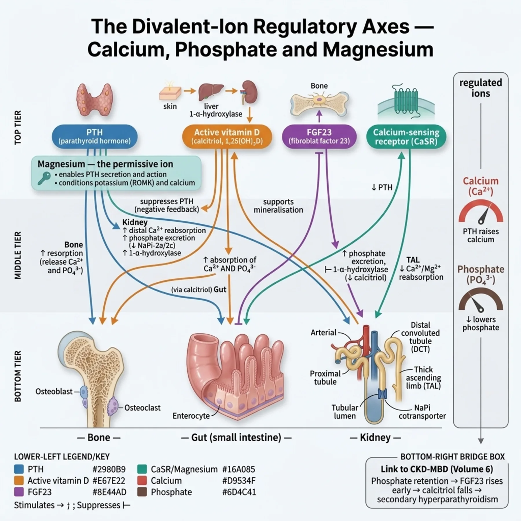
Calcium is held within a narrow range by parathyroid hormone and vitamin D. PTH raises calcium by mobilising it from bone, increasing renal reabsorption, and activating vitamin D, while activated vitamin D (calcitriol) raises calcium chiefly by increasing intestinal absorption; the calcium-sensing receptor on the parathyroid gland reads the calcium and tunes PTH secretion. The active fraction is the ionised calcium, so the total calcium is corrected for albumin (to which much of it is bound) before interpretation. Phosphate is regulated by PTH (phosphaturic) and FGF23 (phosphaturic and suppressing calcitriol), with vitamin D promoting its absorption. Magnesium is regulated mostly by renal reabsorption in the loop and distal tubule, sits largely intracellularly, and — crucially — is required for normal PTH secretion and action. These axes overlap (the CKD mineral-bone disorder of the previous volume is their joint failure), and disorders of one ion frequently disturb another.
— Hypercalcaemia: the PTH splits it
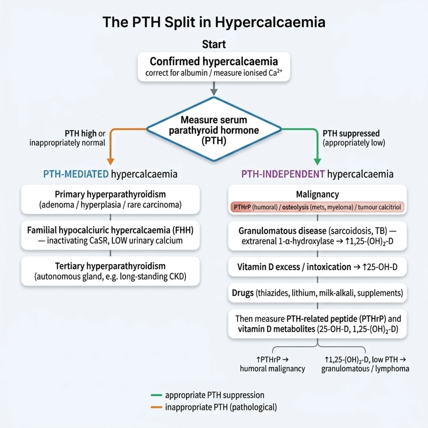
The single most useful step in hypercalcaemia is measuring the PTH, because it cleaves the long differential in two. A high or inappropriately normal PTH means the parathyroid is the driver — PTH-mediated hypercalcaemia, of which primary hyperparathyroidism is the commonest cause (and the commonest cause of hypercalcaemia in outpatients), with familial hypocalciuric hypercalcaemia and tertiary hyperparathyroidism in the group. A suppressed PTH means the hypercalcaemia is PTH-independent, and the parathyroid is appropriately switched off by the high calcium; here malignancy dominates (the commonest inpatient cause, acting via PTH-related peptide, osteolysis, or calcitriol production in lymphoma), alongside granulomatous disease (calcitriol), vitamin D toxicity, thyrotoxicosis, immobilisation, milk-alkali, and drugs such as thiazides and lithium. So one test — the PTH — directs the entire work-up, with PTH-related peptide and vitamin D metabolites measured next when the PTH is suppressed. The symptoms are the classic 'stones, bones, abdominal groans, and psychic moans,' with polyuria (a nephrogenic diabetes insipidus), volume depletion, constipation, and a short QT.
— Treating hypercalcaemia: volume first
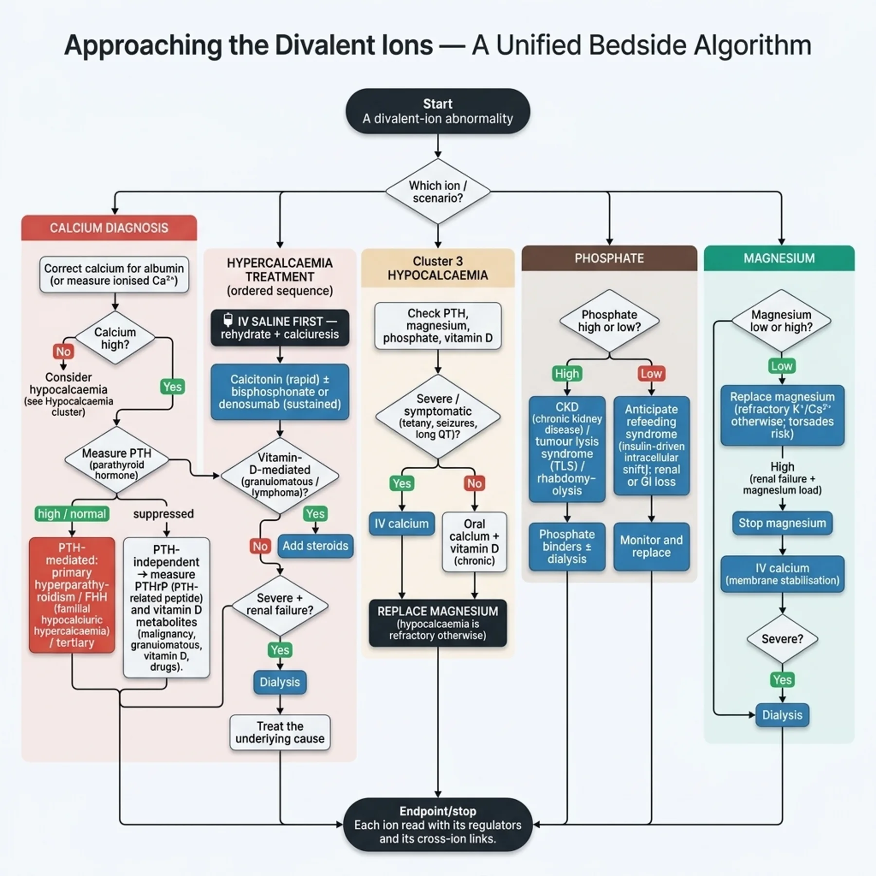
Hypercalcaemia treatment follows a logical order rooted in the physiology. The patient is almost always volume-depleted (the hypercalcaemia causes a nephrogenic diabetes insipidus and the symptoms reduce intake), so the first step is intravenous isotonic saline, which both rehydrates and promotes calciuresis. Calcitonin acts rapidly but transiently (tachyphylaxis), bridging to the slower, more durable agents. Bisphosphonates (zoledronic acid) are the mainstay for malignancy-associated hypercalcaemia, with an onset over two to four days; denosumab is used when bisphosphonates fail or in renal impairment. Glucocorticoids treat the vitamin-D-mediated hypercalcaemias (granulomatous disease, some lymphomas and myeloma). Dialysis is reserved for severe hypercalcaemia with renal failure. Loop diuretics, once routine, are now used only after volume repletion and not as a primary calciuretic — giving a loop diuretic to a volume-depleted hypercalcaemic patient worsens the depletion. And the cause is treated. The order — saline first, then the calcium-lowering agents, then the cause — is the practical heart of acute management.
— Hypocalcaemia: and the magnesium it depends on
Hypocalcaemia produces neuromuscular excitability — perioral and digital paraesthesia, tetany, the Chvostek and Trousseau signs, carpopedal spasm, and, when severe, laryngospasm, seizures, and a prolonged QT with arrhythmia. Its causes are hypoparathyroidism (commonest after thyroid or parathyroid surgery), vitamin D deficiency, CKD, hyperphosphataemia (tumour lysis, rhabdomyolysis, precipitating calcium), acute pancreatitis, citrate (massive transfusion, continuous renal replacement, plasma exchange), and hypomagnesaemia. That last cause is the cross-ion lesson, mirroring hypokalaemia exactly: magnesium is needed for PTH secretion and action, so hypomagnesaemia causes a hypocalcaemia that will not correct with calcium alone until the magnesium is replaced. Treatment is intravenous calcium gluconate for severe or symptomatic hypocalcaemia and oral calcium with vitamin D for chronic disease — but in every refractory case, the magnesium must be checked and replaced, and the cause treated. The work-up measures the corrected or ionised calcium, the PTH, magnesium, phosphate, and vitamin D together.
— Phosphate: the refeeding trap and the CKD context
Phosphate disorders split by direction. Hyperphosphataemia is, overwhelmingly, a CKD problem — the mineral-bone disorder of the previous volume, managed with binders and dialysis — but also follows tumour lysis and rhabdomyolysis (massive cellular release) and hypoparathyroidism. Hypophosphataemia, less familiar but dangerous, comes from three routes: redistribution into cells (the great example being refeeding syndrome, where reintroducing carbohydrate after starvation drives a surge of insulin that shifts phosphate intracellularly, sometimes fatally; also insulin therapy and respiratory alkalosis), renal loss (hyperparathyroidism, Fanconi syndrome, FGF23-driven tumour-induced osteomalacia), and poor intake or gastrointestinal loss (alcoholism, antacids). Severe hypophosphataemia causes muscle weakness, rhabdomyolysis, respiratory failure, haemolysis, and arrhythmia. The clinical imperative is to anticipate refeeding hypophosphataemia in the malnourished or alcoholic patient being fed, monitoring and replacing phosphate before it crashes, because the harm is preventable and the redistribution predictable.
— Magnesium: the ion behind other ions
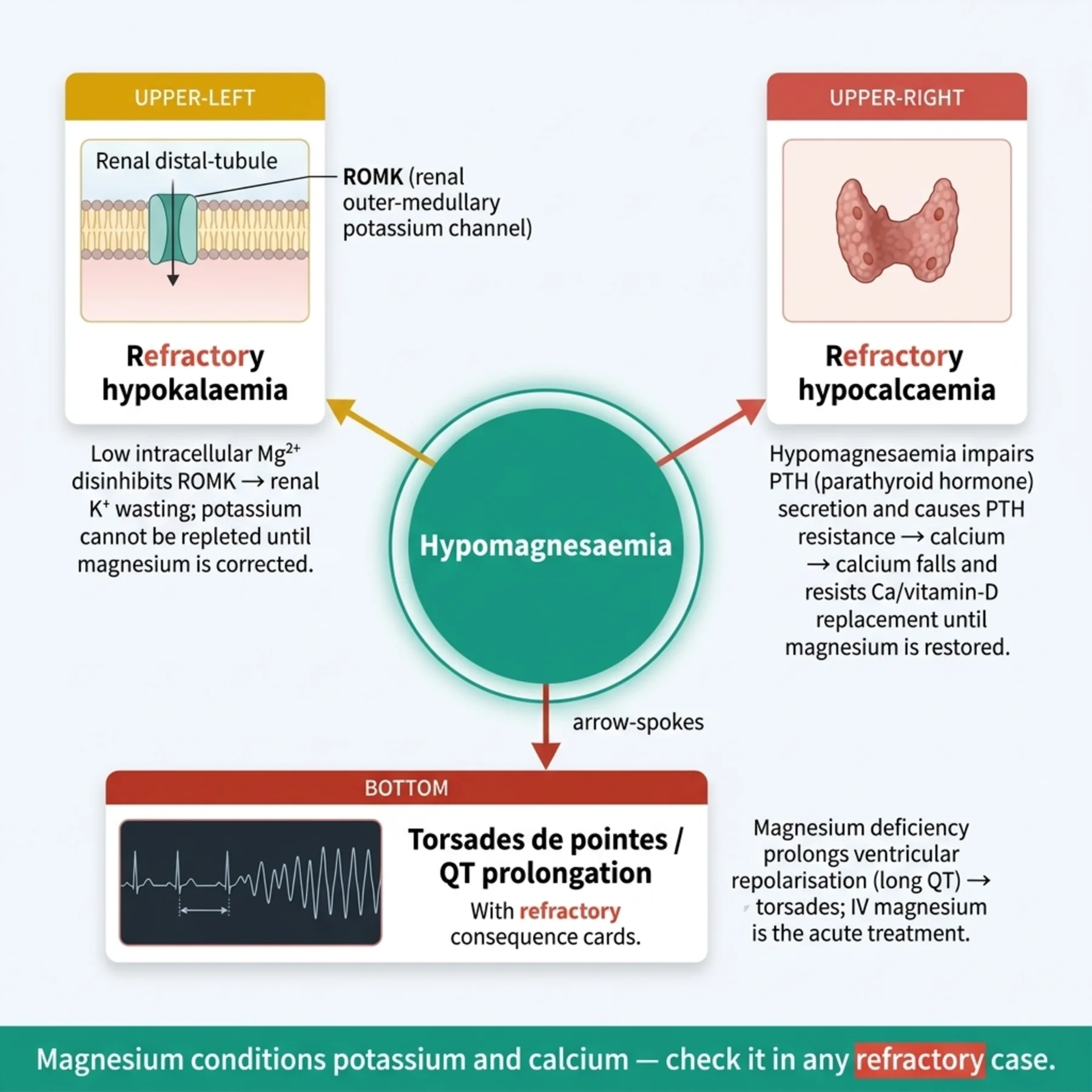
Magnesium disorders matter as much for their effects on other ions as for themselves. Hypomagnesaemia — from gastrointestinal loss (diarrhoea, chronic proton-pump inhibitors), renal loss (loop and thiazide diuretics, alcohol, aminoglycosides, cisplatin, calcineurin inhibitors, EGFR inhibitors, amphotericin), and poor intake — causes neuromuscular excitability (tetany, tremor, seizures) and arrhythmia, classically torsades de pointes with QT prolongation. But its signature is downstream: hypomagnesaemia causes refractory hypokalaemia (via ROMK disinhibition, as the potassium chapter described) and refractory hypocalcaemia (via impaired PTH), so a patient with stubborn potassium or calcium should always have the magnesium checked and replaced. Treatment is intravenous magnesium for severe or symptomatic cases, oral for chronic (limited by diarrhoea), amiloride to reduce renal magnesium loss, and treatment of the cause. Hypermagnesaemia, by contrast, is largely iatrogenic: it requires renal failure plus a magnesium load (the magnesium-containing laxatives and antacids of the CKD volume) or the magnesium given for pre-eclampsia, and it causes dose-related hyporeflexia, hypotension, and — at high levels — respiratory depression, bradycardia, and arrest. Its treatment is to stop the magnesium, give intravenous calcium as a physiological antagonist, and dialyse in renal failure.
— The connective tissue: ions move together
The unifying lesson is that the divalent ions are not independent. The PTH that diagnoses hypercalcaemia also governs phosphate and is itself disabled by low magnesium. The magnesium that seems a minor electrolyte is the hidden cause of refractory hypokalaemia and hypocalcaemia and a precipitant of torsades. The phosphate that crashes in refeeding does so because insulin moves it, just as insulin moves potassium. And in CKD all of these fail together as the mineral-bone disorder. So the practical disciplines are cross-ion: split hypercalcaemia by PTH; in any refractory hypokalaemia or hypocalcaemia, replace magnesium; anticipate refeeding hypophosphataemia; rehydrate before lowering calcium; and remember that a magnesium-containing laxative in renal failure can be lethal. Treating the divalent ions well means reading them as a connected system, with their shared regulators and their mutual dependence kept always in view.
| 04 | PHASE A · LEVEL 4 · ORIENTATION & KNOWLEDGE Reference Tables |
Table 9.1 — The regulatory axes
| Regulator | Action |
| PTH | Raises calcium (bone, renal reabsorption, activates vitamin D); phosphaturic |
| Vitamin D (calcitriol) | Raises calcium (gut absorption) and phosphate absorption |
| FGF23 | Phosphaturic; suppresses calcitriol |
| Calcium-sensing receptor | Senses calcium → tunes PTH |
| Magnesium | Renal regulation; required for PTH secretion/action; affects K and Ca |
Table 9.2 — Hypercalcaemia
| Element | Detail |
| PTH high/normal (PTH-mediated) | Primary hyperparathyroidism (commonest outpatient), FHH, tertiary |
| PTH suppressed (PTH-independent) | Malignancy (commonest inpatient — PTHrP/osteolysis/calcitriol), granulomatous, vit D toxicity, thiazides, lithium |
| Symptoms | Stones, bones, groans, moans; polyuria, dehydration, short QT |
| Treatment | Saline FIRST; calcitonin; bisphosphonate; denosumab; steroids (vit D-mediated); dialysis; treat cause |
Table 9.3 — Hypocalcaemia
| Element | Detail |
| Causes | Hypoparathyroidism (post-surgical), vitamin D deficiency, CKD, hyperphosphataemia, hypomagnesaemia, pancreatitis, citrate |
| Symptoms | Paraesthesia, tetany, Chvostek/Trousseau, laryngospasm, seizures, long QT |
| Treatment (severe) | IV calcium gluconate |
| Treatment (chronic) | Oral calcium + vitamin D |
| Always | Check and replace magnesium — refractory otherwise |
Table 9.4 — Phosphate disorders
| Disorder | Causes / treatment |
| Hyperphosphataemia | CKD (commonest), tumour lysis, rhabdomyolysis — binders, dialysis (Volume 6) |
| Hypophosphataemia — redistribution | Refeeding (major!), insulin, respiratory alkalosis |
| Hypophosphataemia — renal/GI | Hyperparathyroidism, Fanconi, FGF23; alcohol, antacids |
| Severe hypophosphataemia | Weakness, rhabdomyolysis, respiratory failure — replace; anticipate refeeding |
Table 9.5 — Magnesium disorders
| Disorder | Detail |
| Hypomagnesaemia — causes | GI (diarrhoea, PPIs), renal (diuretics, alcohol, aminoglycosides, cisplatin, CNIs) |
| Hypomagnesaemia — effects | Refractory hypokalaemia + hypocalcaemia; torsades, QT prolongation |
| Hypomagnesaemia — treatment | IV (severe), oral (chronic); amiloride reduces renal loss; treat cause |
| Hypermagnesaemia | Renal failure + Mg load (laxatives/antacids), pre-eclampsia — hyporeflexia, hypotension, respiratory/cardiac depression |
| Hypermagnesaemia — treatment | Stop Mg; IV calcium (antagonist); dialysis |
Table 9.6 — The cross-ion links
| Link | Implication |
| Magnesium → potassium | Hypomagnesaemia causes refractory hypokalaemia (replace Mg) |
| Magnesium → calcium | Hypomagnesaemia causes refractory hypocalcaemia (replace Mg) |
| Insulin → phosphate | Refeeding shifts phosphate into cells (anticipate) |
| PTH / vitamin D / FGF23 | Calcium and phosphate move through shared regulators (CKD-MBD) |
| 05 | PHASE B · LEVEL 5 · VISUALISE & MAP Imaging & Flowchart Specifications |
| 06 | PHASE B · LEVEL 6 · VISUALISE & MAP Concept Maps |
Each chain runs from physiology to a named bedside action; read the arrows as “leads to.”
Calcium regulation. PTH (bone/renal/activates vitamin D) + calcitriol (gut) raise calcium; the CaSR tunes PTH → disorders shift calcium → ACTION: correct for albumin and read calcium with its regulators.
The PTH split. Hypercalcaemia → measure PTH → high/normal (PTH-mediated, primary hyperparathyroidism) vs suppressed (PTH-independent, malignancy) → ACTION: let one test direct the whole work-up.
Magnesium hub. Hypomagnesaemia → ROMK disinhibition (refractory hypokalaemia) + impaired PTH (refractory hypocalcaemia) + QT prolongation (torsades) → ACTION: check and replace magnesium in any refractory potassium or calcium problem.
Refeeding phosphate. Carbohydrate refeeding → insulin surge → phosphate shifts into cells → dangerous hypophosphataemia → ACTION: anticipate in the malnourished/alcoholic, monitor, and replace phosphate.
Hypermagnesaemia. Renal failure + magnesium load (laxatives/antacids) → rising magnesium → hyporeflexia → respiratory/cardiac depression → ACTION: stop the magnesium, give calcium as an antagonist, and dialyse in renal failure.
| 07 | PHASE B · LEVEL 7 · VISUALISE & MAP Decision Pathways |
| R1 | IF interpreting a total calcium, THEN correct it for albumin (or measure ionised calcium) before acting. |
| R2 | IF a patient is hypercalcaemic, THEN measure PTH — high/normal indicates PTH-mediated disease, suppressed indicates PTH-independent (then check PTHrP and vitamin D metabolites). |
| R3 | IF treating hypercalcaemia, THEN give intravenous saline first to rehydrate and promote calciuresis, then calcitonin/bisphosphonate/denosumab and treat the cause. |
| R4 | IF hypocalcaemia is refractory to calcium, THEN check and replace magnesium — hypomagnesaemia impairs PTH. |
| R5 | IF severe or symptomatic hypocalcaemia is present, THEN give intravenous calcium gluconate; use oral calcium and vitamin D for chronic disease. |
| R6 | IF feeding a malnourished or alcoholic patient, THEN anticipate refeeding hypophosphataemia — monitor and replace phosphate. |
| R7 | IF potassium or calcium is refractory, THEN replace magnesium — it conditions both. |
| R8 | IF hypermagnesaemia occurs (renal failure plus a magnesium load), THEN stop the magnesium, give intravenous calcium as an antagonist, and dialyse if needed. |
| 08 | PHASE C · LEVEL 8 · CLINICAL REASONING Clinical Cases |
| CASE 1 | ONE TEST SPLITS IT Measure the PTH Hypercalcaemia work-up and treatment |
Presentation
An inpatient with known cancer has a corrected calcium of 3.2 mmol/L, is confused and dehydrated, and has a suppressed PTH. The team is unsure how to work up and treat the hypercalcaemia.
❖ Pause and reflect What does the suppressed PTH tell you, and what is the first treatment? |
Analysis
The suppressed PTH places this firmly in the PTH-independent group, and in an inpatient with cancer the overwhelmingly likely cause is malignancy-associated hypercalcaemia (via PTH-related peptide, osteolysis, or calcitriol) — confirmed by checking PTHrP and vitamin D metabolites. The first treatment is not a calcium-lowering drug but volume: he is dehydrated (the hypercalcaemia causes a nephrogenic diabetes insipidus), so intravenous saline rehydrates and promotes calciuresis, after which a bisphosphonate provides durable control.
Plan
Give intravenous saline first to rehydrate and lower the calcium, add a bisphosphonate (with calcitonin to bridge if severe), check PTHrP and vitamin D metabolites, and treat the malignancy. Avoid a loop diuretic until volume-replete.
Teaching point
Measure PTH to split hypercalcaemia (suppressed = malignancy/PTH-independent), and treat with saline first, then a calcium-lowering agent.
Cross-reference
Exercises rules R2 and R3; the PTH-split concept map; Figure 9.2; Table 9.2.
| CASE 2 | CALCIUM WON'T RISE The magnesium again Refractory hypocalcaemia |
Presentation
A patient with chronic diarrhoea has symptomatic hypocalcaemia with paraesthesia and a positive Trousseau sign. Despite repeated calcium replacement, the calcium will not correct.
❖ Pause and reflect Why won't the calcium correct, and what is the parallel with potassium? |
Analysis
The likely culprit is hypomagnesaemia. Magnesium is required for PTH secretion and action, so a low magnesium causes a hypocalcaemia that is refractory to calcium replacement until the magnesium is corrected — the exact parallel with the magnesium dependence of refractory hypokalaemia. Chronic diarrhoea wastes magnesium, and the calcium will keep failing to rise until the magnesium is replaced. Checking and replacing magnesium is the key step that has been missed.
Plan
Check and replace the magnesium, then continue calcium and vitamin D — the calcium will now correct. Treat the diarrhoea as the shared cause of both deficits.
Teaching point
Refractory hypocalcaemia, like refractory hypokalaemia, is hypomagnesaemia until proven otherwise — replace magnesium.
Cross-reference
Exercises rules R4 and R7; the magnesium-hub concept map; Figure 9.3; Tables 9.3, 9.5, 9.6; refractory hypokalaemia in Chapter 7.
| CASE 3 | THE PHOSPHATE CRASH Anticipate the refeed Refeeding hypophosphataemia |
Presentation
A severely malnourished patient with alcohol-use disorder is started on full nutritional support. Within a day the phosphate falls sharply and the patient develops muscle weakness.
❖ Pause and reflect Why did the phosphate crash, and how should it have been managed? |
Analysis
This is refeeding hypophosphataemia. Reintroducing carbohydrate after starvation triggers a surge of insulin, which drives phosphate (and potassium and magnesium) into cells, dropping the serum phosphate sharply — sometimes fatally, with respiratory and cardiac consequences. In a high-risk patient (severe malnutrition, alcoholism), this is predictable and preventable: feeding should be started cautiously with anticipatory monitoring and replacement of phosphate (and potassium and magnesium), not full support without surveillance.
Plan
Replace the phosphate, slow the feeding, and institute the refeeding protocol — cautious caloric reintroduction with monitoring and replacement of phosphate, potassium, and magnesium. Anticipate refeeding syndrome in high-risk patients from the outset.
Teaching point
Anticipate refeeding hypophosphataemia in the malnourished or alcoholic — insulin shifts phosphate into cells; monitor and replace.
Cross-reference
Exercises rule R6; the refeeding-phosphate concept map; Table 9.4.
| CASE 4 | THE LAXATIVE THAT POISONED Renal failure plus a load Hypermagnesaemia |
Presentation
A patient with advanced CKD given magnesium-containing laxatives for constipation becomes hyporeflexic, hypotensive, and drowsy, with a markedly elevated magnesium.
❖ Pause and reflect How did this hypermagnesaemia arise, and how is it treated? |
Analysis
Hypermagnesaemia requires both renal failure (impaired excretion) and a magnesium load, and here the magnesium-containing laxative provided the load that the failing kidney could not clear — the avoidable, iatrogenic scenario flagged in the CKD volume. The rising magnesium causes dose-related hyporeflexia, hypotension, and drowsiness, progressing at higher levels to respiratory depression and cardiac arrest. Treatment is to stop the magnesium source, give intravenous calcium as a physiological antagonist, and dialyse to remove the magnesium in renal failure.
Plan
Stop all magnesium-containing agents, give intravenous calcium to antagonise the effects, and arrange dialysis to remove the magnesium given the renal failure. Avoid magnesium-containing laxatives and antacids in advanced CKD.
Teaching point
Hypermagnesaemia needs renal failure plus a magnesium load — avoid Mg laxatives/antacids in CKD; treat with stopping Mg, calcium, and dialysis.
Cross-reference
Exercises rule R8; the hypermagnesaemia concept map; Table 9.5; the same caution in Volume 6 Chapter 9.
| 09 | PHASE C · LEVEL 9 · CLINICAL REASONING Clinical Implications |
One triad per mechanism the narrative exposed: the physiology, why it matters, and the bedside move.
MECHANISM PTH and vitamin D raise calcium, and the calcium-sensing receptor tunes PTH to the calcium level. |
WHY IT MATTERS Whether the parathyroid is on or off splits hypercalcaemia into two halves. |
ACTION Measure PTH — high/normal is PTH-mediated, suppressed is PTH-independent. |
MECHANISM Hypercalcaemia causes a nephrogenic diabetes insipidus and volume depletion. |
WHY IT MATTERS The patient is almost always dehydrated when first seen. |
ACTION Give intravenous saline first to rehydrate and promote calciuresis, before calcium-lowering drugs. |
MECHANISM Magnesium is required for PTH secretion and action and conditions ROMK. |
WHY IT MATTERS Hypomagnesaemia causes refractory hypocalcaemia and refractory hypokalaemia. |
ACTION Replace magnesium in any refractory calcium or potassium problem. |
MECHANISM Refeeding triggers an insulin surge that shifts phosphate into cells. |
WHY IT MATTERS Refeeding hypophosphataemia can be severe and fatal but is predictable. |
ACTION Anticipate it in the malnourished/alcoholic — monitor and replace phosphate. |
MECHANISM Magnesium is renally excreted, so it accumulates only when the kidney fails and a load is given. |
WHY IT MATTERS Hypermagnesaemia is therefore an avoidable, iatrogenic event in CKD. |
ACTION Avoid magnesium loads in renal failure; treat with calcium and dialysis. |
| 10 | PHASE C · LEVEL 10 · CLINICAL REASONING Clinical Pearls |
| Divalent ions share regulators (PTH, vitamin D, FGF23) and are interlinked. | Ionised calcium is active — correct total calcium for albumin. |
| Hypercalcaemia: measure PTH first. | PTH high/normal = PTH-mediated (primary hyperparathyroidism — commonest outpatient). |
| PTH suppressed = PTH-independent (malignancy — commonest inpatient). | Hypercalcaemia symptoms: stones, bones, groans, moans; short QT. |
| Treat hypercalcaemia: SALINE FIRST, then calcitonin/bisphosphonate/denosumab. | Steroids for vitamin-D-mediated (granulomatous, lymphoma); dialysis if severe + renal failure. |
| Loop diuretics only after rehydration — not routine. | Hypocalcaemia: tetany, Chvostek/Trousseau, long QT. |
| Hypocalcaemia causes: post-surgical hypoparathyroidism, vit D deficiency, CKD, hyperphosphataemia. | Refractory hypocalcaemia → replace magnesium (like potassium). |
| Hyperphosphataemia: CKD (binders), tumour lysis, rhabdomyolysis. | Hypophosphataemia: refeeding (anticipate!), insulin, renal loss, alcohol. |
| Hypomagnesaemia → refractory hypokalaemia + hypocalcaemia + torsades. | Replace magnesium in refractory K or Ca. |
| Hypermagnesaemia: renal failure + Mg load — avoid Mg laxatives/antacids in CKD. | Hypermagnesaemia: stop Mg, IV calcium (antagonist), dialysis. |
| 11 | PHASE D · LEVEL 11 · SAFETY & EVIDENCE Red Flags & Never-Do |
Panel A — Red flags
| ▲ | Hypercalcaemia with a suppressed PTH — PTH-independent; investigate for malignancy. |
| ▲ | Hypocalcaemia that won't correct with calcium — check and replace magnesium. |
| ▲ | A falling phosphate after starting feeding in a malnourished patient — refeeding; replace and slow the feed. |
| ▲ | Hyporeflexia and hypotension in a CKD patient on magnesium laxatives — hypermagnesaemia; stop Mg, give calcium, dialyse. |
| ▲ | QT prolongation with hypomagnesaemia — torsades risk; replace magnesium urgently. |
Panel B — Never do
| ✖ NEVER — work up hypercalcaemia without measuring PTH. |
| ✖ NEVER — give a loop diuretic for hypercalcaemia before rehydrating. |
| ✖ NEVER — chase a refractory hypocalcaemia (or hypokalaemia) without checking magnesium. |
| ✖ NEVER — give magnesium-containing laxatives or antacids in advanced CKD. |
| 12 | PHASE D · LEVEL 12 · SAFETY & EVIDENCE Common Pitfalls |
Pitfall 1 — Skipping the PTH
| ✖ | WRONG Investigating hypercalcaemia without measuring PTH. |
| ✓ | RIGHT Measuring PTH first to split PTH-mediated from PTH-independent. |
| ✉ | WHY One test directs the entire work-up. |
Pitfall 2 — Diuretic before fluid
| ✖ | WRONG Giving a loop diuretic to a dehydrated hypercalcaemic patient. |
| ✓ | RIGHT Rehydrating with saline first, reserving loops for after repletion. |
| ✉ | WHY A loop diuretic worsens the volume depletion that drives the hypercalcaemia. |
Pitfall 3 — Forgetting magnesium (calcium)
| ✖ | WRONG Repeatedly replacing calcium that will not rise. |
| ✓ | RIGHT Checking and replacing magnesium. |
| ✉ | WHY Hypomagnesaemia impairs PTH and makes hypocalcaemia refractory. |
Pitfall 4 — Unanticipated refeeding
| ✖ | WRONG Starting full feeding in a malnourished patient without monitoring phosphate. |
| ✓ | RIGHT Anticipating refeeding hypophosphataemia with cautious feeding and replacement. |
| ✉ | WHY Insulin shifts phosphate into cells, sometimes fatally. |
Pitfall 5 — Magnesium in CKD
| ✖ | WRONG Giving magnesium-containing laxatives to a CKD patient. |
| ✓ | RIGHT Avoiding magnesium loads in renal failure. |
| ✉ | WHY The failing kidney cannot excrete the magnesium, causing hypermagnesaemia. |
| 13 | PHASE D · LEVEL 13 · SAFETY & EVIDENCE Evidence Grading |
GRADE A HIGH CONFIDENCE The effect is real and the estimate is stable. RCTs at low risk of bias; multiple concordant prospective cohorts; meta-analyses. |
GRADE B MODERATE CONFIDENCE The effect is likely real but may shift with new data. Observational studies, registries, mechanistic human studies. |
GRADE C LOW CONFIDENCE Rests on physiology, reasoning, or consensus rather than outcomes. Pathophysiological reasoning; extrapolation; consensus without outcomes. |
Graded statements (by evidence type)
| Statement | Grade | Basis (evidence type) |
| The PTH level distinguishes PTH-mediated from PTH-independent hypercalcaemia. | A | Established physiology |
| Volume repletion is the first-line treatment of hypercalcaemia. | A | Physiology and clinical consensus |
| Bisphosphonates control malignancy-associated hypercalcaemia. | A | RCTs |
| Hypomagnesaemia causes refractory hypocalcaemia via impaired PTH. | A | Established physiology |
| Refeeding causes insulin-mediated hypophosphataemia. | A | Established physiology and clinical data |
| Hypomagnesaemia predisposes to torsades de pointes. | A | Clinical and electrophysiological data |
| Hypermagnesaemia requires renal failure plus a magnesium load. | A | Established physiology |
| 14 | PHASE E · LEVEL 14 · PATIENT DECISIONS Absolute Risk in Natural Frequency |
Natural-frequency estimates for orientation, from divalent-ion management; they vary with cause and severity. They convey the size of the decisions, expressed per 100 comparable patients.
| Per 100 patients… | Outcome | Roughly how many | See |
| Hypercalcaemia given saline + a bisphosphonate | Achieve control of the calcium | Most | L13 rows 2–3 |
| Refractory hypocalcaemia given magnesium | Finally correct the calcium | Most — if magnesium was the missing factor | L13 row 4 |
| High-risk malnourished patients fed without precaution | Develop refeeding hypophosphataemia | A meaningful share — hence anticipate | L13 row 5 |
| CKD patients given a magnesium load | Develop hypermagnesaemia | More than those without renal failure | L13 row 7 |
★ How to read these Read these as orientation, not promises; outcomes vary with cause and severity. The stable signals: saline and bisphosphonates control hypercalcaemia, magnesium rescues refractory hypocalcaemia, refeeding crashes phosphate, and magnesium loads harm the failing kidney. Communicate them as people out of 100, not as a hazard ratio. |
| 17 | PHASE F · LEVEL 17 · APPLY & TEST Documentation Templates |
Paste-ready notes. Tick the boxes that apply and delete the rest; make the PTH split, the saline-first rule, and the magnesium check explicit.
Template 1 — Hypercalcaemia work-up and treatment
Template 2 — Hypocalcaemia / magnesium
| 18 | PHASE F · LEVEL 18 · APPLY & TEST Cheat Sheet |
| Divalent ions share regulators (PTH/vitamin D/FGF23) — interlinked. | Correct total calcium for albumin; ionised is active. |
| Hypercalcaemia: PTH FIRST. | PTH high/normal = PTH-mediated (primary hyperparathyroidism). |
| PTH suppressed = PTH-independent (malignancy). | Treat hypercalcaemia: SALINE FIRST → calcitonin/bisphosphonate/denosumab. |
| Steroids for vit-D-mediated; dialysis if severe + renal failure. | Loop diuretics only after rehydration. |
| Hypocalcaemia: tetany, Chvostek/Trousseau, long QT. | Refractory hypocalcaemia → replace magnesium. |
| Hyperphosphataemia: CKD/tumour lysis/rhabdomyolysis (binders/dialysis). | Hypophosphataemia: REFEEDING (anticipate!), insulin, renal loss, alcohol. |
| Hypomagnesaemia → refractory K + Ca + torsades. | Replace magnesium in refractory K or Ca. |
| Hypermagnesaemia: renal failure + Mg load (avoid Mg laxatives/antacids in CKD). | Hypermagnesaemia: stop Mg, IV calcium, dialysis. |
| 19 | PHASE F · LEVEL 19 · APPLY & TEST Flashcards |
| CARD 1 | Q. How does the PTH level split hypercalcaemia? A. A high or inappropriately normal PTH indicates PTH-mediated disease (primary hyperparathyroidism, the commonest outpatient cause); a suppressed PTH indicates PTH-independent causes (malignancy, the commonest inpatient cause). DETAILED. One test directs the entire work-up. CLINICAL. Measure PTH first; if suppressed, check PTHrP and vitamin D metabolites. |
| CARD 2 | Q. What is the first-line treatment of hypercalcaemia, and why? A. Intravenous saline — the patient is volume-depleted from the hypercalcaemia-induced nephrogenic diabetes insipidus, and saline rehydrates and promotes calciuresis. DETAILED. Calcitonin, bisphosphonates, and denosumab follow. CLINICAL. Rehydrate first; reserve loop diuretics for after repletion. |
| CARD 3 | Q. Why does hypomagnesaemia cause refractory hypocalcaemia? A. Magnesium is required for PTH secretion and action, so a low magnesium impairs PTH and produces a hypocalcaemia that will not correct with calcium until magnesium is replaced. DETAILED. It mirrors the magnesium dependence of refractory hypokalaemia. CLINICAL. Replace magnesium in any refractory hypocalcaemia. |
| CARD 4 | Q. What causes refeeding hypophosphataemia? A. Reintroducing carbohydrate after starvation triggers an insulin surge that shifts phosphate (and potassium and magnesium) into cells, dropping the serum phosphate sharply. DETAILED. It can be severe and fatal but is predictable in high-risk patients. CLINICAL. Anticipate it in the malnourished/alcoholic — monitor and replace phosphate. |
| CARD 5 | Q. What are the downstream effects of hypomagnesaemia? A. It causes refractory hypokalaemia (ROMK disinhibition), refractory hypocalcaemia (impaired PTH), and predisposes to torsades de pointes with QT prolongation. DETAILED. Magnesium is the ion behind other ions. CLINICAL. Check and replace magnesium in any refractory potassium or calcium problem. |
| CARD 6 | Q. What conditions are needed for hypermagnesaemia? A. Renal failure (impaired excretion) plus a magnesium load — magnesium-containing laxatives or antacids, or the magnesium given for pre-eclampsia. DETAILED. It is largely iatrogenic and avoidable. CLINICAL. Avoid magnesium loads in renal failure. |
| CARD 7 | Q. How is severe hypermagnesaemia treated? A. By stopping the magnesium source, giving intravenous calcium as a physiological antagonist, and dialysing to remove the magnesium in renal failure. DETAILED. The clinical picture is dose-related hyporeflexia, hypotension, and respiratory/cardiac depression. CLINICAL. Stop the magnesium, give calcium, and dialyse. |
| CARD 8 | Q. How is hypocalcaemia treated? A. Intravenous calcium gluconate for severe or symptomatic hypocalcaemia, oral calcium with vitamin D for chronic disease, and replacement of magnesium if low — with the cause treated. DETAILED. Magnesium must be replaced or the hypocalcaemia is refractory. CLINICAL. Match the route to severity and always check magnesium. |
| 20 | PHASE F · LEVEL 20 · APPLY & TEST One-Minute Preceptor |
| SCENE 1 | The intern reaching for furosemide |
GET A COMMITMENT. “You want to give furosemide for this hypercalcaemia — why?”
PROBE FOR EVIDENCE. “To increase calcium excretion” — ask: “Is the patient volume-replete, and what does a loop diuretic do to a dehydrated patient?”
TEACH A GENERAL RULE. Hypercalcaemic patients are volume-depleted, so saline comes first to rehydrate and promote calciuresis; a loop diuretic before repletion worsens the depletion.
REINFORCE WHAT WAS RIGHT. Thinking about calciuresis was reasonable.
CORRECT A MISTAKE. Rehydrate with saline first; reserve the loop for after repletion if needed.
| SCENE 2 | The resident chasing the calcium |
GET A COMMITMENT. “You've replaced calcium several times and it won't rise — what's missing?”
PROBE FOR EVIDENCE. “Maybe more calcium” — ask: “What does magnesium do for PTH, and has it been checked?”
TEACH A GENERAL RULE. Magnesium is needed for PTH, so hypomagnesaemia makes hypocalcaemia refractory — exactly as it does hypokalaemia; replace magnesium.
REINFORCE WHAT WAS RIGHT. Recognising the refractoriness was correct.
CORRECT A MISTAKE. Check and replace the magnesium, then the calcium will correct.
| 22 | PHASE F · LEVEL 22 · APPLY & TEST Board-Style Questions |
| Q 01 | What is the most useful first test in evaluating hypercalcaemia? |
| A | Vitamin D |
| B | PTH — it splits PTH-mediated from PTH-independent causes |
| C | Phosphate |
| D | Magnesium |
Rationale Measuring PTH cleaves the differential: high/normal is PTH-mediated, suppressed is PTH-independent (Figure 9.2, rule R2). A and C are secondary; D is unrelated to the split. |
| Q 02 | An inpatient with cancer has hypercalcaemia and a suppressed PTH. This indicates: |
| A | Primary hyperparathyroidism |
| B | PTH-independent hypercalcaemia — likely malignancy |
| C | Familial hypocalciuric hypercalcaemia |
| D | Tertiary hyperparathyroidism |
Rationale A suppressed PTH places it in the PTH-independent group, dominated by malignancy in inpatients (case 1, Table 9.2). A, C, and D are PTH-mediated. |
| Q 03 | The first-line treatment of symptomatic hypercalcaemia is: |
| A | A loop diuretic |
| B | Intravenous saline to rehydrate and promote calciuresis |
| C | A bisphosphonate alone |
| D | Dialysis |
Rationale Hypercalcaemic patients are volume-depleted, so saline comes first; loops only after repletion (rule R3, case 1). A worsens depletion; C and D follow or are reserved. |
| Q 04 | A hypocalcaemia will not correct despite repeated calcium replacement. The likely missing factor is: |
| A | More calcium |
| B | Hypomagnesaemia impairing PTH |
| C | Vitamin D excess |
| D | Hyperphosphataemia alone |
Rationale Magnesium is required for PTH, so hypomagnesaemia makes hypocalcaemia refractory — the parallel with potassium (case 2, rule R4, Figure 9.3). A repeats the error; C and D don't explain refractoriness to calcium. |
| Q 05 | What causes refeeding hypophosphataemia? |
| A | Renal phosphate wasting |
| B | An insulin surge from carbohydrate refeeding shifting phosphate into cells |
| C | Excess phosphate intake |
| D | Hypoparathyroidism |
Rationale Reintroduced carbohydrate triggers insulin, which drives phosphate intracellularly (case 3, Table 9.4, rule R6). A, C, and D are not the refeeding mechanism. |
| Q 06 | Hypomagnesaemia characteristically causes: |
| A | Refractory hyperkalaemia |
| B | Refractory hypokalaemia and hypocalcaemia, with torsades risk |
| C | Hypercalcaemia |
| D | Hyperphosphataemia |
Rationale Low magnesium disinhibits ROMK (refractory hypokalaemia), impairs PTH (refractory hypocalcaemia), and prolongs the QT (torsades) (Figure 9.3, Table 9.5). A, C, and D are incorrect. |
| Q 07 | Hypermagnesaemia requires: |
| A | Normal renal function |
| B | Renal failure plus a magnesium load |
| C | Hypocalcaemia |
| D | A thiazide diuretic |
Rationale Magnesium is renally excreted, so accumulation needs impaired excretion plus a load (laxatives/antacids) (case 4, rule R8, Table 9.5). A would clear it; C and D are unrelated. |
| Q 08 | The immediate antidote to the membrane effects of severe hypermagnesaemia is: |
| A | More magnesium |
| B | Intravenous calcium |
| C | A loop diuretic |
| D | Insulin |
Rationale Calcium physiologically antagonises magnesium, alongside stopping the magnesium and dialysing (case 4, Table 9.5). A worsens it; C and D are not the antidote. |
| Q 09 | Across 100 patients with refractory hypocalcaemia in whom magnesium was the missing factor, replacing magnesium: |
| A | Helps few |
| B | Allows the calcium to correct in most |
| C | Has no effect |
| D | Worsens the calcium |
Rationale When hypomagnesaemia is the cause of refractoriness, replacing it restores PTH and allows the calcium to correct (L14, L13 row 4). A, C, and D contradict the mechanism. |