Chapter Preamble
Signals declared
Sig-D — Diagnostic (primary). Work through the hyponatraemia algorithm — plasma osmolality, urine osmolality, then volume status and urine sodium — to reach the cause.
Sig-M — Mechanistic (strong). Why hyponatraemia is a water problem of impaired excretion, the role of ADH, and the cerebral adaptation that governs safe treatment.
Levels populated and omitted
Populated (17): L1–L13, L18–L20, L22. The mechanistic signal fires the concept maps (L6) and triads (L9); the diagnostic signal drives the tables, rules, cases, pitfalls, and board items.
L14 absolute-risk — omitted. No Sig-T/E/V; this chapter diagnoses, and the treatment outcomes (and the risks of correction) are quantified in the flagship that follows.
L15 / L16 preference-sensitive map and SDM scripts — omitted. No Sig-E; diagnosing hyponatraemia is a clinical process, not a values-driven choice.
L17 documentation templates — omitted. No Sig-P/T; the treatment templates belong with the treatment chapter (Chapter 5).
| 01 | PHASE A · LEVEL 1 · ORIENTATION & KNOWLEDGE Learning Objectives |
By the end of this chapter you should be able to:
Explain why hyponatraemia is a disorder of water, not sodium, and what two ingredients it requires.
Describe ADH's osmotic and non-osmotic stimuli and its action on the collecting duct.
Use plasma osmolality to separate true hypotonic hyponatraemia from pseudo- and translocational hyponatraemia.
Apply the corrected sodium in hyperglycaemia.
Use urine osmolality to distinguish a suppressed from an active ADH state.
Use volume status and urine sodium to classify hypovolaemic, euvolaemic, and hypervolaemic hyponatraemia.
Diagnose SIADH by its criteria and recognise its causes.
Explain cerebral osmotic adaptation and why it makes the rate of correction critical.
| 02 | PHASE A · LEVEL 2 · ORIENTATION & KNOWLEDGE Executive Summary |
Hyponatraemia is a disorder of water excess relative to solute — a water problem, not a sodium-deficiency problem.
It requires two ingredients: a source of water and an impaired ability to excrete it, the latter usually mediated by ADH.
ADH is released by osmotic stimuli and by non-osmotic stimuli — low effective arterial blood volume, pain, nausea, drugs — and concentrates the urine by inserting aquaporins in the collecting duct.
The diagnostic approach is a fixed sequence: plasma osmolality, then urine osmolality, then volume status with urine sodium.
Plasma osmolality first separates true hypotonic hyponatraemia from pseudohyponatraemia (a lab artifact, normal osmolality) and translocational hyponatraemia (effective osmoles such as glucose or mannitol, high osmolality).
In hyperglycaemia, the sodium is corrected upward for the glucose before interpretation.
Urine osmolality then separates a suppressed-ADH state (dilute urine, under 100 — primary polydipsia or low solute intake) from an active-ADH state (concentrated urine).
With ADH active, volume status and urine sodium classify the disorder.
Hypovolaemic hyponatraemia reflects appropriate ADH for volume depletion, with a low urine sodium in extrarenal losses or higher in renal losses.
Euvolaemic hyponatraemia is most often SIADH, with hypothyroidism, glucocorticoid deficiency, and primary polydipsia to consider.
Hypervolaemic hyponatraemia occurs in the underfill oedema states, where a low effective arterial blood volume drives ADH and avid sodium retention.
SIADH is diagnosed by hypotonic hyponatraemia with inappropriately concentrated urine, a urine sodium above 30, clinical euvolaemia, and exclusion of thyroid, adrenal, and diuretic causes.
The brain adapts to chronic hyponatraemia by extruding osmolytes, which reduces swelling but sets up the risk of osmotic demyelination if the sodium is corrected too fast.
Acuity and symptoms — not just the number — determine the urgency of treatment, the subject of the next chapter.
| 03 | PHASE A · LEVEL 3 · ORIENTATION & KNOWLEDGE Main Narrative |
Hyponatraemia is the commonest electrolyte disorder in hospital and one of the most misunderstood, because its name points at sodium when its problem is water. A low sodium concentration almost never means a shortage of sodium; it means an excess of water that the kidney has failed to excrete. Get that straight, and the diagnostic approach becomes a short, fixed sequence — osmolality, urine osmolality, volume status — that lands on the cause. This chapter builds the water physiology and that algorithm; the next handles treatment.
— Why it is a water problem
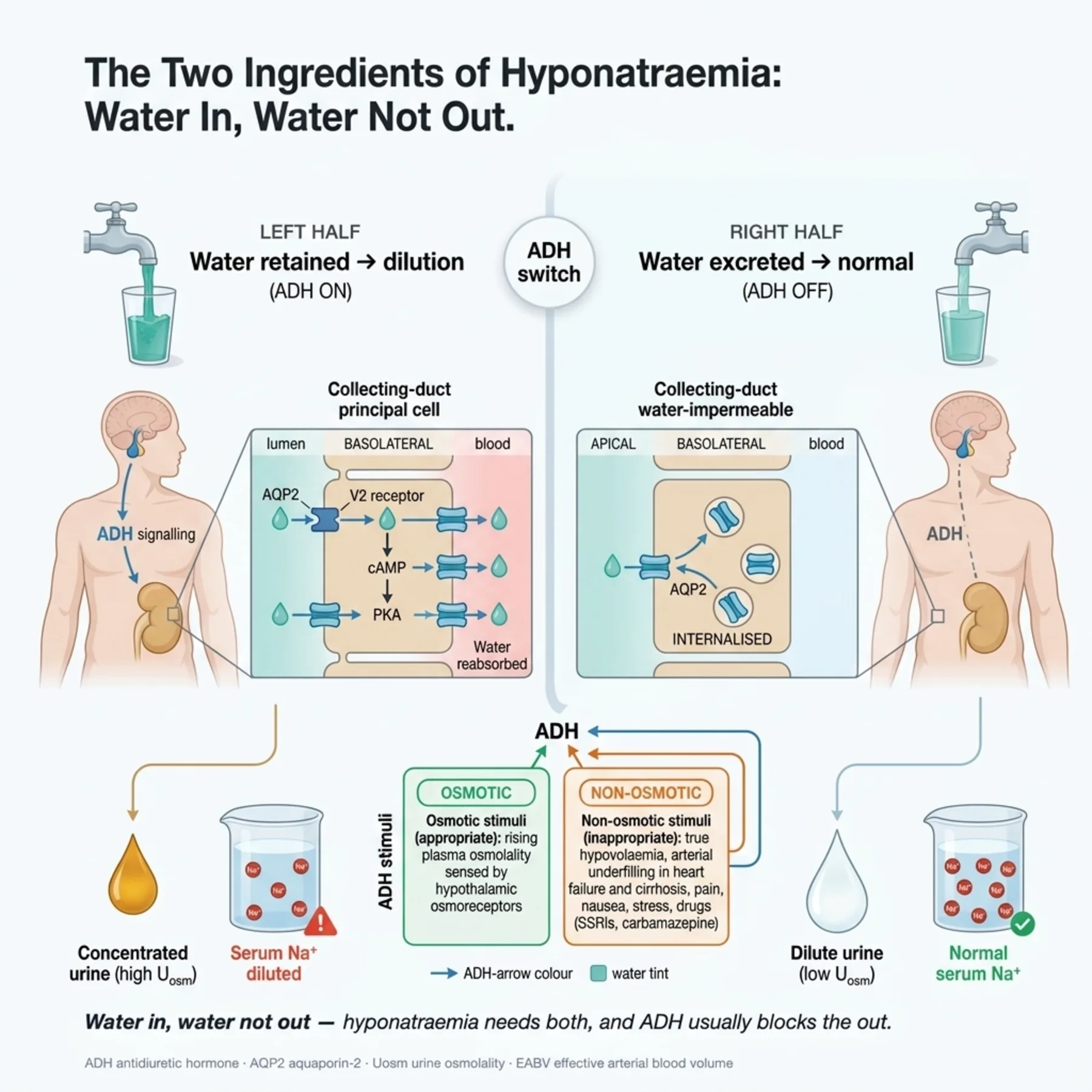
As the opening chapter established, the sodium concentration reflects water balance, not sodium content, so hyponatraemia is dilution — too much water for the solute present. For it to develop, two things must coincide: a source of water (the patient must drink or be given it) and an impaired capacity to excrete that water. The healthy kidney is superb at shedding free water — it can dilute the urine to around 50 to 100 mOsm/kg and excrete many litres a day — so a person with normal excretion is hard to make hyponatraemic by drinking. Hyponatraemia therefore signals a defect in water excretion, and that defect is, in the great majority of cases, the presence of ADH when osmolality says there should be none. The two-ingredient model — water in, water not out — is the lens for every case.
— ADH: the master of water excretion
Antidiuretic hormone, vasopressin, is released from the posterior pituitary and is the switch that determines whether the kidney sheds or retains water. Its physiological stimulus is osmotic: hypothalamic osmoreceptors sense a rising plasma osmolality above a threshold and release ADH, which acts on V2 receptors in the collecting duct to insert aquaporin-2 water channels, reabsorbing water and concentrating the urine. When osmolality falls, ADH is suppressed, aquaporins are withdrawn, and the urine becomes dilute, excreting the excess water — the normal defence against hyponatraemia. But ADH has powerful non-osmotic stimuli that can override the osmotic one: a low effective arterial blood volume (the body prioritises volume over tonicity), and also pain, nausea, the post-operative state, and many drugs. When ADH is present for these non-osmotic reasons despite a low osmolality, the kidney cannot dilute the urine, water is retained, and hyponatraemia results. Most hyponatraemia is, at root, ADH acting when osmolality says it should be silent.
— Step one: plasma osmolality
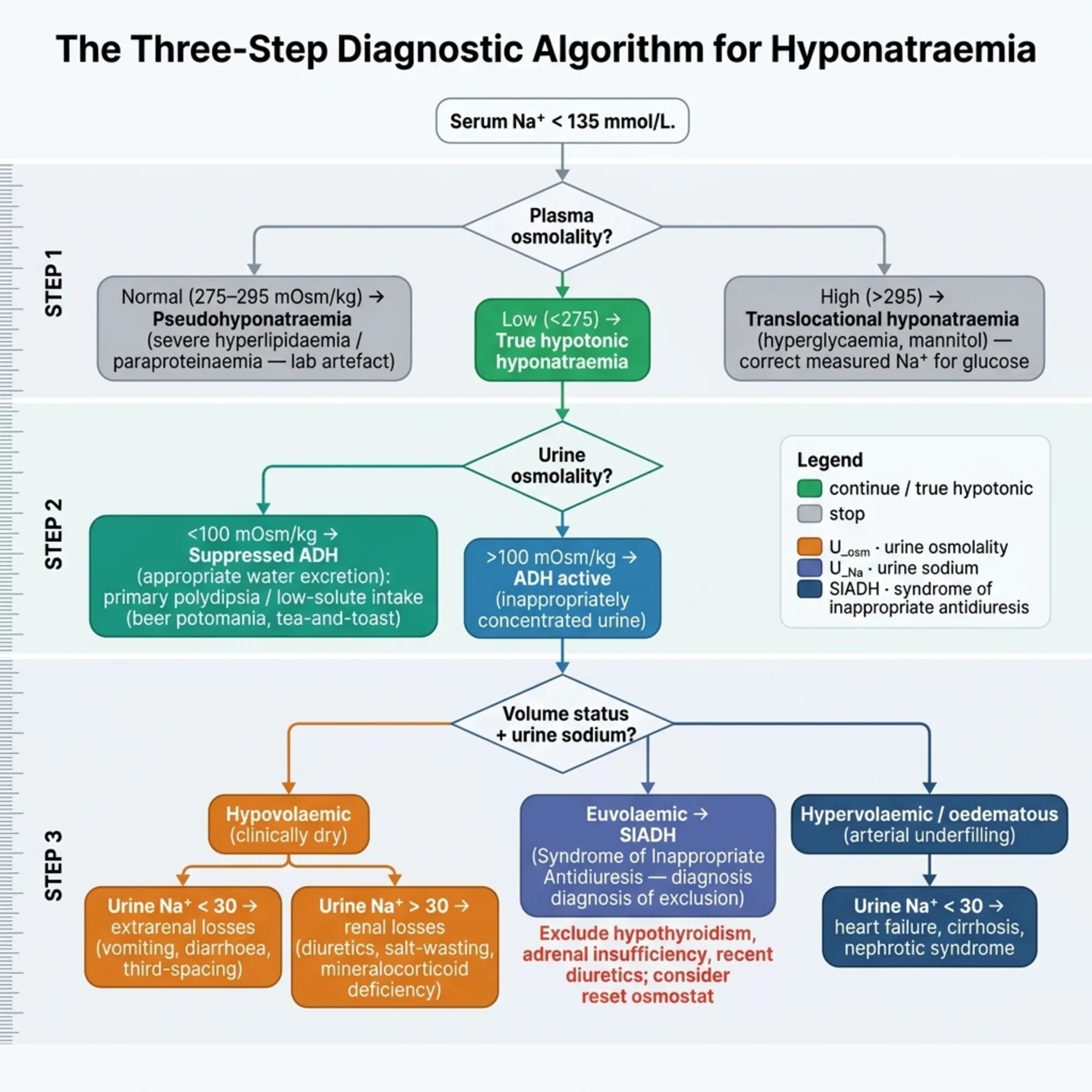
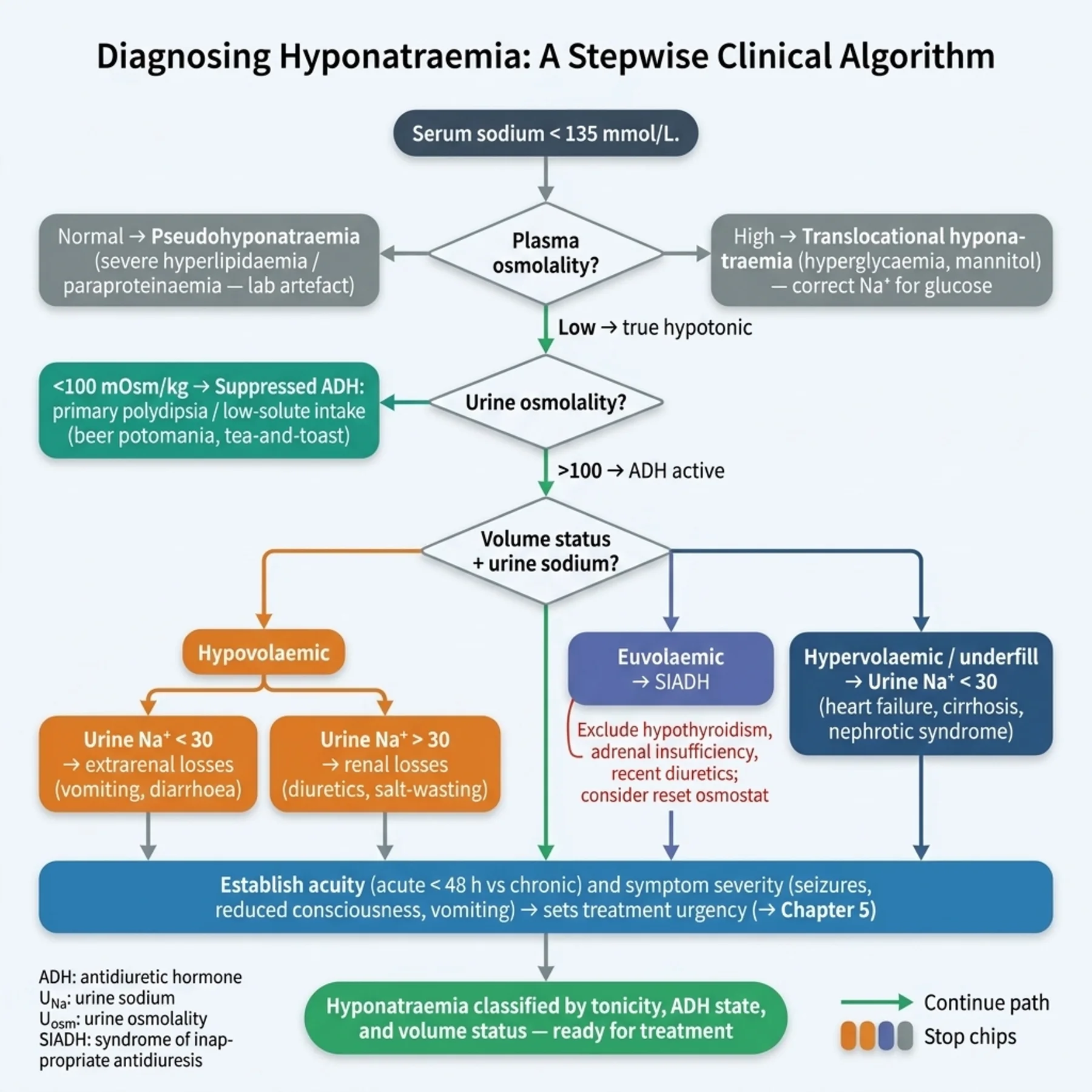
The algorithm begins by confirming that the hyponatraemia is what it appears to be, with the plasma osmolality. True hyponatraemia is hypotonic — the osmolality is low, matching the low sodium. Two impostors must be excluded. Pseudohyponatraemia is a laboratory artifact: very high lipids or proteins occupy plasma volume so that older measurement methods report a falsely low sodium, while the osmolality is normal — there is no real hypotonicity and no water excess. Translocational hyponatraemia is real dilution but from effective osmoles other than sodium: hyperglycaemia or mannitol draws water out of cells into the plasma, diluting the sodium, and the osmolality is high. Here the sodium is corrected for the glucose — adding roughly 2.4 mmol/L of sodium for every 5.5 mmol/L of glucose above normal — to reveal the true tonicity. Only when the osmolality confirms genuine hypotonic hyponatraemia does the rest of the algorithm apply; acting on a pseudo- or translocational low sodium is a classic error.
— Step two: urine osmolality
With true hypotonic hyponatraemia confirmed, the next question is whether ADH is on or off, answered by the urine osmolality. A dilute urine — under about 100 mOsm/kg — means ADH is appropriately suppressed and the kidney is doing its job, so the hyponatraemia must come from a water intake that overwhelms even maximal excretion, or from too little solute to carry water out. Primary polydipsia (drinking enormous volumes) is one cause; the other is low-solute states such as beer potomania or a 'tea and toast' diet, where so little solute is ingested that, even with a maximally dilute urine, the kidney cannot excrete enough water — because water excretion depends on solute to carry it. An inappropriately concentrated urine — over 100 — means ADH is active despite the hypotonicity, and the diagnosis lies in why, which the next step addresses. This single measurement cleanly separates the suppressed-ADH causes from the much larger ADH-active group.
— Step three: volume status and urine sodium
When ADH is active, the cause is classified by the extracellular volume status — the separate axis of the opening chapters — and refined by the urine sodium. The hypovolaemic patient has a low effective arterial blood volume that appropriately drives ADH; the urine sodium is low (under 30) when losses are extrarenal (gastrointestinal, skin, third-space) and higher when they are renal (diuretics, salt-wasting, adrenal insufficiency, cerebral salt wasting). The euvolaemic patient, with no volume stimulus, most often has SIADH — ADH secreted truly inappropriately — with hypothyroidism, glucocorticoid deficiency, and primary polydipsia (which gives a dilute urine) to consider. The hypervolaemic patient is oedematous: the underfill states of heart failure, cirrhosis, and nephrotic syndrome lower the effective arterial blood volume, driving ADH and avid sodium retention, so the urine sodium is low despite the overload. The same volume axis that classified the disorders of volume now classifies the hyponatraemias, and the urine sodium adds the renal-versus-extrarenal detail.
— SIADH, and the cerebral adaptation that governs treatment
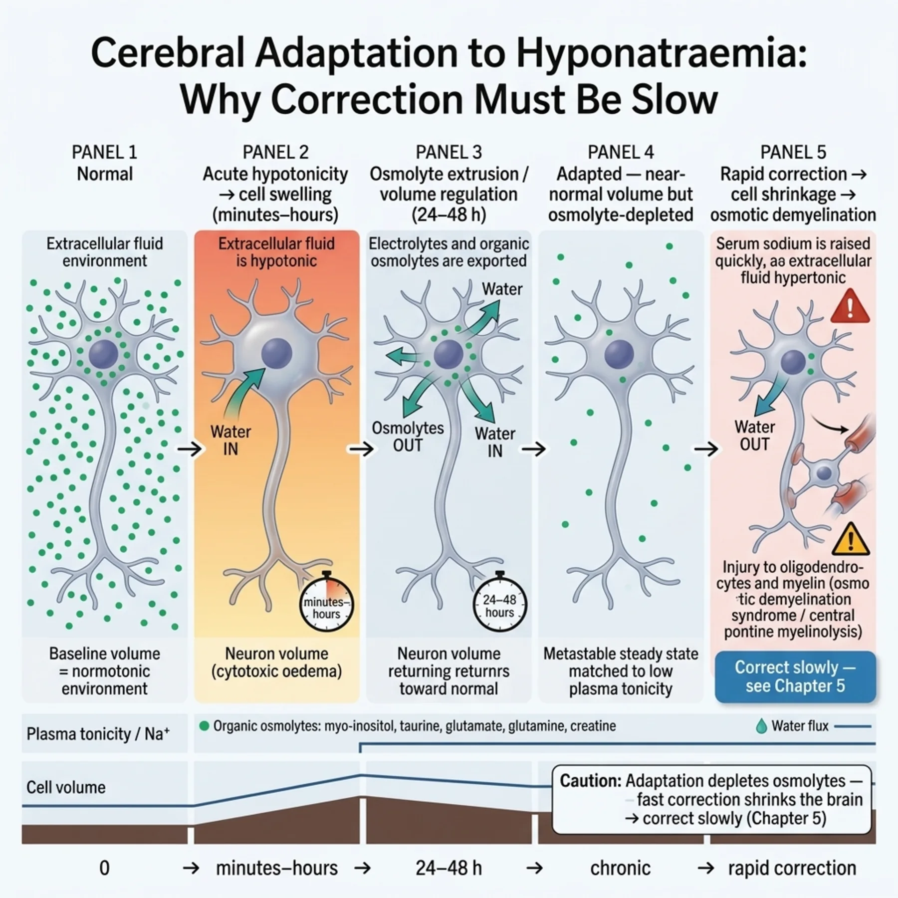
SIADH — the syndrome of inappropriate antidiuresis — deserves its own note because it is the commonest euvolaemic cause and a diagnosis of exclusion with explicit criteria: hypotonic hyponatraemia, an inappropriately concentrated urine (osmolality over 100), a urine sodium above 30 reflecting euvolaemia, clinical euvolaemia, and the exclusion of thyroid disease, adrenal insufficiency, and diuretics. Its causes cluster into central nervous system disease, pulmonary disease, malignancy (classically small-cell lung cancer secreting ectopic ADH), and drugs (SSRIs, carbamazepine, antipsychotics, and others), with pain, nausea, and surgery as transient triggers. Finally, the physiology that dominates the treatment chapter must be introduced here: the brain adapts to hyponatraemia. Acutely, brain cells swell in the hypotonic plasma; over 24 to 48 hours they extrude osmolytes — electrolytes and organic solutes — to shrink back toward normal volume. This adaptation makes chronic hyponatraemia better tolerated, but it sets a trap: if the sodium is then corrected rapidly, the adapted, osmolyte-depleted brain cannot keep pace and shrinks, causing osmotic demyelination. So the acuity of the hyponatraemia — acute (under 48 hours, often symptomatic from cerebral oedema) versus chronic (adapted) — is established during diagnosis precisely because it, and the cerebral adaptation behind it, govern how fast correction may safely proceed — the subject of the next chapter.
— Why the sequence matters
The discipline of the fixed sequence — osmolality, urine osmolality, volume status with urine sodium — is what prevents the common errors. Skipping the osmolality treats a pseudo- or translocational low sodium as if it were real. Skipping the urine osmolality misses the dilute-urine causes and mislabels primary polydipsia as SIADH. Misjudging the volume status — the hardest step, given the insensitivity of clinical signs from Chapter 1 — confuses hypovolaemic hyponatraemia with SIADH, which matters because their treatments differ. And establishing the acuity and the cerebral adaptation at diagnosis is what makes the treatment safe. Diagnosis here is not an end in itself but the foundation for the flagship that follows: classify the hyponatraemia correctly, by water physiology and the algorithm, and the treatment — and its dangers — follow logically.
| 04 | PHASE A · LEVEL 4 · ORIENTATION & KNOWLEDGE Reference Tables |
Table 4.1 — The hyponatraemia algorithm
| Step | Question | Separates |
| 1. Plasma osmolality | True hypotonic? | Pseudo (normal) / translocational (high) / true (low) |
| 2. Urine osmolality | ADH on or off? | Suppressed (<100, dilute) / active (>100) |
| 3. Volume status + urine Na | Which ADH-active cause? | Hypovolaemic / euvolaemic / hypervolaemic |
Table 4.2 — Step 1: types by osmolality
| Type | Osmolality | Note |
| True (hypotonic) | Low | Real water excess — proceed |
| Pseudohyponatraemia | Normal | Lab artifact (high lipids/protein) |
| Translocational | High | Effective osmoles (glucose, mannitol) — correct Na for glucose |
Table 4.3 — Step 3: causes by volume status
| Volume status | Causes | Urine sodium |
| Hypovolaemic | GI/skin/third-space (extrarenal); diuretics/salt-wasting/Addison (renal) | < 30 extrarenal / > 30 renal |
| Euvolaemic | SIADH; hypothyroidism; glucocorticoid deficiency; polydipsia | > 30 (SIADH); polydipsia dilute |
| Hypervolaemic | Heart failure, cirrhosis, nephrotic (underfill); renal failure | < 30 (avid) / higher in renal failure |
Table 4.4 — SIADH: criteria and causes
| Element | Detail |
| Criteria | Hypotonic hyponatraemia; urine osm > 100; urine Na > 30; clinical euvolaemia |
| Exclude | Thyroid disease, adrenal insufficiency, diuretics |
| CNS / pulmonary | Stroke, haemorrhage, infection, trauma; pneumonia, TB |
| Malignancy | Small-cell lung cancer (ectopic ADH) |
| Drugs / triggers | SSRIs, carbamazepine, antipsychotics; pain, nausea, surgery |
Table 4.5 — ADH stimuli
| Stimulus type | Examples |
| Osmotic | Rising plasma osmolality (osmoreceptors) — the physiological stimulus |
| Non-osmotic: volume | Low effective arterial blood volume (overrides osmotic) |
| Non-osmotic: other | Pain, nausea, post-operative state |
| Non-osmotic: drugs | SSRIs, carbamazepine, and others |
Table 4.6 — Cerebral adaptation and acuity
| Aspect | Detail |
| Acute swelling | Brain cells swell in hypotonic plasma |
| Adaptation (24–48 h) | Osmolytes extruded → cells shrink toward normal |
| Consequence | Chronic hyponatraemia tolerated, but rapid correction → demyelination |
| Acuity | Acute (< 48 h, often symptomatic) vs chronic (adapted) — governs treatment (Chapter 5) |
| 05 | PHASE B · LEVEL 5 · VISUALISE & MAP Imaging & Flowchart Specifications |
| 06 | PHASE B · LEVEL 6 · VISUALISE & MAP Concept Maps |
Each chain runs from physiology to a named action or consequence; read the arrows as “leads to.”
Water excess. A water source + impaired excretion (usually ADH) → water retained relative to solute → dilution → hyponatraemia → ACTION: look for both ingredients; hyponatraemia means impaired water excretion, not salt loss.
ADH. Osmotic stimulus (high osmolality) OR non-osmotic (low EABV, pain, nausea, drugs) → ADH → aquaporins → concentrated urine → ACTION: most hyponatraemia is ADH acting when osmolality says it shouldn't — find why.
The algorithm. Plasma osmolality (true vs pseudo/translocational) → urine osmolality (ADH off vs on) → volume status + urine Na (which cause) → ACTION: follow the fixed sequence; skipping a step causes the classic errors.
SIADH. Inappropriate ADH (CNS/pulmonary/malignancy/drugs) → euvolaemic hyponatraemia, concentrated urine, urine Na > 30 → ACTION: diagnose by criteria and exclusion (thyroid/adrenal/diuretic); seek the underlying cause.
Cerebral adaptation. Hypotonicity → brain swelling → osmolyte extrusion over 24–48 h → adapted but osmolyte-depleted → rapid correction shrinks cells (demyelination) → ACTION: establish acuity at diagnosis — it governs safe correction (Chapter 5).
| 07 | PHASE B · LEVEL 7 · VISUALISE & MAP Decision Pathways |
| R1 | IF a patient is hyponatraemic, THEN treat it as a water problem of impaired excretion — not a sodium deficiency — and look for both a water source and an excretion defect. |
| R2 | IF assessing hyponatraemia, THEN follow the fixed sequence: plasma osmolality, then urine osmolality, then volume status with urine sodium. |
| R3 | IF the plasma osmolality is normal or high, THEN diagnose pseudo- or translocational hyponatraemia — not true hypotonic hyponatraemia. |
| R4 | IF hyperglycaemia is present, THEN correct the sodium for glucose before interpreting it. |
| R5 | IF the urine osmolality is below ~100, THEN ADH is suppressed — consider primary polydipsia or a low-solute state, not SIADH. |
| R6 | IF ADH is active (concentrated urine), THEN classify by volume status and urine sodium — hypovolaemic, euvolaemic (SIADH), or hypervolaemic. |
| R7 | IF diagnosing SIADH, THEN confirm the criteria and exclude thyroid disease, adrenal insufficiency, and diuretics — it is a diagnosis of exclusion. |
| R8 | IF hyponatraemia is present, THEN establish its acuity and symptoms at diagnosis, because cerebral adaptation makes the rate of correction critical (Chapter 5). |
| 08 | PHASE C · LEVEL 8 · CLINICAL REASONING Clinical Cases |
| CASE 1 | WORKING THE ALGORITHM Osmolality, urine, volume Reaching SIADH |
Presentation
A patient with small-cell lung cancer has a sodium of 124. The plasma osmolality is low, the urine osmolality is 420, the urine sodium is 50, and the patient is clinically euvolaemic with normal thyroid and adrenal function and no diuretics.
❖ Pause and reflect Where does the algorithm land? |
Analysis
The algorithm lands cleanly on SIADH. The low plasma osmolality confirms true hypotonic hyponatraemia; the urine osmolality of 420 shows ADH is inappropriately active; and with clinical euvolaemia, a urine sodium above 30, and thyroid, adrenal, and diuretic causes excluded, the criteria for SIADH are met. The small-cell lung cancer is the classic cause — ectopic ADH secretion. Each step of the fixed sequence contributed to the diagnosis.
Plan
Diagnose SIADH from the criteria, attribute it to the small-cell carcinoma, and address the underlying malignancy; the specific correction of the hyponatraemia follows the treatment principles of the next chapter, paced for chronicity.
Teaching point
SIADH is reached by the algorithm — low plasma osmolality, concentrated urine, euvolaemia, urine sodium above 30, and exclusion of thyroid/adrenal/diuretic causes.
Cross-reference
Exercises rules R2, R6, R7; the algorithm and SIADH concept maps; Figure 4.2; Tables 4.1, 4.3, 4.4.
| CASE 2 | NOT TRULY LOW Check the osmolality first Translocational hyponatraemia |
Presentation
A patient with diabetic ketoacidosis has a sodium of 128 and a glucose of 44 mmol/L. The team plans to treat the 'hyponatraemia' with hypertonic measures.
❖ Pause and reflect Is this true hypotonic hyponatraemia, or something else? |
Analysis
This is translocational hyponatraemia, not true hypotonic hyponatraemia. The markedly elevated glucose is an effective osmole that draws water out of cells into the plasma, diluting the measured sodium; the plasma osmolality is high, not low. Correcting the sodium for the glucose — adding roughly 2.4 mmol/L per 5.5 mmol/L of glucose above normal — reveals a corrected sodium that is normal or even high. Treating this as hypotonic hyponatraemia would be an error; the disorder is the hyperglycaemia, and the sodium will normalise as the glucose is treated.
Plan
Recognise the translocational hyponatraemia, calculate the corrected sodium, and treat the diabetic ketoacidosis — the measured sodium will rise as the glucose falls. Do not apply hypotonic-hyponatraemia treatment.
Teaching point
Check the plasma osmolality first — a high osmolality with hyperglycaemia is translocational hyponatraemia; correct the sodium for glucose.
Cross-reference
Exercises rules R3 and R4; Figure 4.2; Table 4.2; tonicity in Chapter 1.
| CASE 3 | DEPLETED OR SIADH? The volume step Hypovolaemic versus euvolaemic |
Presentation
Two euvolaemic-looking patients have hypotonic hyponatraemia with concentrated urine. One, on close assessment, has subtle volume depletion from diuretics with a urine sodium of 45; the other is genuinely euvolaemic with a urine sodium of 50 and no diuretics.
❖ Pause and reflect Why does distinguishing hypovolaemic from euvolaemic hyponatraemia matter? |
Analysis
It matters because the treatments differ. Both have ADH active, but in the first patient ADH is appropriate to volume depletion (driven by diuretics), and the hyponatraemia will respond to volume repletion and stopping the diuretic; in the second, ADH is truly inappropriate (SIADH), and volume loading would worsen it. The volume step is the hardest and most consequential, because clinical signs are insensitive (Chapter 1) and the urine sodium is confounded by diuretics. Careful assessment — history, the diuretic, the response to a saline challenge — separates them.
Plan
Distinguish the two carefully: in the depleted patient, stop the diuretic and replete volume; in the euvolaemic patient, diagnose SIADH and manage accordingly. Use the history, the diuretic exposure, and the response to assessment rather than relying on appearance alone.
Teaching point
The volume step is the hardest and most important — hypovolaemic hyponatraemia and SIADH look similar but are treated oppositely.
Cross-reference
Exercises rule R6; the algorithm concept map; Table 4.3; volume assessment in Chapters 1 and 3.
| CASE 4 | TOO LITTLE SOLUTE The dilute-urine clue Low-solute hyponatraemia |
Presentation
An elderly patient with a poor 'tea and toast' diet, and a separate patient drinking large volumes of beer, both present with hyponatraemia. In both, the urine osmolality is low at around 80.
❖ Pause and reflect What does the dilute urine tell you, and what is the mechanism? |
Analysis
The dilute urine (under 100) shows ADH is appropriately suppressed — these are not ADH-driven states, so they are not SIADH. The mechanism is solute-limited water excretion: water excretion requires solute to carry it out, and in a very low-solute diet (tea and toast) or a high-water, low-solute intake (beer potomania), there is too little solute to excrete the ingested water even with a maximally dilute urine, so water is retained and the sodium falls. The clue is entirely in the low urine osmolality, which the algorithm captures at step two.
Plan
Recognise the low-solute mechanism from the dilute urine, restrict water and improve solute (dietary) intake, and avoid mistaking these for SIADH. Correction can be rapid once solute and a water diuresis return, so monitor the rate carefully (Chapter 5).
Teaching point
A dilute urine (under 100) points away from SIADH to primary polydipsia or low-solute states — water excretion needs solute to carry it.
Cross-reference
Exercises rule R5; the water-excess and algorithm concept maps; Tables 4.1 and 4.3.
| 09 | PHASE C · LEVEL 9 · CLINICAL REASONING Clinical Implications |
One triad per mechanism the narrative exposed: the physiology, why it matters, and the action it dictates.
MECHANISM Hyponatraemia requires both a water source and impaired water excretion, the latter usually ADH-mediated. |
WHY IT MATTERS A low sodium reflects water excess, not sodium deficiency. |
ACTION Look for both ingredients and treat the water problem, not a presumed salt shortage. |
MECHANISM ADH is released by osmotic and powerful non-osmotic stimuli, and concentrates the urine. |
WHY IT MATTERS Most hyponatraemia is ADH acting when osmolality says it should be suppressed. |
ACTION Find why ADH is on — volume, SIADH, or another non-osmotic trigger. |
MECHANISM Effective osmoles such as glucose, and lab artifacts from lipids or protein, lower the measured sodium without true hypotonicity. |
WHY IT MATTERS Treating a translocational or pseudo low sodium as real is a classic error. |
ACTION Check the plasma osmolality first and correct the sodium for glucose. |
MECHANISM Water excretion depends on solute to carry water out, so very low solute intake limits it. |
WHY IT MATTERS Low-solute states cause hyponatraemia with a dilute urine, unlike SIADH. |
ACTION Read the low urine osmolality as the clue, and restore solute while restricting water. |
MECHANISM The brain extrudes osmolytes to adapt to chronic hyponatraemia, becoming osmolyte-depleted. |
WHY IT MATTERS Rapid correction then shrinks the adapted brain and causes osmotic demyelination. |
ACTION Establish acuity at diagnosis, because it governs how fast correction may safely proceed. |
| 10 | PHASE C · LEVEL 10 · CLINICAL REASONING Clinical Pearls |
| Hyponatraemia = water excess, not sodium deficiency. | Needs two ingredients: a water source + impaired excretion (usually ADH). |
| The healthy kidney excretes huge free-water loads — hyponatraemia means that failed. | ADH: osmotic + non-osmotic stimuli (low EABV, pain, nausea, drugs). |
| ADH → aquaporin-2 → concentrated urine → water retention. | Algorithm: plasma osmolality → urine osmolality → volume status + urine Na. |
| Step 1: low osm = true; normal = pseudo; high = translocational. | Correct Na for glucose (~2.4 mmol/L per 5.5 mmol/L glucose). |
| Step 2: urine osm < 100 = ADH suppressed (polydipsia/low-solute); > 100 = ADH active. | Step 3 hypovolaemic: urine Na < 30 extrarenal, > 30 renal. |
| Step 3 euvolaemic: SIADH (commonest), hypothyroid, glucocorticoid deficiency, polydipsia. | Step 3 hypervolaemic: underfill (HF/cirrhosis/nephrotic) — urine Na < 30. |
| SIADH = hypotonic, urine osm > 100, urine Na > 30, euvolaemic, exclude thyroid/adrenal/diuretic. | SIADH causes: CNS, pulmonary, malignancy (small-cell), drugs. |
| Brain adapts to chronic hyponatraemia by extruding osmolytes. | Acuity governs correction — establish it at diagnosis (Chapter 5). |
| 11 | PHASE D · LEVEL 11 · SAFETY & EVIDENCE Red Flags & Never-Do |
Panel A — Red flags
| ▲ | A low sodium with a high osmolality and hyperglycaemia — translocational; correct the sodium for glucose, don't treat as hypotonic. |
| ▲ | A dilute urine (< 100) in hyponatraemia — ADH is suppressed; think polydipsia or low solute, not SIADH. |
| ▲ | Hyponatraemia labelled SIADH without excluding thyroid, adrenal, and diuretic causes — reassess. |
| ▲ | Acute, symptomatic hyponatraemia (headache, confusion, seizures) — cerebral oedema; urgent (Chapter 5). |
| ▲ | Chronic hyponatraemia about to be corrected fast — the adapted brain risks demyelination; pace it (Chapter 5). |
Panel B — Never do
| ✖ NEVER — treat hyponatraemia as sodium deficiency without working the water physiology. |
| ✖ NEVER — skip the plasma osmolality — it separates true from pseudo/translocational. |
| ✖ NEVER — diagnose SIADH without checking the urine osmolality and excluding mimics. |
| ✖ NEVER — plan correction without first establishing acuity and the cerebral-adaptation risk. |
| 12 | PHASE D · LEVEL 12 · SAFETY & EVIDENCE Common Pitfalls |
Pitfall 1 — Skipping the osmolality
| ✖ | WRONG Treating any low sodium as true hypotonic hyponatraemia. |
| ✓ | RIGHT Checking plasma osmolality first to exclude pseudo/translocational. |
| ✉ | WHY A normal or high osmolality means it is not true water excess. |
Pitfall 2 — Ignoring hyperglycaemia
| ✖ | WRONG Reading the measured sodium at face value in marked hyperglycaemia. |
| ✓ | RIGHT Correcting the sodium for the glucose. |
| ✉ | WHY Glucose is an effective osmole that dilutes the measured sodium. |
Pitfall 3 — SIADH without the urine
| ✖ | WRONG Labelling euvolaemic hyponatraemia SIADH without the urine osmolality. |
| ✓ | RIGHT Checking urine osmolality — a dilute urine means polydipsia/low solute, not SIADH. |
| ✉ | WHY SIADH requires inappropriately concentrated urine. |
Pitfall 4 — Confusing depletion with SIADH
| ✖ | WRONG Calling subtle hypovolaemic hyponatraemia SIADH. |
| ✓ | RIGHT Assessing volume carefully, including diuretic exposure and response. |
| ✉ | WHY They look similar but are treated oppositely (volume vs water restriction). |
Pitfall 5 — Forgetting the adaptation
| ✖ | WRONG Planning correction without establishing chronicity. |
| ✓ | RIGHT Determining acuity at diagnosis, because adaptation governs safe correction. |
| ✉ | WHY The adapted brain is at risk of demyelination from rapid correction. |
| 13 | PHASE D · LEVEL 13 · SAFETY & EVIDENCE Evidence Grading |
GRADE A HIGH CONFIDENCE The effect is real and the estimate is stable. RCTs at low risk of bias; multiple concordant prospective cohorts; meta-analyses. |
GRADE B MODERATE CONFIDENCE The effect is likely real but may shift with new data. Observational studies, registries, mechanistic human studies. |
GRADE C LOW CONFIDENCE Rests on physiology, reasoning, or consensus rather than outcomes. Pathophysiological reasoning; extrapolation; consensus without outcomes. |
Graded statements (by evidence type)
| Statement | Grade | Basis (evidence type) |
| Hyponatraemia is a disorder of water excess, usually from impaired ADH-mediated excretion. | A | Established physiology |
| Plasma osmolality separates true from pseudo- and translocational hyponatraemia. | A | Established physiology |
| The sodium must be corrected for glucose in hyperglycaemia. | A | Established physiology |
| Urine osmolality distinguishes suppressed from active ADH states. | A | Established physiology |
| SIADH is a diagnosis of exclusion meeting defined criteria. | A | Consensus criteria |
| Low-solute states cause hyponatraemia with a dilute urine. | A | Established physiology |
| Cerebral osmotic adaptation underlies the risk of demyelination with rapid correction. | A | Established physiology and clinical data |
| 18 | PHASE F · LEVEL 18 · APPLY & TEST Cheat Sheet |
| Hyponatraemia = WATER excess (not salt deficiency). | Needs water source + impaired excretion (usually ADH). |
| ADH: osmotic + non-osmotic (low EABV, pain, nausea, drugs). | ADH → aquaporins → concentrated urine. |
| Algorithm: plasma osm → urine osm → volume + urine Na. | Step 1: low osm = true; normal = pseudo; high = translocational. |
| Correct Na for glucose (~2.4/5.5 mmol/L). | Step 2: urine osm < 100 = ADH off (polydipsia/low-solute); > 100 = ADH on. |
| Hypovolaemic: urine Na < 30 extrarenal / > 30 renal. | Euvolaemic: SIADH, hypothyroid, glucocorticoid deficiency, polydipsia. |
| Hypervolaemic: underfill (HF/cirrhosis/nephrotic), urine Na < 30. | SIADH: hypotonic + urine osm > 100 + urine Na > 30 + euvolaemic + exclude thyroid/adrenal/diuretic. |
| SIADH causes: CNS, pulmonary, small-cell ca, drugs (SSRIs, carbamazepine). | Brain adapts (osmolyte extrusion) to chronic hyponatraemia. |
| Rapid correction of adapted brain → demyelination. | Establish acuity + symptoms at diagnosis (sets treatment, Chapter 5). |
| 19 | PHASE F · LEVEL 19 · APPLY & TEST Flashcards |
| CARD 1 | Q. Why is hyponatraemia a water problem? A. A low sodium concentration reflects an excess of water relative to solute — dilution — not a deficiency of sodium; it requires a water source and impaired water excretion. DETAILED. The healthy kidney excretes large free-water loads easily. CLINICAL. Treat the water problem, not a presumed salt shortage. |
| CARD 2 | Q. What stimulates ADH, and what does it do? A. Osmotic stimuli (rising osmolality) and non-osmotic stimuli (low effective arterial blood volume, pain, nausea, drugs) release ADH, which inserts aquaporins in the collecting duct and concentrates the urine. DETAILED. Non-osmotic stimuli can override the osmotic one. CLINICAL. Find why ADH is active when osmolality says it should be off. |
| CARD 3 | Q. What is step one of the algorithm, and why? A. Measure plasma osmolality: low confirms true hypotonic hyponatraemia, normal indicates pseudohyponatraemia (lab artifact), and high indicates translocational hyponatraemia (effective osmoles such as glucose). DETAILED. Acting on a pseudo- or translocational low sodium is a classic error. CLINICAL. Check osmolality first; correct sodium for glucose. |
| CARD 4 | Q. What does the urine osmolality tell you? A. A dilute urine (under 100) means ADH is suppressed — primary polydipsia or a low-solute state; a concentrated urine (over 100) means ADH is active, and the cause is found by volume status. DETAILED. It cleanly separates the two groups. CLINICAL. Use it as step two of the algorithm. |
| CARD 5 | Q. How is ADH-active hyponatraemia classified? A. By volume status and urine sodium: hypovolaemic (appropriate ADH; urine Na low extrarenal, high renal), euvolaemic (SIADH and mimics), and hypervolaemic (underfill states; urine Na low). DETAILED. The volume step is the hardest and most consequential. CLINICAL. Classify carefully — hypovolaemia and SIADH are treated oppositely. |
| CARD 6 | Q. What are the criteria for SIADH? A. Hypotonic hyponatraemia, an inappropriately concentrated urine (osmolality over 100), a urine sodium above 30, clinical euvolaemia, and exclusion of thyroid disease, adrenal insufficiency, and diuretics. DETAILED. It is a diagnosis of exclusion. CLINICAL. Confirm the criteria and seek the cause (CNS, pulmonary, malignancy, drugs). |
| CARD 7 | Q. Why does a low-solute diet cause hyponatraemia? A. Water excretion requires solute to carry water out, so very low solute intake (tea and toast, beer potomania) limits free-water excretion even with a maximally dilute urine, retaining water. DETAILED. The urine osmolality is low — ADH is suppressed. CLINICAL. Read the dilute urine as the clue; restore solute and restrict water. |
| CARD 8 | Q. Why does cerebral adaptation matter for treatment? A. The brain extrudes osmolytes over 24–48 hours to adapt to chronic hyponatraemia, becoming osmolyte-depleted; rapid correction then shrinks the adapted brain and causes osmotic demyelination. DETAILED. Acuity (acute vs chronic) governs the safe correction rate. CLINICAL. Establish acuity at diagnosis (treatment in Chapter 5). |
| 20 | PHASE F · LEVEL 20 · APPLY & TEST One-Minute Preceptor |
| SCENE 1 | The intern treating the number |
GET A COMMITMENT. “You want to give hypertonic measures for this sodium of 128 — have you checked the osmolality?”
PROBE FOR EVIDENCE. “The glucose is 44” — ask: “What does a high glucose do to the measured sodium and the osmolality?”
TEACH A GENERAL RULE. Glucose is an effective osmole that dilutes the measured sodium and raises osmolality — this is translocational, not hypotonic, hyponatraemia; correct the sodium for glucose.
REINFORCE WHAT WAS RIGHT. Noticing the low sodium was correct.
CORRECT A MISTAKE. Check osmolality, correct for glucose, and treat the DKA — not the 'hyponatraemia.'
| SCENE 2 | The resident over-calling SIADH |
GET A COMMITMENT. “You've diagnosed SIADH in this euvolaemic patient — on what basis?”
PROBE FOR EVIDENCE. “The sodium is low and they look euvolaemic” — ask: “What is the urine osmolality, and have you excluded thyroid, adrenal, and diuretics?”
TEACH A GENERAL RULE. SIADH needs an inappropriately concentrated urine and exclusion of mimics — a dilute urine means polydipsia or low solute, not SIADH.
REINFORCE WHAT WAS RIGHT. Assessing volume status was the right step.
CORRECT A MISTAKE. Check the urine osmolality and exclude the mimics before calling it SIADH.
| 22 | PHASE F · LEVEL 22 · APPLY & TEST Board-Style Questions |
| Q 01 | Hyponatraemia is fundamentally a disorder of: |
| A | Sodium deficiency |
| B | Water excess relative to solute |
| C | Potassium balance |
| D | Chloride loss |
Rationale A low sodium concentration reflects dilution — water excess — not a sodium shortage (rule R1, water-excess concept map). A, C, and D misidentify the problem. |
| Q 02 | What is the first step in evaluating hyponatraemia? |
| A | Urine sodium |
| B | Plasma osmolality |
| C | Volume status |
| D | Thyroid function |
Rationale Plasma osmolality first separates true hypotonic from pseudo- and translocational hyponatraemia (Figure 4.2, rule R2). A, C, and D come later in the sequence. |
| Q 03 | A patient with a glucose of 44 mmol/L has a sodium of 128 and a high plasma osmolality. This is: |
| A | True hypotonic hyponatraemia |
| B | Translocational hyponatraemia — correct the sodium for glucose |
| C | Pseudohyponatraemia |
| D | SIADH |
Rationale Glucose is an effective osmole that dilutes the measured sodium with a high osmolality — translocational, requiring correction (case 2, Table 4.2, rule R4). A, C, and D are incorrect. |
| Q 04 | A urine osmolality below 100 in hyponatraemia indicates: |
| A | Active ADH (SIADH) |
| B | Suppressed ADH — primary polydipsia or a low-solute state |
| C | Volume depletion |
| D | Hypervolaemia |
Rationale A dilute urine means ADH is appropriately suppressed, pointing to polydipsia or low solute, not SIADH (rule R5, case 4). A is the opposite; C and D are ADH-active states. |
| Q 05 | Which criteria define SIADH? |
| A | Hypertonic hyponatraemia with dilute urine |
| B | Hypotonic hyponatraemia, urine osmolality > 100, urine sodium > 30, euvolaemia, mimics excluded |
| C | Hypovolaemia with low urine sodium |
| D | Oedema with a high urine sodium |
Rationale SIADH requires hypotonic hyponatraemia with inappropriately concentrated urine, a urine sodium above 30, euvolaemia, and exclusion of thyroid/adrenal/diuretic causes (Table 4.4, rule R7). A, C, and D describe other states. |
| Q 06 | Two patients have hypotonic hyponatraemia with concentrated urine. Distinguishing hypovolaemic from euvolaemic (SIADH) matters because: |
| A | The treatments are the same |
| B | The treatments differ — volume repletion versus water restriction |
| C | Only the euvolaemic one is real |
| D | Volume status is irrelevant |
Rationale Hypovolaemic hyponatraemia responds to volume repletion while SIADH needs water restriction — opposite treatments (case 3, rule R6). A, C, and D are false. |
| Q 07 | Why does a low-solute diet (tea and toast, beer potomania) cause hyponatraemia? |
| A | Excess sodium intake |
| B | Water excretion needs solute to carry it, so low solute limits free-water excretion |
| C | Active ADH |
| D | Volume overload |
Rationale Free-water excretion is solute-limited, so a very low-solute intake retains water despite a dilute urine (case 4, water-excess concept map). A, C, and D are incorrect. |
| Q 08 | Why must acuity be established at the time of diagnosing hyponatraemia? |
| A | It does not matter |
| B | The brain adapts to chronic hyponatraemia, so rapid correction risks osmotic demyelination |
| C | Acute hyponatraemia is always asymptomatic |
| D | Chronic hyponatraemia needs no treatment |
Rationale Cerebral osmotic adaptation makes the safe correction rate depend on chronicity, set up here for the treatment chapter (Figure 4.3, rule R8). A, C, and D are false. |