Chapter Preamble
Signals declared
Sig-D — Diagnostic (primary). Recognise ARPKD, nephronophthisis, acquired cystic disease, and the other cystic diseases, and distinguish them from ADPKD.
Sig-T — Therapeutic (strong). Manage the neonatal and renal-hepatic disease of ARPKD, the progressive CKD of nephronophthisis, and the malignancy risk of acquired cystic disease.
Sig-M — Mechanistic (strong). The fibrocystin ciliopathy of ARPKD, the nephronophthisis ciliopathy, and how each produces its distinctive picture.
Levels populated and omitted
Populated (19): L1–L14, L17–L20, L22. The mechanistic signal fires the concept maps (L6) and triads (L9); the therapeutic signal fires the absolute-risk table (L14) and templates (L17); the diagnostic signal drives the tables, rules, cases, pitfalls, and board items.
L15 / L16 preference-sensitive map and SDM scripts — omitted. No Sig-E; recognising and managing these diseases is established care.
L21 reflective prompts — omitted. No Sig-E/V; the chapter's content is worked through the cases and pitfalls.
| 01 | PHASE A · LEVEL 1 · ORIENTATION & KNOWLEDGE Learning Objectives |
By the end of this chapter you should be able to:
Describe the genetics and renal-hepatic features of ARPKD.
Explain the neonatal presentation of ARPKD and its major cause of mortality.
Explain the congenital hepatic fibrosis and liver-kidney interplay of ARPKD.
Contrast ARPKD with ADPKD.
Recognise nephronophthisis and its place as a cause of childhood ESKD.
Recognise the syndromic (ciliopathy) associations of nephronophthisis.
Recognise acquired cystic kidney disease and its malignancy risk.
Recognise the other cystic diseases — ADTKD, medullary sponge kidney, simple cysts.
| 02 | PHASE A · LEVEL 2 · ORIENTATION & KNOWLEDGE Executive Summary |
ARPKD (autosomal recessive polycystic kidney disease) is caused by PKHD1 mutations encoding fibrocystin, a ciliary protein, and typically presents in the neonate or infant.
It produces bilateral massively enlarged echogenic kidneys with dilated collecting ducts, and the reduced fetal urine output causes oligohydramnios and, through it, pulmonary hypoplasia — the major cause of neonatal death.
Severe early hypertension and progressive CKD follow in survivors.
A defining feature is congenital hepatic fibrosis, which causes portal hypertension (varices, splenomegaly) and may involve the bile ducts (Caroli disease) — so ARPKD is a renal-hepatic disease, and the liver disease dominates in those who survive infancy and in the milder, later-presenting forms.
ARPKD differs from ADPKD in being recessive (affected siblings, unaffected parents), presenting in infancy, affecting the collecting ducts, and causing congenital hepatic fibrosis rather than the liver cysts of ADPKD.
Nephronophthisis is an autosomal recessive ciliopathy and the commonest genetic cause of end-stage kidney disease in children and young adults.
It is a tubulointerstitial disease with small corticomedullary cysts and normal-sized or small kidneys, presenting with a urinary concentrating defect (polyuria, polydipsia), salt wasting, anaemia, and growth failure, with a bland urine.
It has syndromic associations — retinitis pigmentosa, cerebellar and hepatic involvement — reflecting its ciliopathy nature, and does not recur after transplantation.
ADTKD (covered fully later) is the autosomal dominant tubulointerstitial disease, with few or no cysts and a bland urine.
Acquired cystic kidney disease develops in long-standing CKD and dialysis — it is not inherited — and carries an increased risk of renal cell carcinoma, prompting surveillance in long-term dialysis.
Medullary sponge kidney (usually benign, with stones) and simple cysts (common and benign) complete the cystic differential.
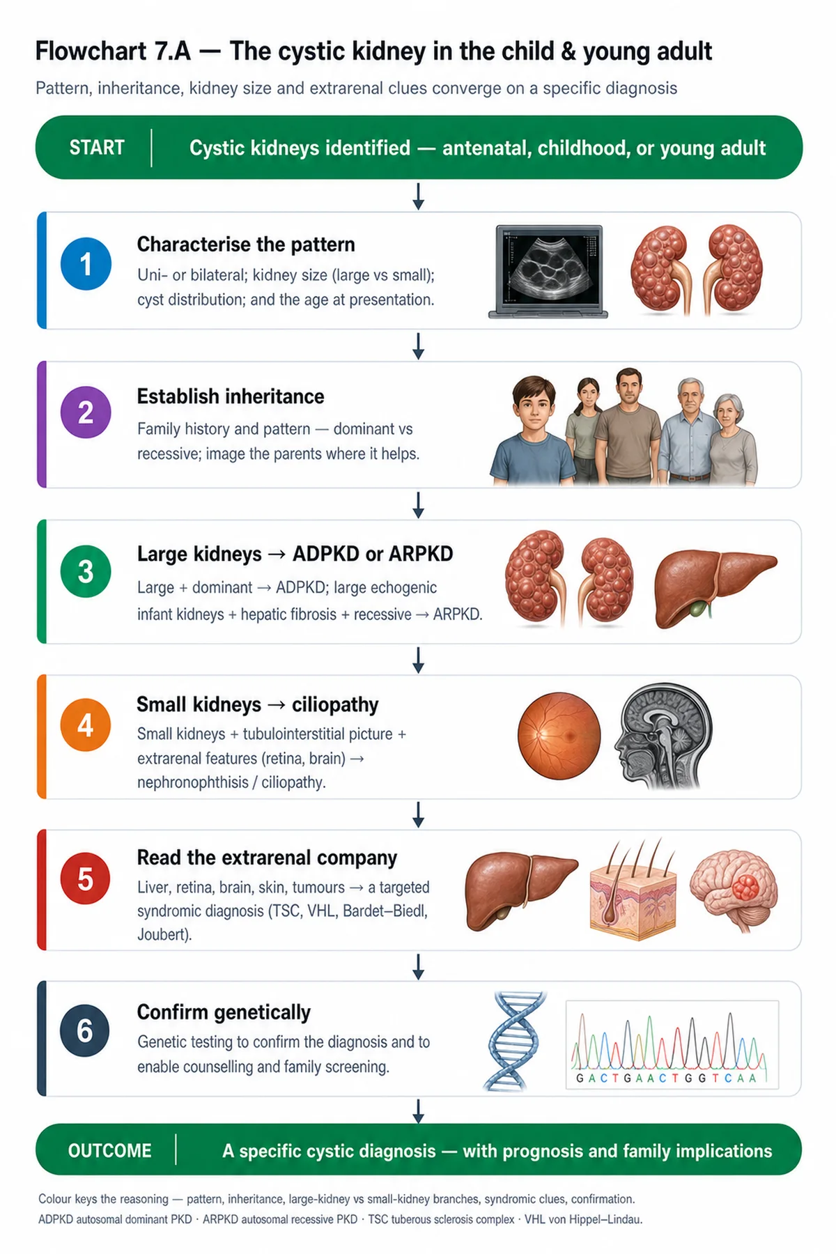
Distinguishing these diseases from ADPKD and from each other — by age, inheritance, cyst location, kidney size, and associated features — is the diagnostic task of the chapter.
| 03 | PHASE A · LEVEL 3 · ORIENTATION & KNOWLEDGE Main Narrative |
Not every cystic kidney is ADPKD. This chapter covers the rest of the cystic diseases — above all ARPKD, the recessive counterpart that presents in infancy and damages both kidney and liver, and nephronophthisis, the commonest genetic cause of kidney failure in children — along with acquired cystic disease and the benign cysts. Each has a distinctive mechanism, presentation, and management, and distinguishing them from ADPKD and from one another is the diagnostic work that completes the cystic part of the volume.
— ARPKD: genetics and the neonatal presentation
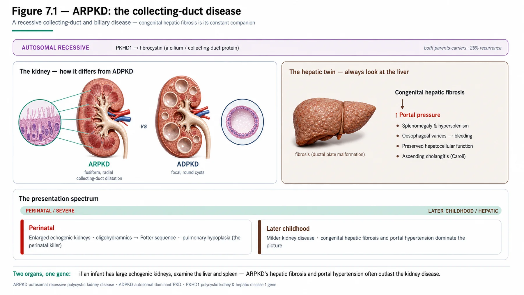
ARPKD is the recessive counterpart of ADPKD, but it is a very different disease. It is caused by mutations in PKHD1, encoding fibrocystin (also called polyductin), another ciliary protein — so it too is a ciliopathy, but recessive, meaning affected siblings with unaffected carrier parents (and the consanguinity that raises recessive risk). It typically presents in the neonate or infant. The kidneys are bilaterally and massively enlarged and echogenic, with dilatation of the collecting ducts (rather than the rounded cysts of ADPKD), and — critically — the diseased fetal kidneys produce too little urine, causing oligohydramnios (too little amniotic fluid). The oligohydramnios in turn causes pulmonary hypoplasia (the lungs need amniotic fluid to develop), and this pulmonary hypoplasia is the major cause of neonatal death and respiratory distress in ARPKD — a baby may die of its lungs, not its kidneys. So the neonatal presentation is dominated by the consequences of poor fetal urine output: enlarged kidneys, oligohydramnios, and a baby in respiratory distress from underdeveloped lungs. Survivors of the neonatal period face severe early hypertension and progressive CKD. The disease has a spectrum, from the severe neonatal (renal-predominant, high mortality) to milder later-childhood forms.
— The liver: congenital hepatic fibrosis
A defining feature of ARPKD, and one that distinguishes it sharply from ADPKD, is its liver involvement: congenital hepatic fibrosis. This arises from a developmental abnormality of the bile ducts (a ductal plate malformation) and produces progressive hepatic fibrosis that causes portal hypertension — with its consequences of oesophageal varices (and the risk of variceal bleeding), splenomegaly, and hypersplenism — and it may involve dilatation of the intrahepatic bile ducts (Caroli disease, or Caroli syndrome when combined with hepatic fibrosis), predisposing to cholangitis. Importantly, this is congenital hepatic fibrosis with portal hypertension, not the simple liver cysts of ADPKD — a different liver lesion entirely. The renal-hepatic interplay is characteristic of ARPKD: the disease affects both organs, and there is often an inverse relationship in which those with the most severe early renal disease (and high neonatal mortality) have less time for the liver disease to manifest, while those who survive infancy, or who present later, have the liver disease come to dominate — so a child who survives the neonatal renal crisis may grow up to face portal hypertension and its complications. Managing ARPKD therefore means managing both the kidney and the liver, and in severe combined disease, combined liver-kidney transplantation may be needed.
— ARPKD versus ADPKD, and management
Contrasting ARPKD with ADPKD consolidates both. ARPKD is recessive (affected siblings, unaffected parents) versus ADPKD's dominant (vertical transmission); ARPKD presents in infancy versus ADPKD's adulthood; ARPKD affects the collecting ducts (fusiform dilatation) versus ADPKD's cysts from all nephron segments; and ARPKD causes congenital hepatic fibrosis with portal hypertension versus ADPKD's liver cysts. The fibrocystin defect of ARPKD versus the polycystin defect of ADPKD underlies the difference, though both are ciliopathies. Management of ARPKD is multi-organ and stage-dependent: intensive neonatal respiratory support for the pulmonary hypoplasia (the immediate threat); aggressive control of the severe hypertension; CKD management toward eventual kidney replacement; surveillance and management of the portal hypertension (varices) and cholangitis; and, in severe combined renal and hepatic disease, consideration of combined liver-kidney transplantation. Because it is recessive, the recurrence risk in siblings is 25%, and genetic counselling is offered to the family (with the reproductive implications of the recessive chapters). ARPKD is thus a disease of childhood spanning two organs, quite distinct from the adult, kidney-and-liver-cyst disease of ADPKD.
— Nephronophthisis
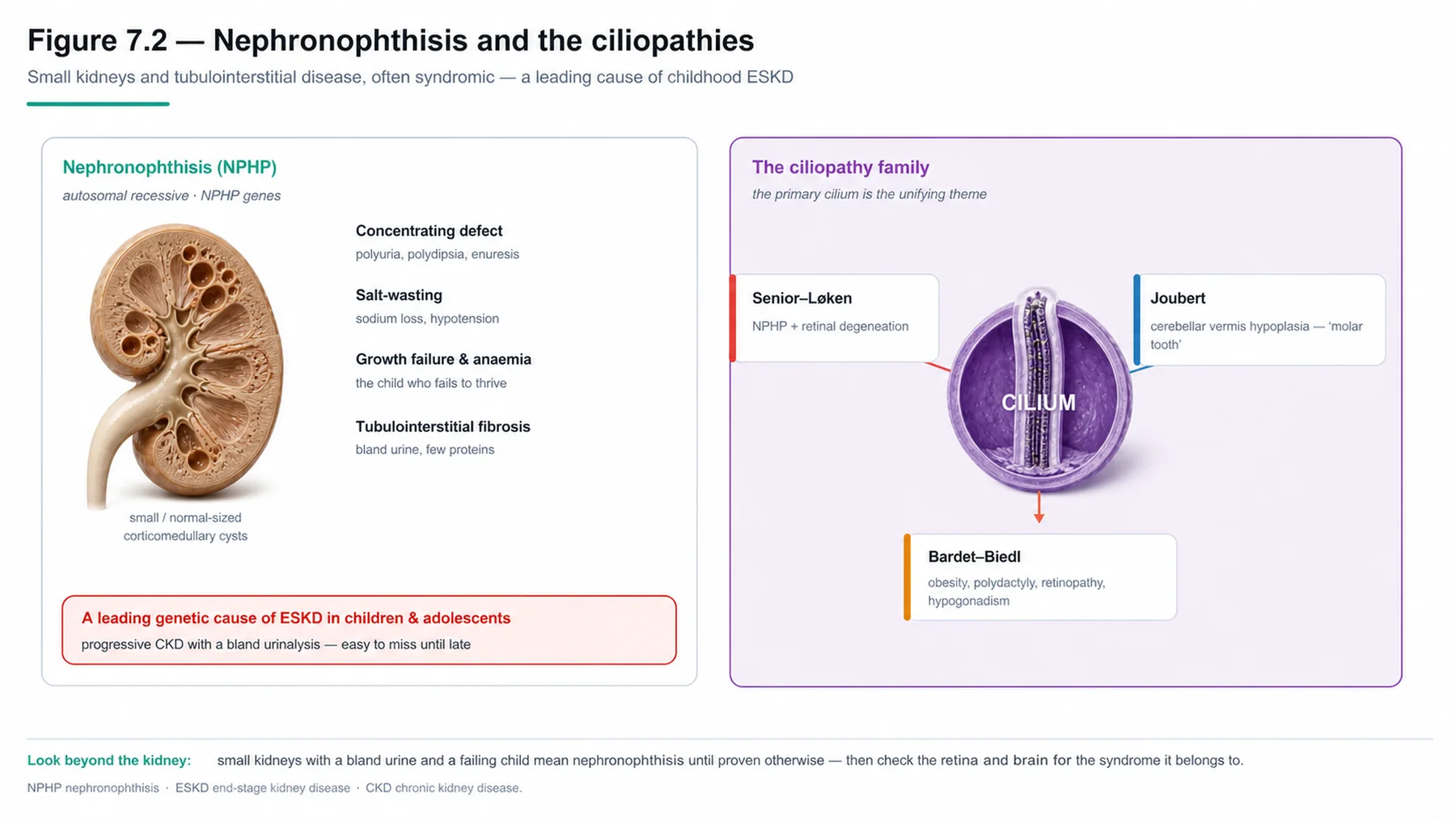
Nephronophthisis is the other major inherited cystic (more precisely, cystic-tubulointerstitial) disease to know, and it is the commonest genetic cause of end-stage kidney disease in children and young adults. It is an autosomal recessive ciliopathy (caused by mutations in the NPHP genes), and its picture is distinctive and quite unlike the polycystic diseases. The kidneys are normal-sized or small (not enlarged), with small cysts characteristically at the corticomedullary junction, and the dominant lesion is tubulointerstitial — tubular atrophy and interstitial fibrosis — so the urine is bland (little proteinuria or haematuria). The clinical presentation reflects the tubulointerstitial damage: an early and prominent urinary concentrating defect (polyuria, polydipsia, and sometimes secondary enuresis), salt wasting, anaemia, and growth failure, with progressive CKD to end-stage disease (the age at which depends on the form — infantile, juvenile, or adolescent). Importantly, nephronophthisis is often syndromic, part of the broader ciliopathy spectrum, with extrarenal associations: retinitis pigmentosa (Senior-Løken syndrome), cerebellar involvement (Joubert syndrome), hepatic fibrosis, and others (Bardet-Biedl, Jeune) — so a young person with a concentrating defect, bland urine, normal-sized kidneys, progressive CKD, and a feature such as retinitis pigmentosa should prompt the diagnosis. It does not recur after transplantation. Nephronophthisis is the disease to think of in a child or young adult with unexplained, bland-urine, normal-kidney-size progressive CKD.
— Acquired cystic disease and the rest
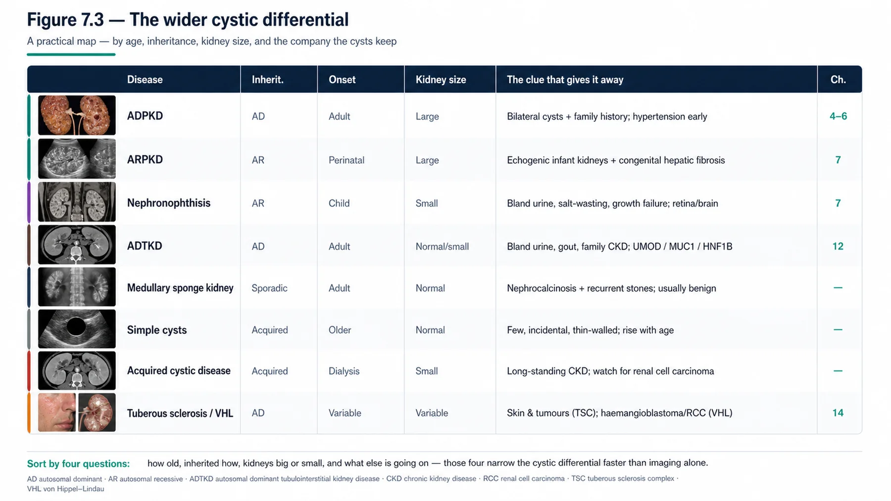
The remaining cystic diseases round out the differential. Acquired cystic kidney disease is important and distinct because it is not inherited: it develops in long-standing CKD, and especially in dialysis patients, who develop multiple bilateral cysts in their (usually small) native kidneys over time — and it carries an increased risk of renal cell carcinoma, which is why surveillance is considered in long-term dialysis patients (a malignancy risk in the native kidneys that outlives the kidneys' function). ADTKD (autosomal dominant tubulointerstitial kidney disease) is the dominant tubulointerstitial disease — with few or no cysts, a bland urine, slowly progressive CKD, and (in the UMOD form) gout — covered fully in its own later chapter. Medullary sponge kidney is a usually sporadic, benign condition of dilated medullary collecting ducts, associated with kidney stones, nephrocalcinosis, and recurrent urinary infections, but generally with a good prognosis. And simple renal cysts are extremely common, age-related, and benign — not to be confused with a cystic disease. So the cystic differential, set against ADPKD, runs: ARPKD (recessive, infantile, renal-hepatic), nephronophthisis (recessive, childhood, tubulointerstitial, syndromic), acquired cystic disease (in CKD/dialysis, malignancy risk), ADTKD (dominant, tubulointerstitial), medullary sponge kidney (benign, stones), and simple cysts (benign). Distinguishing them — by age and mode of onset, inheritance, cyst number and location, kidney size, and associated features — is the diagnostic skill, and it completes the cystic part of the volume before it turns to the inherited glomerular diseases.
| 04 | PHASE A · LEVEL 4 · ORIENTATION & KNOWLEDGE Reference Tables |
Table 7.1 — ARPKD: genetics and renal disease
| Aspect | Detail |
| Gene / protein | PKHD1 → fibrocystin (polyductin) — a ciliary protein; autosomal recessive |
| Presentation | Neonate/infant — bilateral massively enlarged echogenic kidneys; collecting-duct dilatation |
| Neonatal threat | Oligohydramnios → pulmonary hypoplasia (major cause of neonatal death) |
| Survivors | Severe early hypertension; progressive CKD |
Table 7.2 — ARPKD: the liver and the spectrum
| Aspect | Detail |
| Congenital hepatic fibrosis | Ductal plate malformation → portal hypertension (varices, splenomegaly) |
| Caroli disease/syndrome | Intrahepatic bile-duct dilatation → cholangitis |
| Renal-hepatic interplay | Both organs affected; liver dominates in survivors/later forms |
| Severe combined disease | May need combined liver-kidney transplantation |
Table 7.3 — ARPKD versus ADPKD
| Feature | ARPKD | ADPKD |
| Inheritance | Recessive (siblings affected) | Dominant (vertical) |
| Onset / kidney | Infancy; collecting-duct dilatation | Adulthood; cysts from all segments |
| Liver | Congenital hepatic fibrosis (portal HTN) | Liver cysts |
| Gene | PKHD1 (fibrocystin) | PKD1/PKD2 (polycystins) |
Table 7.4 — Nephronophthisis
| Aspect | Detail |
| Genetics / significance | Autosomal recessive ciliopathy (NPHP genes); commonest genetic ESKD in children |
| Kidney / urine | Normal-sized/small; corticomedullary cysts; tubulointerstitial; bland urine |
| Presentation | Concentrating defect (polyuria/polydipsia), salt wasting, anaemia, growth failure, progressive CKD |
| Syndromic / transplant | Retinitis pigmentosa (Senior-Løken), cerebellar (Joubert), hepatic; does not recur after transplant |
Table 7.5 — ADTKD and the benign cystic conditions
| Condition | Detail |
| ADTKD | Autosomal dominant tubulointerstitial; few/no cysts; bland urine; gout (UMOD) — Chapter 12 |
| Medullary sponge kidney | Usually sporadic, benign; dilated medullary collecting ducts; stones, nephrocalcinosis, UTIs |
| Simple cysts | Common, age-related, benign — not a cystic disease |
| Approach | Distinguish by age, inheritance, cyst location, kidney size, associated features |
Table 7.6 — Acquired cystic kidney disease
| Aspect | Detail |
| What | Multiple cysts in native kidneys of long-standing CKD / dialysis patients |
| Not inherited | Acquired — develops with duration of CKD/dialysis |
| Risk | Increased renal cell carcinoma in the native kidneys |
| Implication | Consider surveillance in long-term dialysis patients |
| 05 | PHASE B · LEVEL 5 · VISUALISE & MAP Imaging & Flowchart Specifications |
| 06 | PHASE B · LEVEL 6 · VISUALISE & MAP Concept Maps |
Each chain runs from mechanism to a named clinical insight; read the arrows as “leads to.”
ARPKD genetics/mechanism. PKHD1/fibrocystin ciliopathy (recessive) → collecting-duct dilatation + congenital hepatic fibrosis → a renal-hepatic disease → ACTION: recognise ARPKD in the infant with enlarged kidneys and liver involvement.
ARPKD neonatal threat. Diseased fetal kidneys → ↓ urine → oligohydramnios → pulmonary hypoplasia → the major neonatal killer → ACTION: anticipate and support the lungs in the ARPKD neonate.
ARPKD liver-kidney interplay. Congenital hepatic fibrosis → portal hypertension (varices); dominates in survivors/later forms → ACTION: manage both organs; consider combined liver-kidney transplant in severe disease.
Nephronophthisis. Recessive ciliopathy → tubulointerstitial disease, corticomedullary cysts, normal-sized kidneys, bland urine, concentrating defect → commonest genetic childhood ESKD → ACTION: suspect it in young, bland-urine, normal-kidney CKD; look for syndromic features.
Acquired cystic disease. Long-standing CKD/dialysis → acquired native-kidney cysts → renal cell carcinoma risk → ACTION: consider surveillance in long-term dialysis patients.
| 07 | PHASE B · LEVEL 7 · VISUALISE & MAP Decision Pathways |
| R1 | IF a neonate or infant has bilateral enlarged echogenic kidneys with liver involvement and recessive inheritance, THEN suspect ARPKD (PKHD1/fibrocystin). |
| R2 | IF an ARPKD neonate presents, THEN anticipate pulmonary hypoplasia from oligohydramnios — the major cause of neonatal death — and support respiration. |
| R3 | IF a child with ARPKD survives infancy, THEN watch for congenital hepatic fibrosis and portal hypertension, which come to dominate. |
| R4 | IF severe combined renal and hepatic disease occurs in ARPKD, THEN consider combined liver-kidney transplantation. |
| R5 | IF a child or young adult has bland-urine, normal-kidney-size progressive CKD with a concentrating defect, THEN suspect nephronophthisis — the commonest genetic childhood ESKD. |
| R6 | IF nephronophthisis is suspected, THEN look for syndromic ciliopathy features (retinitis pigmentosa, cerebellar, hepatic). |
| R7 | IF a long-standing CKD or dialysis patient has native-kidney cysts, THEN recognise acquired cystic kidney disease and consider renal cell carcinoma surveillance. |
| R8 | IF distinguishing cystic diseases, THEN use age, inheritance, cyst number/location, kidney size, and associated features. |
| 08 | PHASE C · LEVEL 8 · CLINICAL REASONING Clinical Cases |
| CASE 1 | THE NEONATE'S LUNGS Oligohydramnios and pulmonary hypoplasia ARPKD neonatal presentation |
Presentation
A neonate is born with bilateral massively enlarged echogenic kidneys (on antenatal and postnatal imaging), a history of oligohydramnios, and severe respiratory distress. The team is focused on the kidneys.
❖ Pause and reflect What is the immediate threat to this neonate's life? |
Analysis
The lungs, not the kidneys — this is ARPKD, and the immediate threat is pulmonary hypoplasia. The bilateral massively enlarged echogenic kidneys and recessive inheritance fit ARPKD (PKHD1/fibrocystin); the diseased fetal kidneys produce too little urine, causing oligohydramnios, and the oligohydramnios causes pulmonary hypoplasia because the developing lungs need amniotic fluid — so the baby is in respiratory distress from underdeveloped lungs, and pulmonary hypoplasia is the major cause of neonatal death in ARPKD. The team's focus on the kidneys, while understandable, misses that the baby's survival depends first on respiratory support for the hypoplastic lungs. Recognising the chain — enlarged kidneys → low fetal urine → oligohydramnios → pulmonary hypoplasia — directs the immediate priority to the lungs.
Plan
Recognise ARPKD and prioritise intensive neonatal respiratory support for the pulmonary hypoplasia (the immediate threat to life), alongside managing the severe hypertension and renal disease. Anticipate pulmonary hypoplasia as the major neonatal threat in ARPKD.
Teaching point
In the ARPKD neonate, oligohydramnios causes pulmonary hypoplasia — the major cause of neonatal death; the lungs, not the kidneys, are the immediate threat.
Cross-reference
Exercises rules R1 and R2; the ARPKD genetics and neonatal concept maps; Figure 7.1; Table 7.1.
| CASE 2 | THE LIVER CATCHES UP Congenital hepatic fibrosis ARPKD liver-kidney interplay |
Presentation
A child who survived neonatal ARPKD, now with stable CKD, develops splenomegaly and an episode of upper-gastrointestinal bleeding. The team is puzzled, having focused only on the kidneys.
❖ Pause and reflect What explains the splenomegaly and bleeding in this ARPKD survivor? |
Analysis
Congenital hepatic fibrosis with portal hypertension — the liver disease of ARPKD coming to dominate. ARPKD is a renal-hepatic disease: alongside the renal disease, it causes congenital hepatic fibrosis (from a ductal plate malformation), which produces portal hypertension with splenomegaly (and hypersplenism) and oesophageal varices that can bleed — explaining the splenomegaly and the upper-gastrointestinal bleed here. The renal-hepatic interplay means that those who survive the neonatal renal crisis often have the liver disease emerge and dominate over time, so a child who has done well from the kidney standpoint may grow up to face portal hypertension and variceal bleeding. Focusing only on the kidneys misses this defining second organ. The bleeding is likely variceal, from portal hypertension, and the management must now include the hepatic disease (variceal management, surveillance), with combined liver-kidney transplantation a consideration in severe combined disease.
Plan
Recognise the congenital hepatic fibrosis with portal hypertension (splenomegaly, variceal bleeding), manage the portal hypertension and varices, and address both organs — considering combined liver-kidney transplantation in severe combined disease. Remember ARPKD is a renal-hepatic disease.
Teaching point
ARPKD is a renal-hepatic disease — congenital hepatic fibrosis causes portal hypertension (splenomegaly, varices) that dominates in survivors; manage both organs.
Cross-reference
Exercises rules R3 and R4; the liver-kidney-interplay concept map; Figure 7.1; Tables 7.2, 7.3.
| CASE 3 | SMALL KIDNEYS, BLAND URINE The commonest childhood genetic ESKD Nephronophthisis |
Presentation
A teenager has progressive CKD with a bland urine, normal-sized kidneys on ultrasound, a long history of polyuria and polydipsia, anaemia, and poor growth — and, on examination, retinitis pigmentosa. The cause is unclear to the team.
❖ Pause and reflect What diagnosis ties these features together? |
Analysis
Nephronophthisis — the commonest genetic cause of end-stage kidney disease in children and young adults. The picture is characteristic: progressive CKD with a bland urine (little proteinuria or haematuria), normal-sized or small (not enlarged) kidneys, and a prominent urinary concentrating defect (the polyuria and polydipsia), with salt wasting, anaemia, and growth failure — all from the tubulointerstitial disease (tubular atrophy, interstitial fibrosis, corticomedullary cysts) of this recessive ciliopathy. The retinitis pigmentosa is the clincher: nephronophthisis is often syndromic, part of the ciliopathy spectrum, and its association with retinitis pigmentosa is Senior-Løken syndrome (other associations include cerebellar and hepatic involvement). So the bland-urine, normal-kidney-size, concentrating-defect CKD in a young person, with retinitis pigmentosa, points firmly to nephronophthisis. It does not recur after transplantation.
Plan
Diagnose nephronophthisis (recessive ciliopathy) from the bland-urine, normal-kidney-size progressive CKD with a concentrating defect and the syndromic retinitis pigmentosa; confirm genetically, manage the CKD, and plan for transplantation (which does not recur). Suspect nephronophthisis in young, bland-urine, normal-kidney CKD with syndromic features.
Teaching point
Nephronophthisis — the commonest genetic childhood ESKD — gives bland-urine, normal-kidney-size CKD with a concentrating defect; look for syndromic features (retinitis pigmentosa, Senior-Løken).
Cross-reference
Exercises rules R5 and R6; the nephronophthisis concept map; Figure 7.3; Table 7.4.
| CASE 4 | CYSTS ON DIALYSIS The malignancy risk Acquired cystic kidney disease |
Presentation
A patient on long-term dialysis is found to have developed multiple cysts in their small native kidneys, which were not cystic at the start of dialysis. A clinician assumes this is an inherited cystic disease that was missed.
❖ Pause and reflect Is this an inherited cystic disease, and what is the concern? |
Analysis
No — this is acquired cystic kidney disease, not an inherited disease. Acquired cystic kidney disease develops in long-standing CKD and especially in dialysis patients, who develop multiple bilateral cysts in their (usually small) native kidneys over the duration of dialysis — the key being that the kidneys were not cystic before and the cysts are acquired with time on dialysis, not inherited. The important concern is the increased risk of renal cell carcinoma in these acquired cysts: a malignancy risk in the native kidneys that persists (indeed rises) even though the kidneys no longer function. This is why surveillance for renal cell carcinoma is considered in long-term dialysis patients with acquired cystic disease. Mistaking it for a missed inherited disease misdirects the concern; recognising it as acquired prompts the appropriate worry about malignancy and the consideration of surveillance.
Plan
Recognise the cysts as acquired cystic kidney disease (developing with dialysis duration, not inherited), and consider renal cell carcinoma surveillance given the increased malignancy risk in the native kidneys. Recognise acquired cystic disease and its malignancy risk in long-term dialysis.
Teaching point
Acquired cystic kidney disease develops with long-standing CKD/dialysis (not inherited) and carries an increased renal cell carcinoma risk — consider surveillance.
Cross-reference
Exercises rule R7; the acquired-cystic concept map; Figure 7.3; Table 7.6.
| 09 | PHASE C · LEVEL 9 · CLINICAL REASONING Clinical Implications |
One triad per mechanism the narrative exposed: the physiology, why it matters, and the clinical move.
MECHANISM ARPKD is a recessive fibrocystin ciliopathy affecting the collecting ducts and the liver. |
WHY IT MATTERS It is a renal-hepatic disease, unlike the adult cyst disease of ADPKD. |
ACTION Recognise ARPKD in the infant with enlarged kidneys and liver involvement. |
MECHANISM Diseased fetal kidneys cause oligohydramnios and so pulmonary hypoplasia. |
WHY IT MATTERS Pulmonary hypoplasia is the major cause of neonatal death in ARPKD. |
ACTION Anticipate and support the lungs in the ARPKD neonate. |
MECHANISM Congenital hepatic fibrosis causes portal hypertension in ARPKD. |
WHY IT MATTERS The liver disease comes to dominate in survivors and later forms. |
ACTION Manage both organs and consider combined liver-kidney transplantation. |
MECHANISM Nephronophthisis is a recessive ciliopathy causing tubulointerstitial disease. |
WHY IT MATTERS It is the commonest genetic cause of childhood end-stage kidney disease. |
ACTION Suspect it in young, bland-urine, normal-kidney CKD; look for syndromic features. |
MECHANISM Acquired cystic disease develops in long-standing CKD and dialysis. |
WHY IT MATTERS It carries an increased risk of renal cell carcinoma. |
ACTION Consider surveillance in long-term dialysis patients. |
| 10 | PHASE C · LEVEL 10 · CLINICAL REASONING Clinical Pearls |
| ARPKD: autosomal recessive; PKHD1 → fibrocystin (ciliopathy). | Presents in neonate/infant: bilateral massively enlarged echogenic kidneys. |
| Oligohydramnios → pulmonary hypoplasia — the major cause of neonatal death. | Renal: collecting-duct dilatation; severe early hypertension; progressive CKD. |
| Congenital hepatic fibrosis → portal hypertension (varices, splenomegaly). | Caroli disease → cholangitis; ARPKD is a RENAL-HEPATIC disease. |
| Liver disease dominates in survivors / later forms. | ARPKD vs ADPKD: recessive vs dominant; infant vs adult; hepatic fibrosis vs liver cysts. |
| Severe combined disease → combined liver-kidney transplant. | Nephronophthisis: recessive ciliopathy; commonest genetic childhood ESKD. |
| Tubulointerstitial; normal-sized/small kidneys; corticomedullary cysts; bland urine. | Concentrating defect (polyuria/polydipsia), salt wasting, anaemia, growth failure. |
| Syndromic: retinitis pigmentosa (Senior-Løken), cerebellar (Joubert), hepatic. | Acquired cystic kidney disease: in long-standing CKD/dialysis (NOT inherited). |
| Acquired cystic disease → renal cell carcinoma risk → consider surveillance. | Also: ADTKD (Ch 12), medullary sponge kidney (stones, benign), simple cysts (benign). |
| 11 | PHASE D · LEVEL 11 · SAFETY & EVIDENCE Red Flags & Never-Do |
Panel A — Red flags
| ▲ | An ARPKD neonate with respiratory distress — pulmonary hypoplasia is the major threat; support the lungs. |
| ▲ | An ARPKD survivor with splenomegaly or GI bleeding — portal hypertension from congenital hepatic fibrosis. |
| ▲ | A child/young adult with bland-urine, normal-kidney-size CKD and a concentrating defect — think nephronophthisis. |
| ▲ | Retinitis pigmentosa with CKD — a syndromic ciliopathy (Senior-Løken / nephronophthisis). |
| ▲ | New native-kidney cysts in a long-term dialysis patient — acquired cystic disease with RCC risk. |
Panel B — Never do
| ✖ NEVER — focus on the kidneys alone in the ARPKD neonate — the lungs are the immediate threat. |
| ✖ NEVER — forget the liver in ARPKD — it is a renal-hepatic disease. |
| ✖ NEVER — overlook nephronophthisis in young, bland-urine, normal-kidney CKD. |
| ✖ NEVER — mistake acquired cystic disease for inherited disease, or ignore its malignancy risk. |
| 12 | PHASE D · LEVEL 12 · SAFETY & EVIDENCE Common Pitfalls |
Pitfall 1 — Ignoring the neonate's lungs
| ✖ | WRONG Focusing on the kidneys in the ARPKD neonate. |
| ✓ | RIGHT Prioritising respiratory support for pulmonary hypoplasia. |
| ✉ | WHY Pulmonary hypoplasia (from oligohydramnios) is the major cause of neonatal death. |
Pitfall 2 — Forgetting the ARPKD liver
| ✖ | WRONG Managing ARPKD as a renal-only disease. |
| ✓ | RIGHT Recognising and managing the congenital hepatic fibrosis/portal hypertension. |
| ✉ | WHY ARPKD is a renal-hepatic disease; the liver dominates in survivors. |
Pitfall 3 — Missing nephronophthisis
| ✖ | WRONG Leaving young bland-urine, normal-kidney CKD unexplained. |
| ✓ | RIGHT Suspecting nephronophthisis and looking for syndromic features. |
| ✉ | WHY It is the commonest genetic cause of childhood ESKD. |
Pitfall 4 — Misclassifying acquired cysts
| ✖ | WRONG Calling acquired cystic disease a missed inherited disease. |
| ✓ | RIGHT Recognising it as acquired (CKD/dialysis) with a malignancy risk. |
| ✉ | WHY Acquired cystic disease develops with dialysis duration and risks RCC. |
Pitfall 5 — Lumping the cystic diseases
| ✖ | WRONG Treating all cystic kidneys as one entity. |
| ✓ | RIGHT Distinguishing by age, inheritance, cyst location, kidney size, and features. |
| ✉ | WHY The cystic diseases differ greatly in cause, course, and management. |
| 13 | PHASE D · LEVEL 13 · SAFETY & EVIDENCE Evidence Grading |
GRADE A HIGH CONFIDENCE The effect is real and the estimate is stable. RCTs at low risk of bias; multiple concordant prospective cohorts; meta-analyses. |
GRADE B MODERATE CONFIDENCE The effect is likely real but may shift with new data. Observational studies, registries, mechanistic human studies. |
GRADE C LOW CONFIDENCE Rests on physiology, reasoning, or consensus rather than outcomes. Pathophysiological reasoning; extrapolation; consensus without outcomes. |
Graded statements (by evidence type)
| Statement | Grade | Basis (evidence type) |
| ARPKD is caused by PKHD1 mutations (fibrocystin). | A | Molecular genetics |
| Pulmonary hypoplasia is the major cause of neonatal death in ARPKD. | A | Clinical cohort data |
| ARPKD causes congenital hepatic fibrosis with portal hypertension. | A | Clinical and pathological data |
| Nephronophthisis is the commonest genetic cause of childhood ESKD. | A | Epidemiological data |
| Nephronophthisis has syndromic ciliopathy associations. | A | Clinical genetics |
| Acquired cystic kidney disease carries an increased renal cell carcinoma risk. | A | Cohort data |
| Nephronophthisis does not recur after transplantation. | A | Transplant outcome data |
| 14 | PHASE E · LEVEL 14 · PATIENT DECISIONS Absolute Risk in Natural Frequency |
Natural-frequency estimates for orientation, from the non-ADPKD cystic diseases; they vary with the population. They convey the size of the decisions, expressed per 100 comparable patients.
| Per 100 patients… | Outcome | Roughly how many | See |
| ARPKD neonates | Die of pulmonary hypoplasia in the neonatal period | A substantial share — the major killer | L13 row 2 |
| ARPKD survivors of infancy | Develop portal hypertension over time | Many — the liver catches up | L13 row 3 |
| Children/young adults with genetic ESKD | Have nephronophthisis | A leading share — the commonest genetic cause | L13 row 4 |
| Long-term dialysis patients | Develop acquired cystic disease (with RCC risk) | Many with time — hence surveillance | L13 row 6 |
★ How to read these Read these as orientation, not promises; proportions vary with the population. The stable signals: pulmonary hypoplasia kills ARPKD neonates, the liver disease emerges in survivors, nephronophthisis is the leading genetic childhood ESKD, and acquired cystic disease (with malignancy risk) accumulates on dialysis. Communicate them as people out of 100, not as a hazard ratio. |
| 17 | PHASE F · LEVEL 17 · APPLY & TEST Documentation Templates |
Paste-ready notes. Tick the boxes that apply and delete the rest; make the disease, its two organs (where relevant), and the management explicit.
Template 1 — ARPKD assessment
Template 2 — Other cystic diseases
| 18 | PHASE F · LEVEL 18 · APPLY & TEST Cheat Sheet |
| ARPKD: recessive; PKHD1 → fibrocystin (ciliopathy). | Neonate: bilateral enlarged echogenic kidneys. |
| Oligohydramnios → pulmonary hypoplasia (major neonatal killer). | Renal: collecting-duct dilatation; severe HTN; CKD. |
| Liver: congenital hepatic fibrosis → portal hypertension. | Caroli → cholangitis; ARPKD = RENAL-HEPATIC disease. |
| Liver dominates in survivors/later forms. | ARPKD vs ADPKD: recessive/infant/hepatic fibrosis vs dominant/adult/liver cysts. |
| Severe combined → combined liver-kidney transplant. | Nephronophthisis: recessive ciliopathy; commonest genetic childhood ESKD. |
| Tubulointerstitial; normal/small kidneys; corticomedullary cysts; bland urine. | Concentrating defect, salt wasting, anaemia, growth failure. |
| Syndromic: retinitis pigmentosa (Senior-Løken), cerebellar (Joubert), hepatic. | Acquired cystic disease: CKD/dialysis (NOT inherited) → RCC risk. |
| Consider RCC surveillance in long-term dialysis. | Also: ADTKD (Ch 12), medullary sponge kidney, simple cysts. |
| 19 | PHASE F · LEVEL 19 · APPLY & TEST Flashcards |
| CARD 1 | Q. What causes ARPKD and how does it typically present? A. Autosomal recessive mutations in PKHD1, encoding the ciliary protein fibrocystin; it typically presents in the neonate or infant with bilateral massively enlarged echogenic kidneys (collecting-duct dilatation), and the reduced fetal urine output causes oligohydramnios. DETAILED. It is recessive — affected siblings, unaffected parents. CLINICAL. Suspect ARPKD in the infant with enlarged echogenic kidneys. |
| CARD 2 | Q. Why is pulmonary hypoplasia the major neonatal threat in ARPKD? A. Because the diseased fetal kidneys produce too little urine, causing oligohydramnios, and the developing lungs need amniotic fluid — so the oligohydramnios causes pulmonary hypoplasia, which is the major cause of neonatal death and respiratory distress in ARPKD. DETAILED. The baby may die of its lungs, not its kidneys. CLINICAL. Prioritise respiratory support in the ARPKD neonate. |
| CARD 3 | Q. What is the liver disease of ARPKD, and why does it matter? A. Congenital hepatic fibrosis (from a ductal plate malformation), causing portal hypertension (varices, splenomegaly) and sometimes bile-duct dilatation (Caroli disease) with cholangitis — making ARPKD a renal-hepatic disease, with the liver disease dominating in survivors of infancy and in milder, later forms. DETAILED. It is congenital hepatic fibrosis, not the liver cysts of ADPKD. CLINICAL. Manage both organs; consider combined liver-kidney transplant in severe disease. |
| CARD 4 | Q. How does ARPKD differ from ADPKD? A. ARPKD is recessive (affected siblings) versus ADPKD's dominant (vertical transmission); it presents in infancy versus adulthood; it affects the collecting ducts (fusiform dilatation) versus cysts from all nephron segments; and it causes congenital hepatic fibrosis with portal hypertension versus liver cysts — with the PKHD1/fibrocystin defect versus the PKD1/PKD2 polycystins. DETAILED. Both are ciliopathies. CLINICAL. Contrast them by inheritance, age, renal lesion, liver disease, and gene. |
| CARD 5 | Q. What is nephronophthisis and how does it present? A. An autosomal recessive ciliopathy and the commonest genetic cause of end-stage kidney disease in children and young adults; it is a tubulointerstitial disease with small corticomedullary cysts and normal-sized or small kidneys, presenting with a urinary concentrating defect (polyuria, polydipsia), salt wasting, anaemia, growth failure, and progressive CKD, with a bland urine. DETAILED. It does not recur after transplantation. CLINICAL. Suspect it in young, bland-urine, normal-kidney CKD with a concentrating defect. |
| CARD 6 | Q. What are the syndromic associations of nephronophthisis? A. As part of the ciliopathy spectrum, it associates with retinitis pigmentosa (Senior-Løken syndrome), cerebellar involvement (Joubert syndrome), hepatic fibrosis, and others (Bardet-Biedl, Jeune) — so an extrarenal feature such as retinitis pigmentosa with the renal picture points to the diagnosis. DETAILED. It is often more than a kidney disease. CLINICAL. Look for syndromic features when nephronophthisis is suspected. |
| CARD 7 | Q. What is acquired cystic kidney disease? A. Multiple bilateral cysts that develop in the native kidneys of patients with long-standing CKD, and especially on dialysis — it is acquired, not inherited — and it carries an increased risk of renal cell carcinoma in those native kidneys, prompting consideration of surveillance in long-term dialysis patients. DETAILED. The kidneys were not cystic before. CLINICAL. Recognise it as acquired and consider malignancy surveillance. |
| CARD 8 | Q. How are the cystic kidney diseases distinguished? A. By age and mode of onset, inheritance pattern, the number and location of cysts, kidney size (enlarged in ADPKD/ARPKD, normal/small in nephronophthisis), and associated features (congenital hepatic fibrosis in ARPKD, retinitis pigmentosa in nephronophthisis, malignancy in acquired cystic disease). DETAILED. ADPKD is the commonest, but not the only, cystic disease. CLINICAL. Use age, inheritance, cyst location, kidney size, and features to distinguish them. |
| 20 | PHASE F · LEVEL 20 · APPLY & TEST One-Minute Preceptor |
| SCENE 1 | The intern focused on the kidneys |
GET A COMMITMENT. “This ARPKD neonate has huge kidneys and respiratory distress — you're focused on the kidneys. What's the immediate threat?”
PROBE FOR EVIDENCE. “The kidneys are massive” — ask: “What did the oligohydramnios do to the lungs, and what kills these babies?”
TEACH A GENERAL RULE. Diseased fetal kidneys → oligohydramnios → pulmonary hypoplasia, the major cause of neonatal death in ARPKD — the lungs, not the kidneys, are the immediate threat.
REINFORCE WHAT WAS RIGHT. Recognising the enlarged kidneys/ARPKD was correct.
CORRECT A MISTAKE. Prioritise respiratory support for the pulmonary hypoplasia.
| SCENE 2 | The resident missing nephronophthisis |
GET A COMMITMENT. “This teenager has progressive CKD with a bland urine and normal-sized kidneys, and you've no diagnosis — what fits?”
PROBE FOR EVIDENCE. “Nothing obvious” — ask: “What is the commonest genetic cause of childhood ESKD, and what about the polyuria, growth failure, and that retinitis pigmentosa?”
TEACH A GENERAL RULE. Nephronophthisis — a recessive ciliopathy — gives bland-urine, normal-kidney-size CKD with a concentrating defect, and is often syndromic (retinitis pigmentosa = Senior-Løken); it is the commonest genetic childhood ESKD.
REINFORCE WHAT WAS RIGHT. Noting the bland urine and normal kidney size was the key observation.
CORRECT A MISTAKE. Suspect nephronophthisis and pursue genetic testing.
| 22 | PHASE F · LEVEL 22 · APPLY & TEST Board-Style Questions |
| Q 01 | ARPKD is caused by mutations in: |
| A | PKD1 |
| B | PKHD1 (fibrocystin) |
| C | NPHP genes |
| D | COL4A5 |
Rationale ARPKD is caused by PKHD1 (fibrocystin) (Table 7.1). A is ADPKD; C is nephronophthisis; D is Alport. |
| Q 02 | The major cause of neonatal death in ARPKD is: |
| A | Hyperkalaemia |
| B | Pulmonary hypoplasia (from oligohydramnios) |
| C | Liver failure |
| D | Sepsis |
Rationale Oligohydramnios causes pulmonary hypoplasia, the major neonatal killer (case 1, Table 7.1). A, C, and D are not the major cause. |
| Q 03 | The characteristic liver lesion of ARPKD is: |
| A | Liver cysts (as in ADPKD) |
| B | Congenital hepatic fibrosis with portal hypertension |
| C | Cirrhosis from alcohol |
| D | Hepatocellular carcinoma |
Rationale ARPKD causes congenital hepatic fibrosis, not the liver cysts of ADPKD (case 2, Tables 7.2, 7.3). A, C, and D are wrong. |
| Q 04 | Compared with ADPKD, ARPKD is: |
| A | Dominant and adult-onset |
| B | Recessive, infantile, with collecting-duct disease and congenital hepatic fibrosis |
| C | Identical |
| D | Non-genetic |
Rationale ARPKD is recessive, infantile, collecting-duct, with hepatic fibrosis (Table 7.3). A describes ADPKD; C and D are wrong. |
| Q 05 | The commonest genetic cause of end-stage kidney disease in children is: |
| A | ADPKD |
| B | Nephronophthisis |
| C | Alport syndrome |
| D | Acquired cystic disease |
Rationale Nephronophthisis is the commonest genetic childhood ESKD (case 3, Table 7.4). A, C, and D are not. |
| Q 06 | Nephronophthisis characteristically shows: |
| A | Massively enlarged kidneys |
| B | Normal-sized/small kidneys, bland urine, and a concentrating defect |
| C | Heavy proteinuria |
| D | Liver cysts |
Rationale It is tubulointerstitial with normal/small kidneys and a bland urine (case 3, Table 7.4). A, C, and D are wrong. |
| Q 07 | Retinitis pigmentosa with nephronophthisis is known as: |
| A | Joubert syndrome |
| B | Senior-Løken syndrome |
| C | Caroli syndrome |
| D | Bardet-Biedl syndrome |
Rationale Nephronophthisis with retinitis pigmentosa is Senior-Løken (Table 7.4). A is cerebellar; C is hepatic biliary; D is a different ciliopathy. |
| Q 08 | Acquired cystic kidney disease in a long-term dialysis patient: |
| A | Is inherited |
| B | Is acquired and carries an increased renal cell carcinoma risk |
| C | Is benign with no follow-up |
| D | Improves kidney function |
Rationale It is acquired with dialysis and risks RCC, prompting surveillance (case 4, Table 7.6). A, C, and D are incorrect. |