Chapter Preamble
Signals declared
Sig-D — Diagnostic (primary). Recognise tuberous sclerosis and von Hippel-Lindau from their renal lesions and extrarenal features, and make the diagnosis.
Sig-T — Therapeutic (strong). Use mTOR inhibitors in TSC, nephron-sparing surgery and surveillance in VHL, and the emerging targeted therapies.
Sig-M — Mechanistic (strong). The TSC1/2-mTOR axis and the VHL-HIF-VEGF axis — the tumour-suppressor pathways that produce the renal and systemic lesions.
Levels populated and omitted
Populated (19): L1–L14, L17–L20, L22. The mechanistic signal fires the concept maps (L6) and triads (L9); the therapeutic signal fires the absolute-risk table (L14) and templates (L17); the diagnostic signal drives the tables, rules, cases, pitfalls, and board items.
L15 / L16 preference-sensitive map and SDM scripts — omitted. No Sig-E; recognition, surveillance, and treatment here are established care.
L21 reflective prompts — omitted. No Sig-E/V; the content is worked through the cases and pitfalls.
| 01 | PHASE A · LEVEL 1 · ORIENTATION & KNOWLEDGE Learning Objectives |
By the end of this chapter you should be able to:
Define the phakomatoses and the two with major renal involvement.
Explain the TSC1/2-mTOR mechanism of tuberous sclerosis.
Describe the renal lesions of TSC (angiomyolipomas, cysts) and the extrarenal features.
Use mTOR inhibitors and manage angiomyolipomas in TSC.
Explain the VHL-HIF-VEGF mechanism of von Hippel-Lindau.
Describe the renal lesions of VHL (cysts, clear-cell RCC) and the extrarenal tumours.
Manage VHL with surveillance, nephron-sparing surgery, and targeted therapy.
Recognise the renal lesion within the syndrome and screen for the extrarenal features.
| 02 | PHASE A · LEVEL 2 · ORIENTATION & KNOWLEDGE Executive Summary |
The phakomatoses are inherited tumour-suppressor syndromes with multisystem manifestations; two have major renal involvement — tuberous sclerosis complex (TSC) and von Hippel-Lindau (VHL).
TSC results from loss of TSC1 (hamartin) or TSC2 (tuberin), which normally restrain mTOR; their loss unleashes mTOR signalling and produces hamartomatous tumours in many organs.
Its renal lesions are angiomyolipomas (benign tumours that can haemorrhage when large) and renal cysts (more with TSC2, and severe when a contiguous deletion involves PKD1), with a small risk of renal cell carcinoma.
Its extrarenal features include cortical tubers (epilepsy, intellectual disability), subependymal giant cell astrocytoma, skin lesions (facial angiofibromas, ash-leaf macules), cardiac rhabdomyomas, and pulmonary lymphangioleiomyomatosis.
mTOR inhibitors (everolimus, sirolimus) are disease-modifying — they shrink angiomyolipomas, the giant cell astrocytoma, and lymphangioleiomyomatosis.
Large or bleeding angiomyolipomas are managed with surveillance, mTOR inhibitors, embolisation, or nephron-sparing surgery, avoiding total nephrectomy.
VHL results from loss of the VHL gene, which normally degrades hypoxia-inducible factor (HIF); HIF then accumulates and drives VEGF and angiogenesis, producing highly vascular tumours.
Its renal lesions are cysts and clear-cell renal cell carcinoma — often multiple, bilateral, and recurrent, and a leading cause of death in VHL.
Its extrarenal tumours include CNS and retinal haemangioblastomas, phaeochromocytoma, and pancreatic and other lesions.
VHL is managed by lifelong surveillance for the tumours, nephron-sparing partial nephrectomy with a size threshold for renal cell carcinoma (to preserve kidney function given recurrent bilateral disease), and the newer HIF-2-alpha inhibitor belzutifan.
Both syndromes are diagnosed by their clinical features and genetic testing, with multidisciplinary care, surveillance, and genetic counselling.
The unifying theme is that a renal lesion — angiomyolipoma or clear-cell carcinoma — may be the clue to a multisystem tumour-suppressor syndrome that demands screening and targeted therapy.
| 03 | PHASE A · LEVEL 3 · ORIENTATION & KNOWLEDGE Main Narrative |
The phakomatoses are inherited tumour-suppressor syndromes in which the loss of a single gene unleashes tumours across many organs — and two of them, tuberous sclerosis and von Hippel-Lindau, prominently involve the kidney. Each has an elegant mechanism (a tumour-suppressor pathway), a characteristic renal lesion (angiomyolipoma in TSC, clear-cell carcinoma in VHL), and a targeted therapy. The lesson is that the renal lesion may be the clue to a whole-body syndrome demanding surveillance and treatment.
— Tuberous sclerosis: the mTOR mechanism
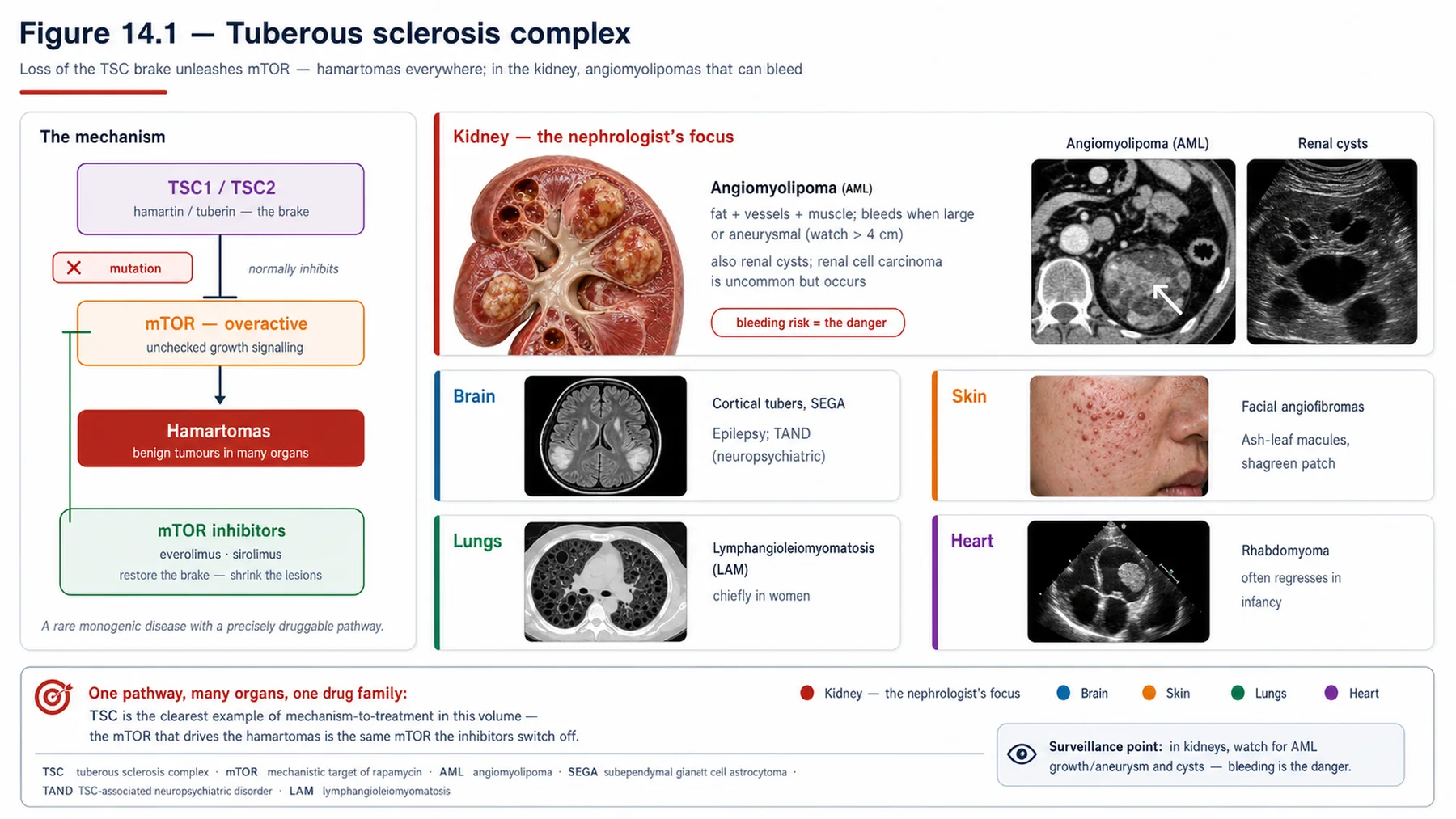
Tuberous sclerosis complex is an autosomal dominant tumour-suppressor syndrome with a clean mechanistic story. It is caused by loss-of-function of TSC1 (encoding hamartin) or TSC2 (encoding tuberin), whose proteins form the TSC1-TSC2 complex that normally restrains the mTOR signalling pathway — a master regulator of cell growth. When the complex is lost, mTOR signalling runs unrestrained, driving excessive cell growth and the formation of hamartomas (benign tumour-like overgrowths) in many organs. This single mechanism — unrestrained mTOR — explains the multisystem hamartomas of TSC and, crucially, points directly to the treatment: mTOR inhibitors. The renal manifestations of TSC are two. Angiomyolipomas are benign tumours composed of fat, smooth muscle, and abnormal blood vessels, often multiple and bilateral, whose main danger is haemorrhage — large angiomyolipomas (above about 4 cm) and those with aneurysms can bleed catastrophically. Renal cysts also occur (more with TSC2), and when a TSC2 mutation also involves the adjacent PKD1 gene (a contiguous gene deletion), the patient develops severe, early polycystic kidney disease. There is also a small increased risk of renal cell carcinoma. The renal lesions, especially the angiomyolipomas, are a major cause of morbidity in TSC.
— TSC: the extrarenal features and mTOR-inhibitor therapy
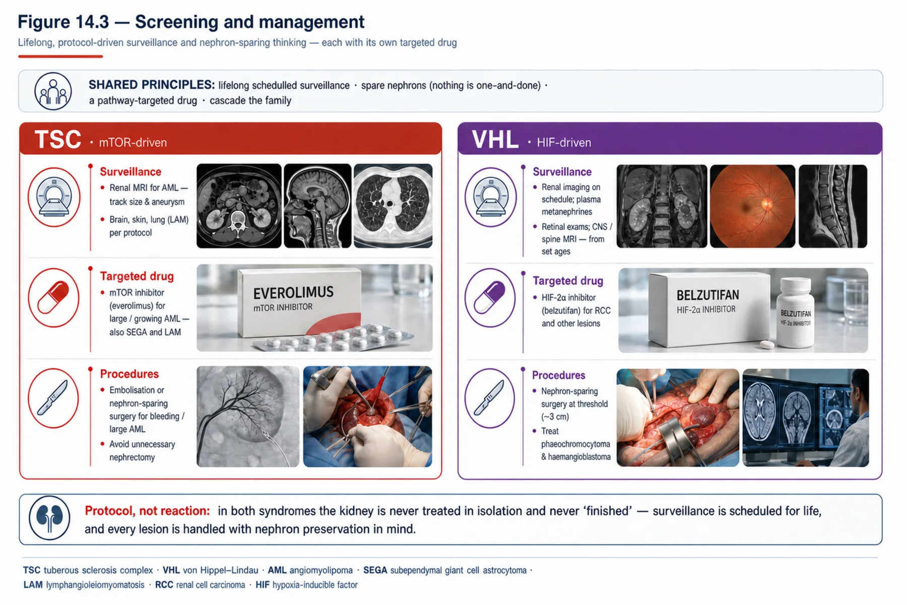
Because unrestrained mTOR causes hamartomas everywhere, TSC is a multisystem disease, and recognising the extrarenal features supports the diagnosis and the surveillance. The brain shows cortical tubers (causing epilepsy, intellectual disability, and autism) and subependymal giant cell astrocytomas (SEGA, which can obstruct cerebrospinal fluid flow and cause hydrocephalus). The skin shows characteristic lesions — facial angiofibromas, hypomelanotic 'ash-leaf' macules, shagreen patches, and ungual fibromas. The heart shows rhabdomyomas (often in infancy), the eye retinal hamartomas, and the lungs (in women) lymphangioleiomyomatosis (LAM). The therapeutic breakthrough, flowing directly from the mechanism, is the mTOR inhibitor: drugs such as everolimus and sirolimus inhibit the now-unrestrained mTOR pathway and are disease-modifying — they shrink the renal angiomyolipomas (reducing the bleeding risk), the subependymal giant cell astrocytomas, and the pulmonary lymphangioleiomyomatosis. So the management of TSC renal disease is: surveillance of angiomyolipomas; an mTOR inhibitor for angiomyolipomas above a size threshold (or growing or symptomatic), which shrinks them and reduces bleeding; and, for an acutely bleeding or very large angiomyolipoma, selective arterial embolisation or nephron-sparing surgery — avoiding total nephrectomy, because the disease is bilateral and nephrons must be preserved. The mechanism (mTOR) and the treatment (mTOR inhibitor) are beautifully linked.
— Von Hippel-Lindau: the HIF mechanism
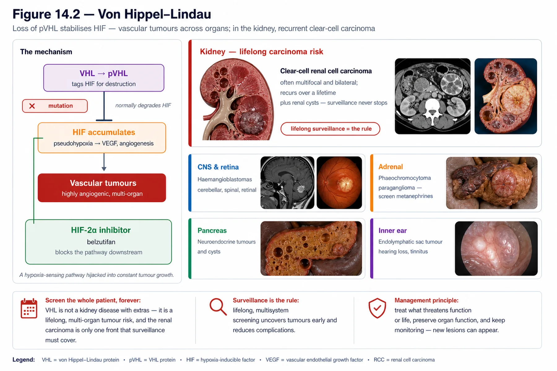
Von Hippel-Lindau is the other major renal phakomatosis, autosomal dominant, with an equally elegant mechanism. It is caused by loss of the VHL tumour-suppressor gene, whose protein normally targets hypoxia-inducible factor (HIF) for degradation. When VHL is lost, HIF is not degraded and accumulates — a state of 'pseudohypoxia' in which the cell behaves as though starved of oxygen — driving the upregulation of vascular endothelial growth factor (VEGF) and other pro-angiogenic and pro-growth factors. The result is a tendency to form highly vascular tumours throughout the body. The renal manifestations are renal cysts and, the major threat, clear-cell renal cell carcinoma — which in VHL is characteristically multiple, bilateral, and recurrent, arising over the patient's lifetime, and is a leading cause of death in the syndrome. This is quite different from the benign angiomyolipoma of TSC: in VHL the renal lesion is a malignancy, and a recurring one. The VHL-HIF-VEGF mechanism also points to therapy: because the tumours are driven by HIF and VEGF, agents targeting this axis (VEGF-pathway inhibitors, and the newer HIF-2-alpha inhibitor) are effective.
— VHL: the extrarenal tumours and management
Like TSC, VHL is multisystem, and its tumours, all reflecting the vascular, HIF-driven mechanism, define both the diagnosis and the surveillance. Beyond the renal cell carcinoma and cysts, VHL causes central-nervous-system and retinal haemangioblastomas (highly vascular tumours of the brain, spinal cord, and retina), phaeochromocytoma and paraganglioma (catecholamine-secreting tumours — the alpha-before-beta rule of the hypertension and endocrine chapters applies), pancreatic cysts and neuroendocrine tumours, endolymphatic sac tumours of the ear, and epididymal cystadenomas. The management of VHL rests on two pillars. First, lifelong surveillance: because the tumours arise and recur over a lifetime, patients undergo regular screening — imaging for renal tumours, brain and spine MRI for haemangioblastomas, and biochemical screening for phaeochromocytoma — to detect and treat tumours early. Second, organ-preserving treatment of the renal cell carcinoma: because the carcinoma is multiple, bilateral, and recurrent, the goal is to preserve kidney function over a lifetime of tumours, so the approach is nephron-sparing partial nephrectomy (not total nephrectomy) and active surveillance of small tumours with intervention at a size threshold (commonly around 3 cm) — removing tumours when they reach a size that risks metastasis while sparing as much kidney as possible. The newer HIF-2-alpha inhibitor belzutifan, targeting the mechanism directly, reduces tumour burden across the VHL lesions (renal, CNS, pancreatic) and is a major systemic advance. Phaeochromocytomas are managed with alpha-blockade then resection. So VHL is a disease of lifelong surveillance and nephron-sparing, mechanism-targeted treatment.
— Diagnosis, and the renal lesion as the clue
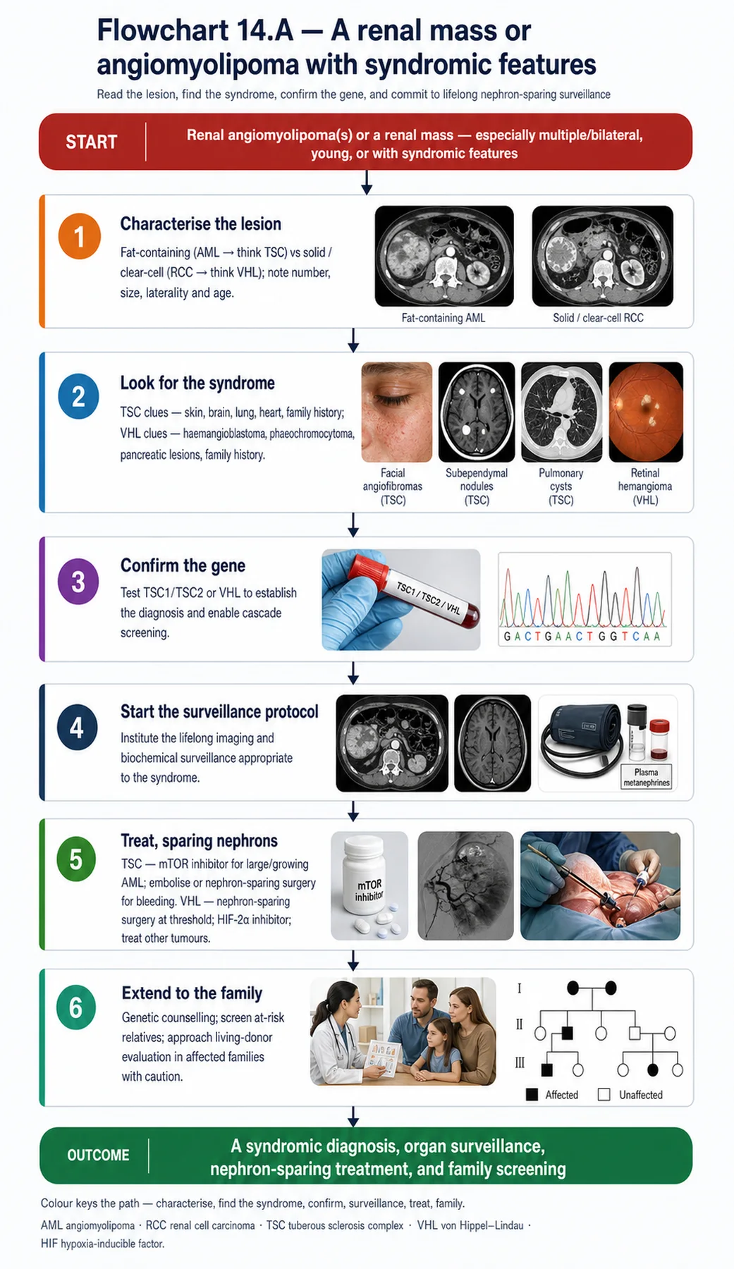
Diagnosing the phakomatoses brings together the renal lesion, the extrarenal features, and genetics — and the practical message is that the renal lesion may be the clue to the whole syndrome. Tuberous sclerosis is diagnosed by clinical diagnostic criteria (a constellation of major and minor features across skin, brain, kidney, heart, and lung) and/or genetic testing for TSC1 or TSC2 (with about two-thirds of cases sporadic, from de novo mutations, so a negative family history is common). Von Hippel-Lindau is diagnosed by its characteristic tumours, the family history, and genetic testing for the VHL gene. In both, the renal finding can be the presenting or recognising feature: multiple bilateral angiomyolipomas (especially with renal cysts) should prompt consideration of TSC and a search for the skin, brain, and other features; and clear-cell renal cell carcinoma — particularly if multiple, bilateral, recurrent, or in a young patient — should prompt consideration of VHL and a search for haemangioblastomas, phaeochromocytoma, and the other tumours. Recognising the renal lesion within its syndromic context (rather than as an isolated finding) triggers the genetic diagnosis, the screening for extrarenal disease, the targeted therapy (mTOR inhibitor or HIF inhibitor), the lifelong surveillance, and the genetic counselling and family screening. The unifying theme of the chapter is that, in these tumour-suppressor syndromes, an angiomyolipoma or a clear-cell carcinoma in the kidney is not just a renal problem but a window onto a multisystem disease — and the clinician who recognises it serves the patient and the family far beyond the kidney.
| 04 | PHASE A · LEVEL 4 · ORIENTATION & KNOWLEDGE Reference Tables |
Table 14.1 — The two renal phakomatoses
| Feature | Tuberous sclerosis (TSC) | von Hippel-Lindau (VHL) |
| Gene / pathway | TSC1/TSC2 → unrestrained mTOR | VHL → HIF accumulation → VEGF |
| Renal lesion | Angiomyolipomas (benign) + cysts | Clear-cell RCC (malignant) + cysts |
| Targeted therapy | mTOR inhibitors (everolimus/sirolimus) | VEGF-pathway / HIF-2α inhibitor (belzutifan) |
Table 14.2 — TSC: mechanism and renal lesions
| Aspect | Detail |
| Mechanism | TSC1 (hamartin)/TSC2 (tuberin) loss → unrestrained mTOR → hamartomas |
| Angiomyolipomas | Benign (fat/muscle/vessel); multiple/bilateral; bleed if large (>~4 cm)/aneurysmal |
| Cysts | More with TSC2; TSC2/PKD1 contiguous deletion → severe early polycystic disease |
| Also | Small increased renal cell carcinoma risk |
Table 14.3 — TSC: extrarenal features and treatment
| Aspect | Detail |
| Brain | Cortical tubers (epilepsy, intellectual disability); subependymal giant cell astrocytoma |
| Skin / heart / lung | Facial angiofibromas, ash-leaf macules, shagreen; cardiac rhabdomyomas; LAM (women) |
| mTOR inhibitors | Everolimus/sirolimus — shrink angiomyolipomas, SEGA, LAM (disease-modifying) |
| AML management | Surveillance; mTOR inhibitor (size threshold); embolisation/nephron-sparing surgery for bleeding/large — avoid total nephrectomy |
Table 14.4 — VHL: mechanism and renal lesions
| Aspect | Detail |
| Mechanism | VHL loss → HIF not degraded → 'pseudohypoxia' → VEGF/angiogenesis → vascular tumours |
| Renal cell carcinoma | Clear-cell; multiple, bilateral, recurrent; a leading cause of death |
| Cysts | Renal cysts also occur |
| Contrast with TSC | VHL renal lesion is MALIGNANT and recurrent (vs benign AML in TSC) |
Table 14.5 — VHL: extrarenal tumours and management
| Aspect | Detail |
| Extrarenal tumours | CNS/retinal haemangioblastomas; phaeochromocytoma; pancreatic cysts/tumours; endolymphatic sac |
| Surveillance | Lifelong — renal imaging, brain/spine MRI, biochemical phaeochromocytoma screening |
| Renal cell carcinoma | Nephron-sparing partial nephrectomy; active surveillance, intervene at ~3 cm (preserve function) |
| Targeted / phaeo | HIF-2α inhibitor (belzutifan) reduces tumour burden; phaeochromocytoma — alpha-block then resect |
Table 14.6 — Diagnosis: the renal lesion as the clue
| Point | Detail |
| TSC diagnosis | Clinical criteria (major/minor features) and/or TSC1/TSC2 genetics; ~2/3 sporadic (de novo) |
| VHL diagnosis | Characteristic tumours, family history, VHL genetics |
| Renal clue (TSC) | Multiple bilateral angiomyolipomas (+ cysts) → search for skin/brain/other features |
| Renal clue (VHL) | Multiple/bilateral/recurrent or young-onset clear-cell RCC → search for haemangioblastoma/phaeo |
| 05 | PHASE B · LEVEL 5 · VISUALISE & MAP Imaging & Flowchart Specifications |
| 06 | PHASE B · LEVEL 6 · VISUALISE & MAP Concept Maps |
Each chain runs from mechanism to a named clinical action; read the arrows as “leads to.”
Phakomatoses overview. Tumour-suppressor loss → multisystem tumours, including renal → TSC (angiomyolipomas) and VHL (clear-cell RCC) → ACTION: recognise the renal lesion as a clue to a whole-body syndrome.
TSC mechanism. TSC1/TSC2 loss → unrestrained mTOR → hamartomas (angiomyolipomas, cysts, tubers, LAM) → ACTION: recognise TSC and use the mTOR node as the treatment target.
TSC treatment. mTOR inhibitors shrink angiomyolipomas/SEGA/LAM → reduce bleeding risk → ACTION: use an mTOR inhibitor for AMLs above threshold; embolise/spare nephrons for bleeding, avoid total nephrectomy.
VHL mechanism. VHL loss → HIF accumulates → VEGF/angiogenesis → vascular tumours including clear-cell RCC (multiple/bilateral/recurrent) → ACTION: recognise VHL and the malignant, recurrent renal lesion.
VHL surveillance/treatment. Lifelong recurrent tumours → surveillance + nephron-sparing surgery (threshold ~3 cm) + HIF-2α inhibitor → ACTION: screen lifelong, spare nephrons, and use targeted therapy.
| 07 | PHASE B · LEVEL 7 · VISUALISE & MAP Decision Pathways |
| R1 | IF a patient has multiple or bilateral renal angiomyolipomas (especially with cysts), THEN suspect tuberous sclerosis and search for the skin, brain, heart, and lung features. |
| R2 | IF TSC is suspected, THEN diagnose by the clinical criteria and/or TSC1/TSC2 genetics (remembering ~2/3 are sporadic). |
| R3 | IF a TSC angiomyolipoma is large (above ~4 cm), growing, or symptomatic, THEN treat with an mTOR inhibitor (which shrinks it and reduces bleeding). |
| R4 | IF a TSC angiomyolipoma is acutely bleeding or very large, THEN use embolisation or nephron-sparing surgery — avoid total nephrectomy. |
| R5 | IF a patient has clear-cell renal cell carcinoma that is multiple, bilateral, recurrent, or young-onset, THEN suspect von Hippel-Lindau and search for haemangioblastomas, phaeochromocytoma, and pancreatic lesions. |
| R6 | IF VHL is confirmed, THEN institute lifelong surveillance for the renal, CNS, and adrenal tumours. |
| R7 | IF VHL renal cell carcinoma is treated, THEN use nephron-sparing partial nephrectomy with a size threshold (~3 cm), and consider the HIF-2α inhibitor — to preserve kidney function over a lifetime of recurrent tumours. |
| R8 | IF a renal lesion suggests a phakomatosis, THEN provide multidisciplinary care, genetic counselling, and cascade family screening. |
| 08 | PHASE C · LEVEL 8 · CLINICAL REASONING Clinical Cases |
| CASE 1 | FAT AND VESSELS, AND MORE mTOR drives it Tuberous sclerosis |
Presentation
A young adult is found to have multiple bilateral renal angiomyolipomas and some renal cysts, and on examination has facial angiofibromas and a history of epilepsy. One large angiomyolipoma is growing.
❖ Pause and reflect What syndrome unites these findings, and how should the growing angiomyolipoma be managed? |
Analysis
Tuberous sclerosis complex — and the growing angiomyolipoma calls for an mTOR inhibitor. The combination is classic TSC: multiple bilateral renal angiomyolipomas with cysts (the renal lesions), facial angiofibromas (the skin lesion), and epilepsy (from cortical tubers) — reflecting the unrestrained mTOR signalling from loss of TSC1 or TSC2 that drives hamartomas across organs. The renal angiomyolipomas matter because large ones (above about 4 cm) and those that are growing can haemorrhage catastrophically. The mechanism points to the treatment: an mTOR inhibitor (everolimus or sirolimus) inhibits the unrestrained pathway and shrinks the angiomyolipomas, reducing the bleeding risk — so the growing, large angiomyolipoma here should be treated with an mTOR inhibitor (rather than waiting for it to bleed). Acute bleeding or a very large lesion would call for embolisation or nephron-sparing surgery, avoiding total nephrectomy (the disease is bilateral). The patient should also be assessed for the other TSC features and have genetic confirmation and counselling.
Plan
Diagnose TSC from the angiomyolipomas, cysts, skin lesions, and epilepsy; treat the large, growing angiomyolipoma with an mTOR inhibitor (shrinks it, reduces bleeding); reserve embolisation/nephron-sparing surgery for bleeding/very large lesions; and assess the other features with genetic confirmation. Use an mTOR inhibitor for the large/growing angiomyolipoma.
Teaching point
Multiple bilateral angiomyolipomas with skin and brain features are tuberous sclerosis — unrestrained mTOR; treat large/growing angiomyolipomas with an mTOR inhibitor (avoid total nephrectomy).
Cross-reference
Exercises rules R1, R3, and R4; the TSC mechanism and treatment concept maps; Figure 14.1; Tables 14.2, 14.3.
| CASE 2 | RECURRENT CANCER, SPARE THE KIDNEY Nephron-sparing surgery von Hippel-Lindau |
Presentation
A young patient has clear-cell renal cell carcinoma that is bilateral and has recurred after a previous resection, along with a cerebellar haemangioblastoma. A surgeon proposes a radical (total) nephrectomy of the more affected kidney.
❖ Pause and reflect Is total nephrectomy the right approach in this recurrent bilateral disease? |
Analysis
No — this is von Hippel-Lindau, and the approach should be nephron-sparing, not total nephrectomy. The picture — young-onset, bilateral, recurrent clear-cell renal cell carcinoma with a cerebellar haemangioblastoma — is classic VHL, in which loss of the VHL gene lets HIF accumulate and drive VEGF-mediated vascular tumours, with the renal cell carcinoma characteristically multiple, bilateral, and recurrent over the patient's lifetime. Because the carcinoma keeps recurring in both kidneys, the management goal is to preserve kidney function over a lifetime of tumours: this means nephron-sparing partial nephrectomy (removing the tumour while sparing the kidney), with active surveillance of small tumours and intervention at a size threshold (commonly around 3 cm, balancing metastasis risk against nephron preservation) — not a total nephrectomy, which would sacrifice a kidney that will face more tumours and hasten kidney failure. The newer HIF-2-alpha inhibitor belzutifan can reduce the tumour burden across the VHL lesions. The patient also needs lifelong surveillance and genetic counselling.
Plan
Recognise VHL, use nephron-sparing partial nephrectomy with a size threshold (not total nephrectomy) to preserve function over recurrent bilateral disease, consider the HIF-2α inhibitor, and institute lifelong surveillance and counselling. Spare nephrons in VHL renal cell carcinoma.
Teaching point
VHL renal cell carcinoma is multiple, bilateral, and recurrent — use nephron-sparing partial nephrectomy (threshold ~3 cm), not total nephrectomy, to preserve function; consider belzutifan.
Cross-reference
Exercises rules R5 and R7; the VHL mechanism and surveillance concept maps; Figure 14.2; Tables 14.4, 14.5.
| CASE 3 | A WINDOW ONTO THE WHOLE BODY The renal lesion as the clue Recognising the syndrome |
Presentation
A young patient presents with a single clear-cell renal cell carcinoma, managed as an isolated tumour with no consideration of an underlying syndrome or family screening.
❖ Pause and reflect Should a young patient's clear-cell carcinoma be treated as an isolated tumour? |
Analysis
Not without considering an underlying syndrome — the renal lesion may be the clue to von Hippel-Lindau. Clear-cell renal cell carcinoma in a young patient (or that is multiple, bilateral, or recurrent) should prompt consideration of VHL rather than being managed as an isolated sporadic tumour, because in these syndromic settings the carcinoma is the renal manifestation of a multisystem tumour-suppressor disease. Recognising it as a possible VHL clue triggers the search for the extrarenal tumours (CNS and retinal haemangioblastomas, phaeochromocytoma, pancreatic lesions), genetic testing of the VHL gene, lifelong surveillance, and — critically — cascade screening of relatives, who may also carry the mutation and need surveillance. Treating the carcinoma as an isolated tumour misses all of this: the patient's other (and future) tumours, and the at-risk family. The broader lesson, shared with TSC (where multiple bilateral angiomyolipomas are the clue), is that a renal lesion in these syndromes is a window onto a whole-body disease, and recognising it serves the patient and family far beyond the kidney.
Plan
Consider VHL behind the young-onset clear-cell carcinoma, search for the extrarenal tumours, perform VHL genetic testing, institute surveillance, and cascade-screen the family — rather than managing it as an isolated tumour. Treat the renal lesion as a possible clue to a syndrome.
Teaching point
A young or multiple/bilateral/recurrent clear-cell carcinoma is a clue to VHL (as multiple bilateral angiomyolipomas are to TSC) — recognise the syndrome, screen the body, and cascade-screen the family.
Cross-reference
Exercises rules R5 and R8; the phakomatoses-overview concept map; Figure 14.3; Table 14.6; the renal-lesion-as-clue principle in Chapter 1.
| CASE 4 | WATCH FOR A LIFETIME Surveillance and the extrarenal tumours VHL surveillance |
Presentation
A patient with established VHL is followed only for their kidneys, with no systematic surveillance for the CNS, retinal, or adrenal tumours. They develop a symptomatic cerebellar haemangioblastoma and an undetected phaeochromocytoma.
❖ Pause and reflect What is missing from this patient's VHL care? |
Analysis
Lifelong, systematic, multi-organ surveillance — the cornerstone of VHL care. VHL causes tumours that arise and recur throughout the body over a lifetime, so following only the kidneys leaves the other tumours to present late and dangerously, as here: a symptomatic cerebellar haemangioblastoma (which surveillance brain MRI would have detected earlier) and an undetected phaeochromocytoma (which biochemical screening would have caught — and which is dangerous, especially if unrecognised before surgery or anaesthesia, given the alpha-before-beta rule). Proper VHL surveillance is lifelong and multi-organ: regular imaging for renal tumours, brain and spine MRI for haemangioblastomas, biochemical screening for phaeochromocytoma, and ophthalmological screening for retinal haemangioblastomas — to detect and treat each tumour early. The phaeochromocytoma, once found, is managed with alpha-blockade before beta-blockade and then resection. Restricting follow-up to the kidneys, in a multisystem syndrome, is a serious gap; surveillance must cover the whole body.
Plan
Institute lifelong multi-organ VHL surveillance (renal imaging, brain/spine MRI, biochemical phaeochromocytoma screening, retinal screening), treat the haemangioblastoma, and manage the phaeochromocytoma with alpha-blockade then resection. Surveil the whole body, not just the kidneys, in VHL.
Teaching point
VHL needs lifelong multi-organ surveillance (renal, CNS/spine, retinal, biochemical for phaeochromocytoma) — following only the kidneys misses the other recurrent tumours.
Cross-reference
Exercises rule R6; the VHL surveillance concept map; Table 14.5; phaeochromocytoma in Chapter 9 and Volume 8.
| 09 | PHASE C · LEVEL 9 · CLINICAL REASONING Clinical Implications |
One triad per mechanism the narrative exposed: the physiology, why it matters, and the clinical move.
MECHANISM TSC1/2 loss unleashes mTOR, driving hamartomas including renal angiomyolipomas. |
WHY IT MATTERS Large angiomyolipomas can haemorrhage catastrophically. |
ACTION Use an mTOR inhibitor for large/growing angiomyolipomas; avoid total nephrectomy. |
MECHANISM TSC is multisystem (skin, brain, heart, lung) from the same mTOR mechanism. |
WHY IT MATTERS The extrarenal features support the diagnosis and surveillance. |
ACTION Search for the skin, brain, and lung features when angiomyolipomas are found. |
MECHANISM VHL loss lets HIF accumulate, driving VEGF and vascular tumours. |
WHY IT MATTERS The renal cell carcinoma is multiple, bilateral, and recurrent — a leading cause of death. |
ACTION Spare nephrons (partial nephrectomy, threshold ~3 cm) and consider a HIF-2α inhibitor. |
MECHANISM VHL tumours arise and recur across organs over a lifetime. |
WHY IT MATTERS Following only the kidneys lets other tumours present late. |
ACTION Institute lifelong multi-organ surveillance. |
MECHANISM A renal angiomyolipoma or clear-cell carcinoma may be the first sign of the syndrome. |
WHY IT MATTERS It is a window onto a multisystem, heritable disease. |
ACTION Recognise the renal lesion as a clue, screen the body, and cascade-screen the family. |
| 10 | PHASE C · LEVEL 10 · CLINICAL REASONING Clinical Pearls |
| Phakomatoses = tumour-suppressor syndromes with renal involvement: TSC and VHL. | TSC: autosomal dominant; TSC1 (hamartin)/TSC2 (tuberin) loss → unrestrained mTOR. |
| TSC renal: angiomyolipomas (benign, bleed if large) + cysts. | TSC2/PKD1 contiguous deletion → severe early polycystic disease. |
| TSC extrarenal: tubers/epilepsy, SEGA, skin lesions, rhabdomyomas, LAM. | mTOR inhibitors (everolimus/sirolimus) shrink AMLs, SEGA, LAM (disease-modifying). |
| Large/growing AML → mTOR inhibitor; bleeding/large → embolisation/nephron-sparing surgery. | Avoid total nephrectomy in TSC (bilateral disease). |
| VHL: autosomal dominant; VHL loss → HIF accumulates → VEGF → vascular tumours. | VHL renal: clear-cell RCC (multiple, bilateral, recurrent) + cysts. |
| VHL RCC is MALIGNANT and recurrent (vs benign AML in TSC). | VHL extrarenal: CNS/retinal haemangioblastomas, phaeochromocytoma, pancreatic tumours. |
| VHL: lifelong multi-organ surveillance. | VHL RCC: nephron-sparing partial nephrectomy, intervene at ~3 cm (preserve function). |
| HIF-2α inhibitor (belzutifan) reduces VHL tumour burden. | The renal lesion (AML or clear-cell RCC) is the clue to the syndrome — screen body + family. |
| 11 | PHASE D · LEVEL 11 · SAFETY & EVIDENCE Red Flags & Never-Do |
Panel A — Red flags
| ▲ | Multiple bilateral renal angiomyolipomas (± cysts) — suspect tuberous sclerosis; search skin/brain/lung. |
| ▲ | A large (>~4 cm) or growing angiomyolipoma — bleeding risk; treat with an mTOR inhibitor. |
| ▲ | Multiple/bilateral/recurrent or young-onset clear-cell renal cell carcinoma — suspect von Hippel-Lindau. |
| ▲ | A VHL patient followed only for the kidneys — missing CNS, retinal, and adrenal tumours; surveil the whole body. |
| ▲ | An undetected phaeochromocytoma in VHL — dangerous before surgery; screen and apply alpha-before-beta. |
Panel B — Never do
| ✖ NEVER — perform total nephrectomy for a TSC angiomyolipoma when nephron-sparing options exist. |
| ✖ NEVER — treat VHL renal cell carcinoma with total nephrectomy when partial nephrectomy preserves function. |
| ✖ NEVER — manage a young or recurrent clear-cell carcinoma as an isolated tumour without considering VHL. |
| ✖ NEVER — restrict VHL follow-up to the kidneys — surveil the whole body. |
| 12 | PHASE D · LEVEL 12 · SAFETY & EVIDENCE Common Pitfalls |
Pitfall 1 — Missing TSC behind angiomyolipomas
| ✖ | WRONG Treating multiple angiomyolipomas as an isolated renal finding. |
| ✓ | RIGHT Recognising the TSC syndrome and searching for the other features. |
| ✉ | WHY Multiple bilateral angiomyolipomas point to tuberous sclerosis. |
Pitfall 2 — Total nephrectomy in TSC
| ✖ | WRONG Removing a whole kidney for a large angiomyolipoma. |
| ✓ | RIGHT Using an mTOR inhibitor or embolisation/nephron-sparing surgery. |
| ✉ | WHY The disease is bilateral — nephrons must be preserved. |
Pitfall 3 — Total nephrectomy in VHL
| ✖ | WRONG Performing total nephrectomy for VHL renal cell carcinoma. |
| ✓ | RIGHT Using nephron-sparing partial nephrectomy with a size threshold. |
| ✉ | WHY The carcinoma is recurrent and bilateral; function must be preserved. |
Pitfall 4 — Isolated-tumour thinking
| ✖ | WRONG Managing a young clear-cell carcinoma as a sporadic isolated tumour. |
| ✓ | RIGHT Considering VHL, screening the body, and cascade-screening the family. |
| ✉ | WHY The renal lesion may be the clue to a multisystem syndrome. |
Pitfall 5 — Kidney-only VHL follow-up
| ✖ | WRONG Following a VHL patient for the kidneys alone. |
| ✓ | RIGHT Lifelong multi-organ surveillance (CNS, retina, adrenal). |
| ✉ | WHY VHL tumours arise and recur throughout the body. |
| 13 | PHASE D · LEVEL 13 · SAFETY & EVIDENCE Evidence Grading |
GRADE A HIGH CONFIDENCE The effect is real and the estimate is stable. RCTs at low risk of bias; multiple concordant prospective cohorts; meta-analyses. |
GRADE B MODERATE CONFIDENCE The effect is likely real but may shift with new data. Observational studies, registries, mechanistic human studies. |
GRADE C LOW CONFIDENCE Rests on physiology, reasoning, or consensus rather than outcomes. Pathophysiological reasoning; extrapolation; consensus without outcomes. |
Graded statements (by evidence type)
| Statement | Grade | Basis (evidence type) |
| TSC results from TSC1/TSC2 loss with unrestrained mTOR signalling. | A | Molecular genetics |
| mTOR inhibitors shrink angiomyolipomas and reduce bleeding risk. | A | RCTs |
| VHL results from VHL loss with HIF accumulation and VEGF upregulation. | A | Molecular biology |
| VHL renal cell carcinoma is multiple, bilateral, and recurrent. | A | Cohort data |
| Nephron-sparing surgery with a size threshold preserves function in VHL. | A | Clinical cohort data |
| The HIF-2α inhibitor belzutifan reduces VHL tumour burden. | A | RCT |
| Lifelong surveillance improves outcomes in VHL. | A | Guidelines and cohort data |
| 14 | PHASE E · LEVEL 14 · PATIENT DECISIONS Absolute Risk in Natural Frequency |
Natural-frequency estimates for orientation, from phakomatosis cohorts; they vary with the syndrome. They convey the size of the decisions, expressed per 100 comparable patients.
| Per 100 patients… | Outcome | Roughly how many | See |
| TSC patients | Have renal angiomyolipomas | Most — the major renal lesion | L13 row 1 |
| TSC angiomyolipomas treated with an mTOR inhibitor | Shrink (reduced bleeding risk) | Many | L13 row 2 |
| VHL patients | Develop clear-cell renal cell carcinoma over a lifetime | Most — a leading cause of death | L13 row 4 |
| VHL renal cell carcinoma managed nephron-sparing vs total nephrectomy | Preserve kidney function | More with nephron-sparing surgery | L13 row 5 |
★ How to read these Read these as orientation, not promises; outcomes vary with the syndrome. The stable signals: angiomyolipomas are the major TSC renal lesion and shrink with mTOR inhibitors, VHL carcinoma is lifelong and recurrent, and nephron-sparing surgery preserves function. Communicate them as people out of 100, not as a hazard ratio. |
| 17 | PHASE F · LEVEL 17 · APPLY & TEST Documentation Templates |
Paste-ready notes. Tick the boxes that apply and delete the rest; make the syndrome, the renal lesion, and the surveillance explicit.
Template 1 — Tuberous sclerosis
Template 2 — von Hippel-Lindau
| 18 | PHASE F · LEVEL 18 · APPLY & TEST Cheat Sheet |
| Phakomatoses with renal involvement: TSC and VHL. | TSC: TSC1/TSC2 loss → unrestrained mTOR → hamartomas. |
| TSC renal: angiomyolipomas (benign, bleed if large) + cysts. | TSC2/PKD1 deletion → severe early polycystic disease. |
| TSC extrarenal: tubers/epilepsy, SEGA, skin, rhabdomyomas, LAM. | mTOR inhibitors shrink AMLs/SEGA/LAM. |
| Large/growing AML → mTOR inhibitor; bleeding → embolise/spare nephrons. | Avoid total nephrectomy in TSC. |
| VHL: VHL loss → HIF accumulates → VEGF → vascular tumours. | VHL renal: clear-cell RCC (multiple/bilateral/recurrent) + cysts. |
| VHL RCC is malignant and recurrent (vs benign AML). | VHL extrarenal: haemangioblastomas, phaeochromocytoma, pancreatic tumours. |
| VHL: lifelong multi-organ surveillance. | VHL RCC: nephron-sparing partial nephrectomy, threshold ~3 cm. |
| HIF-2α inhibitor (belzutifan) reduces VHL tumour burden. | The renal lesion is the clue — screen body + family. |
| 19 | PHASE F · LEVEL 19 · APPLY & TEST Flashcards |
| CARD 1 | Q. What are the phakomatoses, and which two involve the kidney? A. Inherited tumour-suppressor syndromes with multisystem (neurocutaneous and visceral) manifestations; the two with major renal involvement are tuberous sclerosis complex (TSC) and von Hippel-Lindau (VHL). DETAILED. Each has a characteristic renal lesion. CLINICAL. Recognise the renal lesion as a clue to the syndrome. |
| CARD 2 | Q. What is the mechanism and the renal lesions of tuberous sclerosis? A. Autosomal dominant loss of TSC1 (hamartin) or TSC2 (tuberin), which normally restrain mTOR; their loss unleashes mTOR signalling, producing hamartomas — in the kidney, angiomyolipomas (benign, with a haemorrhage risk when large) and cysts (with severe polycystic disease if a TSC2/PKD1 contiguous deletion). DETAILED. The same mechanism causes the extrarenal hamartomas. CLINICAL. Recognise TSC and target the mTOR pathway. |
| CARD 3 | Q. How are angiomyolipomas in tuberous sclerosis treated? A. By surveillance, with mTOR inhibitors (everolimus, sirolimus) for large (above ~4 cm), growing, or symptomatic angiomyolipomas — which shrink them and reduce the bleeding risk — and selective arterial embolisation or nephron-sparing surgery for acute bleeding or very large lesions, avoiding total nephrectomy (the disease is bilateral). DETAILED. The treatment follows the mTOR mechanism. CLINICAL. Use an mTOR inhibitor for large angiomyolipomas; spare nephrons. |
| CARD 4 | Q. What is the mechanism and the renal lesion of von Hippel-Lindau? A. Autosomal dominant loss of the VHL gene, whose protein normally degrades hypoxia-inducible factor (HIF); loss lets HIF accumulate ('pseudohypoxia'), driving VEGF and angiogenesis and producing highly vascular tumours — in the kidney, cysts and clear-cell renal cell carcinoma that is multiple, bilateral, and recurrent. DETAILED. The renal lesion is malignant and recurrent (unlike the benign angiomyolipoma of TSC). CLINICAL. Recognise VHL and the recurrent malignant renal lesion. |
| CARD 5 | Q. How is renal cell carcinoma managed in von Hippel-Lindau? A. With nephron-sparing partial nephrectomy and active surveillance of small tumours, intervening at a size threshold (commonly around 3 cm) to balance metastasis risk against nephron preservation — because the carcinoma is multiple, bilateral, and recurrent over a lifetime — and the HIF-2-alpha inhibitor belzutifan reduces tumour burden. DETAILED. Total nephrectomy would sacrifice a kidney that will face more tumours. CLINICAL. Spare nephrons (threshold ~3 cm) and consider a HIF-2α inhibitor. |
| CARD 6 | Q. What are the extrarenal tumours of von Hippel-Lindau, and what surveillance is needed? A. CNS and retinal haemangioblastomas, phaeochromocytoma, pancreatic cysts and neuroendocrine tumours, endolymphatic sac tumours, and epididymal cystadenomas; surveillance is lifelong and multi-organ — renal imaging, brain and spine MRI, biochemical phaeochromocytoma screening, and retinal screening. DETAILED. The tumours arise and recur over a lifetime. CLINICAL. Institute lifelong multi-organ surveillance. |
| CARD 7 | Q. How are the phakomatoses diagnosed? A. Tuberous sclerosis by clinical diagnostic criteria (major and minor features across skin, brain, kidney, heart, and lung) and/or genetic testing (TSC1/TSC2), with about two-thirds sporadic; von Hippel-Lindau by its characteristic tumours, family history, and VHL genetic testing. DETAILED. The renal lesion may be the presenting clue. CLINICAL. Use the clinical features and genetics, prompted by the renal lesion. |
| CARD 8 | Q. Why is the renal lesion important beyond the kidney in these syndromes? A. Because an angiomyolipoma (TSC) or a clear-cell carcinoma (VHL) may be the first sign of a multisystem, heritable tumour-suppressor syndrome — so recognising it triggers the search for extrarenal tumours, genetic testing, lifelong surveillance, targeted therapy, and cascade screening of relatives. DETAILED. The renal lesion is a window onto a whole-body disease. CLINICAL. Recognise the renal lesion as a clue, screen the body, and cascade-screen the family. |
| 20 | PHASE F · LEVEL 20 · APPLY & TEST One-Minute Preceptor |
| SCENE 1 | The intern removing a kidney for an angiomyolipoma |
GET A COMMITMENT. “You want a total nephrectomy for this large angiomyolipoma in a TSC patient — why?”
PROBE FOR EVIDENCE. “It's big and might bleed” — ask: “What does the mTOR mechanism offer as a treatment, and what's the problem with total nephrectomy in TSC?”
TEACH A GENERAL RULE. TSC angiomyolipomas shrink with mTOR inhibitors (reducing bleeding); embolisation or nephron-sparing surgery handles bleeding/large lesions — avoid total nephrectomy, because the disease is bilateral.
REINFORCE WHAT WAS RIGHT. Recognising the bleeding risk was correct.
CORRECT A MISTAKE. Use an mTOR inhibitor / nephron-sparing approach, not total nephrectomy.
| SCENE 2 | The resident following only the kidneys in VHL |
GET A COMMITMENT. “You're following this VHL patient just for the kidneys — is that enough?”
PROBE FOR EVIDENCE. “The kidneys are the renal problem” — ask: “Where else do VHL tumours arise, and what happens if they're not surveilled?”
TEACH A GENERAL RULE. VHL tumours arise and recur throughout the body — CNS/retinal haemangioblastomas, phaeochromocytoma, pancreatic tumours — so surveillance must be lifelong and multi-organ, not kidney-only.
REINFORCE WHAT WAS RIGHT. Following the kidneys was necessary but not sufficient.
CORRECT A MISTAKE. Institute lifelong multi-organ surveillance.
| 22 | PHASE F · LEVEL 22 · APPLY & TEST Board-Style Questions |
| Q 01 | Tuberous sclerosis results from loss of TSC1/TSC2, leading to: |
| A | HIF accumulation |
| B | Unrestrained mTOR signalling → hamartomas |
| C | Collagen IV deficiency |
| D | Vasopressin excess |
Rationale TSC1/2 loss unleashes mTOR, producing hamartomas (Figure 14.1, Table 14.2). A is VHL; C is Alport; D is unrelated. |
| Q 02 | The characteristic renal lesion of tuberous sclerosis is: |
| A | Clear-cell renal cell carcinoma |
| B | Angiomyolipoma (benign; bleeds if large) |
| C | Membranous nephropathy |
| D | Medullary sponge kidney |
Rationale Angiomyolipomas are the TSC renal lesion (Table 14.2). A is VHL; C and D are different. |
| Q 03 | A large, growing angiomyolipoma in tuberous sclerosis is best treated with: |
| A | Total nephrectomy |
| B | An mTOR inhibitor (everolimus/sirolimus) |
| C | Observation only |
| D | Chemotherapy |
Rationale mTOR inhibitors shrink AMLs and reduce bleeding; avoid total nephrectomy (case 1, Table 14.3, rules R3–R4). A, C, and D are wrong. |
| Q 04 | Von Hippel-Lindau results from loss of VHL, leading to: |
| A | Unrestrained mTOR |
| B | HIF accumulation → VEGF → vascular tumours |
| C | ENaC overactivity |
| D | Uromodulin misfolding |
Rationale VHL loss lets HIF accumulate, driving VEGF and vascular tumours (Figure 14.2, Table 14.4). A is TSC; C is Liddle; D is ADTKD-UMOD. |
| Q 05 | The characteristic renal lesion of von Hippel-Lindau is: |
| A | Benign angiomyolipoma |
| B | Clear-cell renal cell carcinoma (multiple, bilateral, recurrent) |
| C | Thin basement membrane |
| D | Cystinuria |
Rationale VHL causes recurrent clear-cell carcinoma, a leading cause of death (case 2, Table 14.4). A is TSC; C and D are different. |
| Q 06 | Renal cell carcinoma in von Hippel-Lindau is best managed with: |
| A | Total nephrectomy |
| B | Nephron-sparing partial nephrectomy with a size threshold (~3 cm) |
| C | Observation indefinitely |
| D | Dialysis |
Rationale Nephron-sparing surgery preserves function given recurrent bilateral disease (case 2, Table 14.5, rule R7). A, C, and D are wrong. |
| Q 07 | Surveillance in von Hippel-Lindau should be: |
| A | Kidney-only |
| B | Lifelong and multi-organ (renal, CNS/spine, retinal, phaeochromocytoma) |
| C | One-time |
| D | Unnecessary |
Rationale VHL tumours arise across organs over a lifetime (case 4, Table 14.5, rule R6). A, C, and D miss tumours. |
| Q 08 | A young patient with a clear-cell renal cell carcinoma should prompt consideration of: |
| A | No syndrome — treat as isolated |
| B | Von Hippel-Lindau — screen the body and the family |
| C | Tuberous sclerosis |
| D | ADPKD |
Rationale Young/recurrent/bilateral clear-cell carcinoma is a VHL clue (case 3, Figure 14.3, Table 14.6, rule R5). A misses it; C and D have different renal lesions. |