Chapter Preamble
Signals declared
Sig-D — Diagnostic (primary). Diagnose stones and their type, perform the metabolic evaluation, and recognise inherited stone disease.
Sig-T — Therapeutic (strong). Manage the acute stone, prevent recurrence by type, and treat the inherited stone diseases.
Sig-M — Mechanistic (strong). How supersaturation, urine pH, citrate, and the specific metabolic drivers produce each stone type.
Levels populated and omitted
Populated (19): L1–L14, L17–L20, L22. The mechanistic signal fires the concept maps (L6) and triads (L9); the therapeutic signal fires the absolute-risk table (L14) and templates (L17); the diagnostic signal drives the tables, rules, cases, pitfalls, and board items.
L15 / L16 preference-sensitive map and SDM scripts — omitted. No Sig-E; stone diagnosis, prevention, and treatment are established care.
L21 reflective prompts — omitted. No Sig-E/V; the content is worked through the cases and pitfalls.
| 01 | PHASE A · LEVEL 1 · ORIENTATION & KNOWLEDGE Learning Objectives |
By the end of this chapter you should be able to:
Explain stone formation — supersaturation, promoters, and the citrate inhibitor.
Describe the stone types and their metabolic and pH drivers.
Explain why uric acid stones form in acid urine and can be dissolved by alkalinisation.
Recognise struvite (infection) stones and their management.
Perform a metabolic evaluation and apply type-specific prevention.
Explain why dietary calcium should be normal, not low, in calcium stone-formers.
Recognise the inherited stone diseases (cystinuria, primary hyperoxaluria, APRT deficiency).
Manage the acute stone and the inherited stone diseases.
| 02 | PHASE A · LEVEL 2 · ORIENTATION & KNOWLEDGE Executive Summary |
Stones form when the urine is supersaturated with a stone-forming salt, allowing crystals to nucleate, grow, and aggregate — promoted by a high solute load, low urine volume, and an abnormal pH, and inhibited chiefly by citrate.
Calcium stones (calcium oxalate and calcium phosphate) are the commonest, driven by hypercalciuria (the commonest metabolic abnormality), hyperoxaluria, hypocitraturia, and low urine volume.
Uric acid stones form in persistently acidic urine, are radiolucent, are associated with gout and metabolic syndrome, and can be dissolved by alkalinising the urine.
Struvite (infection) stones are caused by urease-producing bacteria that alkalinise the urine; they form staghorn calculi and require complete surgical removal and eradication of the infection.
Cystine stones arise from the inherited disease cystinuria.
The inherited stone diseases include cystinuria (defective cystine reabsorption), primary hyperoxaluria (hepatic oxalate overproduction causing recurrent stones, nephrocalcinosis, and systemic oxalosis), and APRT deficiency (2,8-dihydroxyadenine stones, treated with allopurinol).
Primary hyperoxaluria now has disease-modifying RNA-interference therapies (lumasiran) that reduce oxalate production, in addition to the combined liver-kidney transplantation used historically for end-stage type 1 disease.
Acute stones are diagnosed by non-contrast CT, and stone composition analysis guides prevention.
A metabolic evaluation — a 24-hour urine collection and serum tests — is performed in recurrent, multiple, young, or high-risk stone-formers.
Inherited stone disease should be suspected with young onset, recurrence, a family history, nephrocalcinosis, or a specific stone composition.
Acute management is analgesia, hydration, medical expulsive therapy for small ureteric stones, and urological intervention for obstruction or large, infected, or refractory stones.
Prevention centres on a high fluid intake — the single most important measure — with type-specific additions: a thiazide for hypercalciuria, citrate for hypocitraturia and to alkalinise, and so on.
Crucially, dietary calcium should be normal, not low — low calcium intake increases oxalate absorption and stone risk.
The unifying theme is to identify the stone type and its metabolic driver, then to prevent recurrence accordingly — with the inherited diseases each carrying a specific treatment.
| 03 | PHASE A · LEVEL 3 · ORIENTATION & KNOWLEDGE Main Narrative |
Kidney stones are common, painful, and recurrent — and largely preventable once their type and metabolic driver are identified. The principle is straightforward: stones form from supersaturated urine, so prevention dilutes the urine and corrects the specific abnormality. This chapter covers stone formation, the stone types and their drivers, the metabolic evaluation and prevention, and the inherited stone diseases that the kidney specialist must recognise — each with its own mechanism and, increasingly, its own targeted treatment.
— How stones form
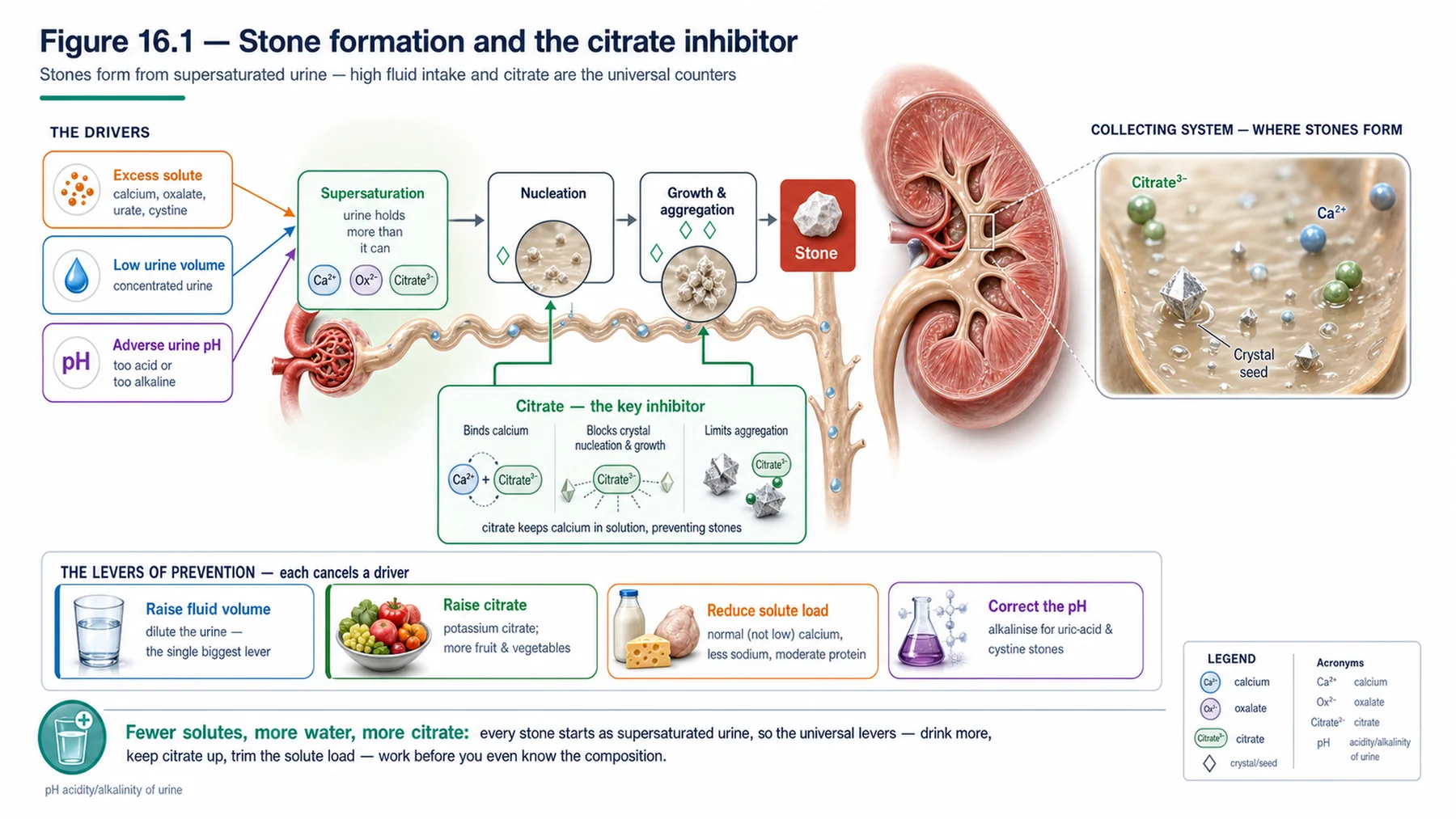
Stone formation begins with supersaturation: when the urine contains more of a stone-forming salt than it can hold in solution, crystals nucleate, grow, and aggregate, and — if retained — form a stone. Three factors push toward supersaturation: a high load of the stone-forming solute (calcium, oxalate, uric acid, cystine), a low urine volume (which concentrates everything), and an abnormal urine pH (each salt has a pH at which it is least soluble). Against these, the urine contains inhibitors of crystallisation, of which citrate is the most important — it binds calcium and inhibits crystal formation and aggregation, so a low urine citrate (hypocitraturia) promotes stones, and citrate supplementation is a mainstay of prevention. This framework — supersaturation from excess solute, low volume, and adverse pH, opposed by citrate — explains every stone type and every preventive measure: raising the urine volume (dilution) and the citrate, and correcting the solute and the pH, are the levers of prevention. The single most powerful and universal measure is simply a high fluid intake, which dilutes the urine and lowers the supersaturation of every salt.
— The stone types and their drivers
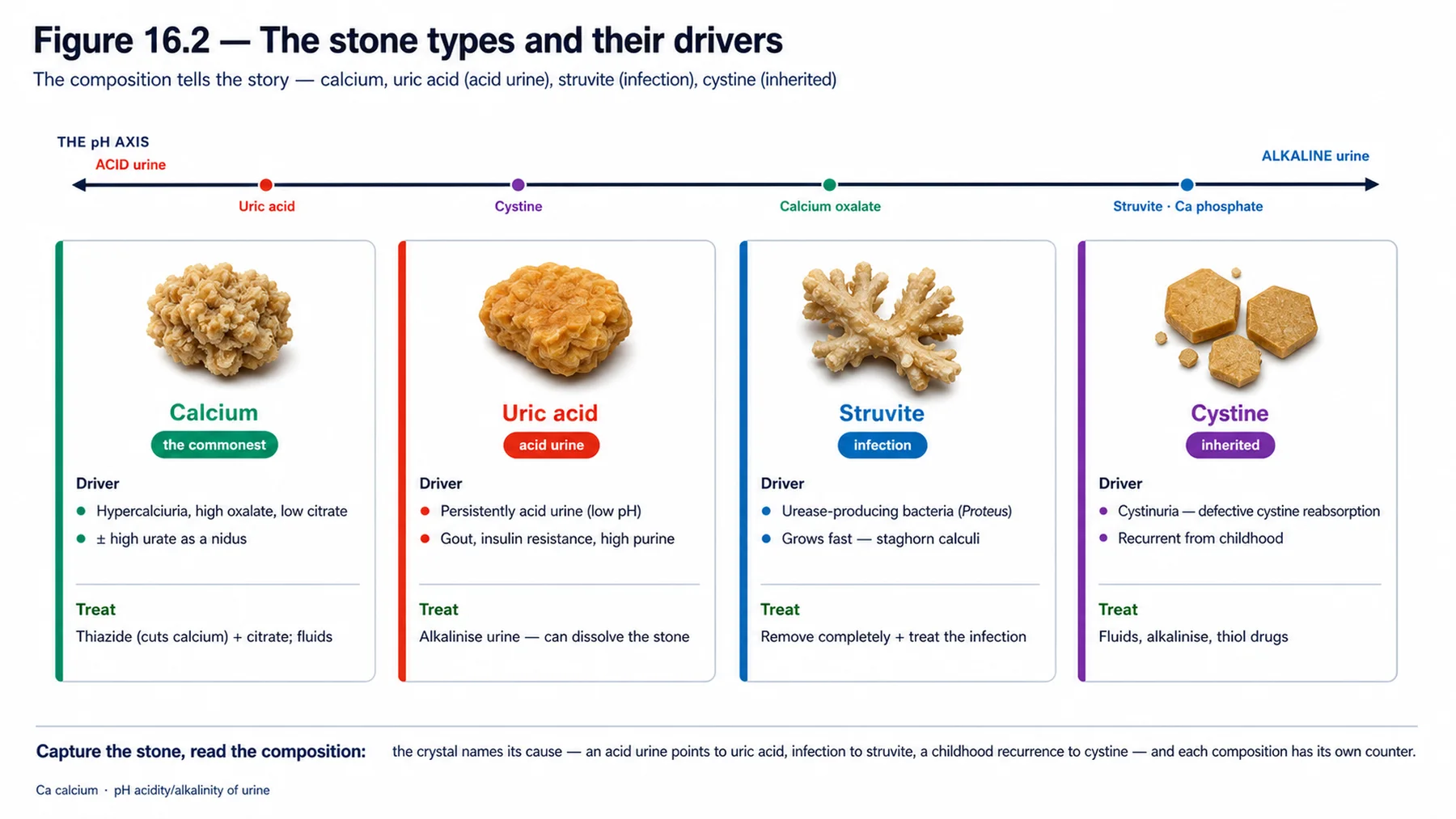
Stones are classified by composition, and each type has characteristic drivers that determine its prevention. Calcium stones are the commonest (about 80%), comprising calcium oxalate (the single commonest stone type) and calcium phosphate; their metabolic drivers are hypercalciuria (the commonest metabolic abnormality in stone-formers), hyperoxaluria, hypocitraturia, hyperuricosuria, and low urine volume, with calcium phosphate favoured by an alkaline urine (and associated with distal renal tubular acidosis). Uric acid stones have one dominant driver: a persistently acidic urine, in which uric acid is insoluble (so they form even with normal urate if the urine is acid enough), along with hyperuricosuria and low volume; they are associated with gout and the metabolic syndrome (insulin resistance impairs ammonium excretion, acidifying the urine), are radiolucent on plain film, and — uniquely — can be dissolved by alkalinising the urine with citrate. Struvite (magnesium ammonium phosphate, or 'infection') stones are caused by urease-producing bacteria (such as Proteus) that split urea to ammonia, alkalinising the urine and precipitating struvite; they form large staghorn calculi and require complete surgical removal plus eradication of the infection (any residual fragment perpetuates the stone and the infection). Cystine stones arise from cystinuria, an inherited disease. So the composition tells the story: calcium (the common metabolic stones), uric acid (acid urine), struvite (infection), and cystine (inherited) — each pointing to its own prevention.
— The metabolic evaluation and prevention
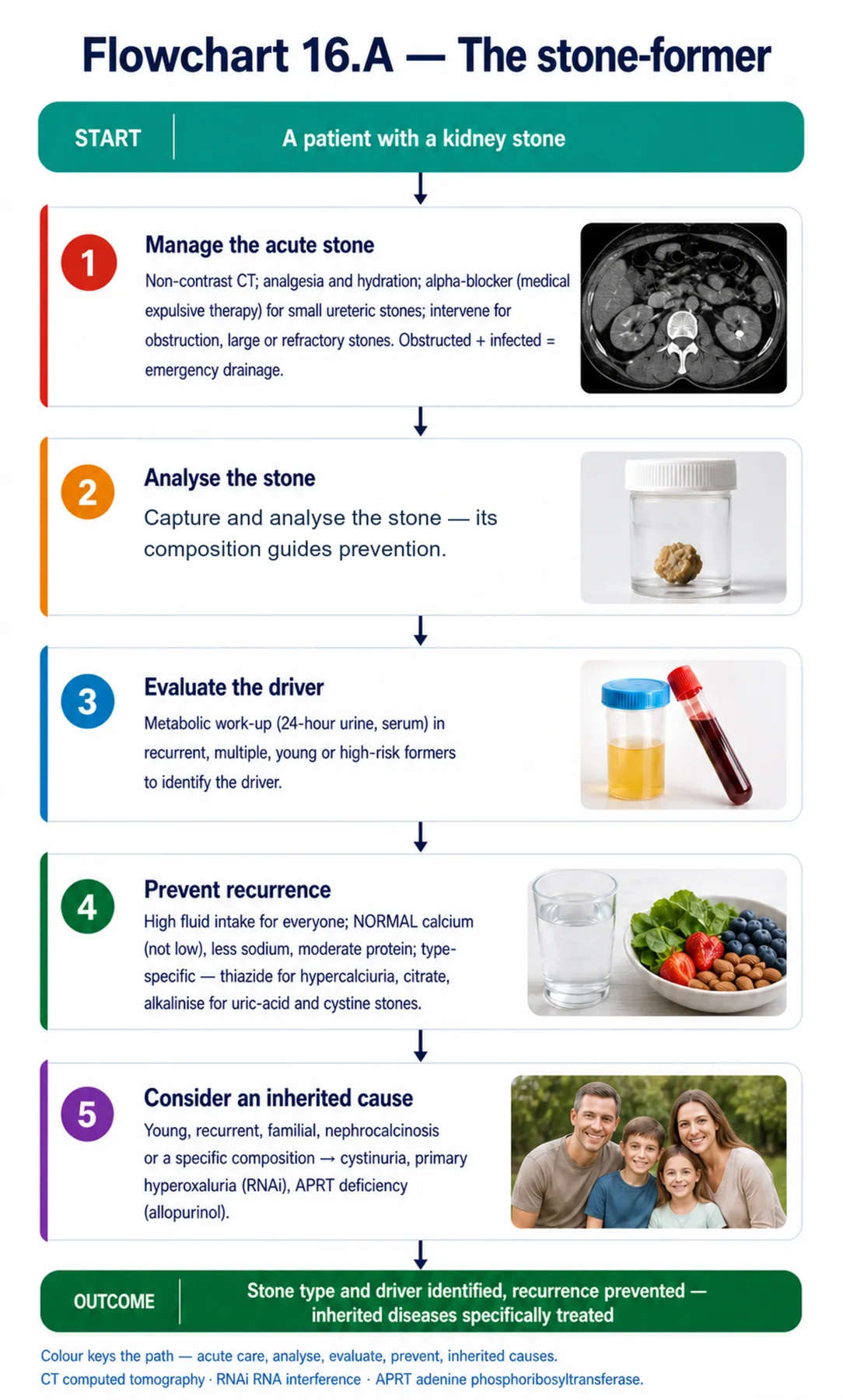
Because stones are recurrent and preventable, the stone-former — especially one with recurrent, multiple, young-onset, or otherwise high-risk stones — warrants a metabolic evaluation to identify the driver and target prevention. The evaluation has two parts: stone composition analysis (the captured stone is analysed, as the composition guides everything) and a metabolic work-up — a 24-hour urine collection measuring volume, calcium, oxalate, citrate, uric acid, pH, and cystine, with serum tests (calcium, parathyroid hormone, electrolytes) — which reveals the metabolic abnormality (low volume, hypercalciuria, hyperoxaluria, hypocitraturia, acid urine). Prevention then combines a universal measure with type-specific ones. The universal, and most important, measure is a high fluid intake to keep the urine dilute (a high urine volume is the single most effective preventive). The dietary advice carries one crucial, counter-intuitive point: dietary calcium should be normal, not low — restricting calcium increases intestinal oxalate absorption (calcium normally binds oxalate in the gut) and so raises urinary oxalate and stone risk, so low-calcium diets are harmful for stone-formers; instead, sodium is reduced (high sodium raises urinary calcium), oxalate is reduced if there is hyperoxaluria, and animal protein is moderated. The type-specific measures follow the driver: a thiazide diuretic for hypercalciuria (it lowers urinary calcium), potassium citrate for hypocitraturia and to alkalinise the urine (for uric acid and cystine stones, and it can dissolve uric acid stones), and urate-lowering therapy where appropriate. The principle is: find the metabolic driver and correct it, on a foundation of high fluid intake and the (normal-calcium) dietary measures.
— Acute management
Alongside prevention, the acute stone needs managing. Acute renal (ureteric) colic is diagnosed by non-contrast CT, the gold standard for detecting stones (showing size, site, and obstruction, and even radiolucent uric acid stones that a plain film misses). Acute management is analgesia and hydration; for small ureteric stones likely to pass, medical expulsive therapy with an alpha-blocker (relaxing the ureter) facilitates passage; and urological intervention — extracorporeal shock wave lithotripsy, ureteroscopy, or percutaneous nephrolithotomy — is needed for obstruction (especially with infection, an emergency requiring urgent decompression), large stones, or those that do not pass. An obstructing stone with infection (an infected, obstructed kidney) is a urological emergency requiring urgent drainage and antibiotics, as it can lead to sepsis and kidney loss. After the acute episode, the captured stone is analysed and the metabolic evaluation performed to prevent recurrence — because, untreated, stones recur. So acute management (diagnose by CT, relieve pain, treat obstruction/infection, remove when needed) gives way to prevention (analyse the stone, evaluate the metabolism, treat the driver).
— The inherited stone diseases
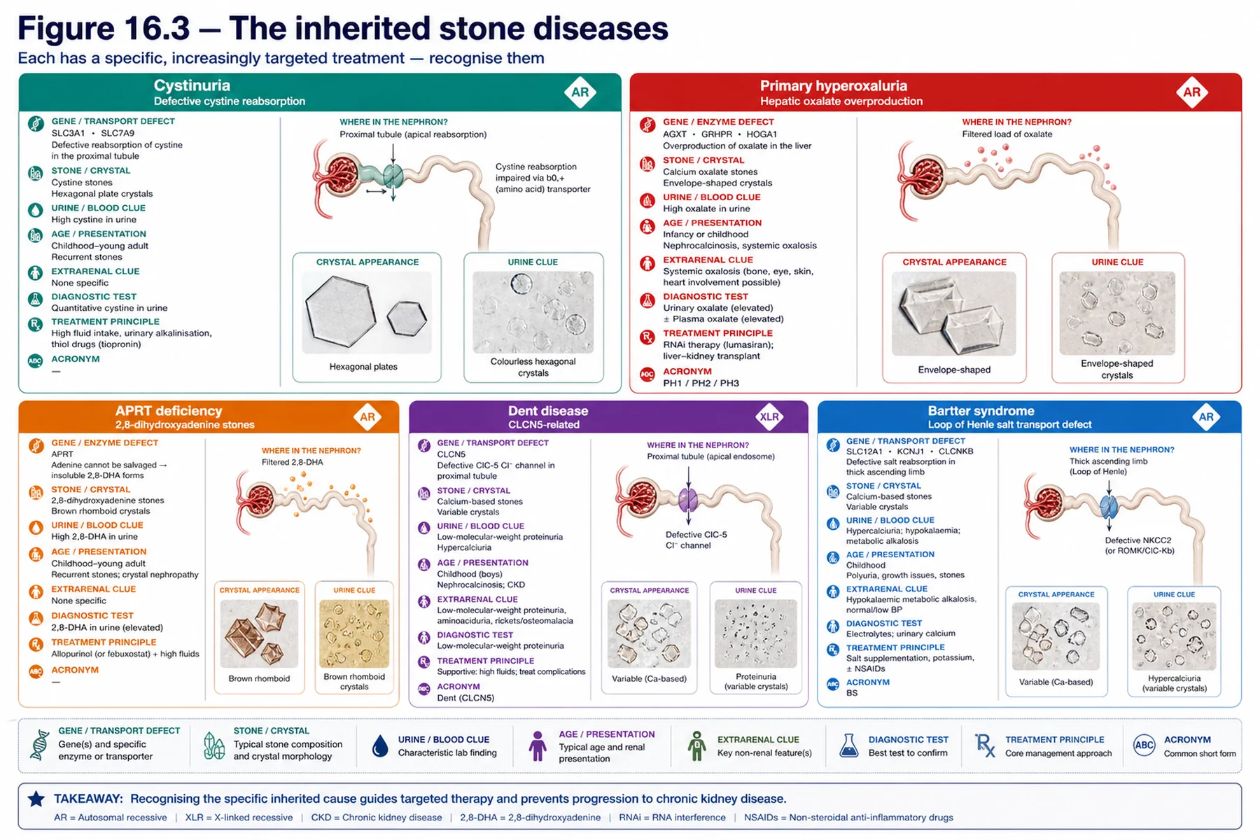
The kidney specialist must recognise the inherited stone diseases, because they are recurrent, often severe, and carry specific treatments. Cystinuria (autosomal recessive, from defects in cystine transporters) impairs the proximal reabsorption of cystine, which precipitates into cystine stones — recurrent, often from childhood or young adulthood — treated with high fluid intake, urinary alkalinisation (citrate), and, if needed, thiol drugs (tiopronin, penicillamine) that increase cystine solubility. Primary hyperoxaluria (autosomal recessive) is a hepatic enzyme defect in glyoxylate metabolism (type 1, from AGXT, being the commonest and most severe) causing endogenous oxalate overproduction, with recurrent calcium oxalate stones, nephrocalcinosis, progressive CKD to end-stage disease, and — once the GFR falls and oxalate cannot be excreted — systemic oxalosis (oxalate deposition in bones, heart, and other organs); its treatment has been transformed by RNA-interference therapies (lumasiran, nedosiran) that reduce hepatic oxalate production (disease-modifying), alongside high fluid, citrate, and pyridoxine (which some type 1 patients respond to), with combined liver-kidney transplantation used historically for end-stage type 1 disease (replacing the liver that is the source of the defect). APRT deficiency (adenine phosphoribosyltransferase) causes 2,8-dihydroxyadenine stones and crystalluria that can damage the kidney, and — importantly — it is treated with a xanthine oxidase inhibitor (allopurinol or febuxostat) that blocks the formation of dihydroxyadenine, and it can recur in a transplanted kidney if undiagnosed. Dent disease (the tubulopathy chapter) is another inherited stone-former. Inherited stone disease should be suspected with young-onset, recurrent, or familial stones, nephrocalcinosis, bilateral stones, or a specific composition (cystine, 2,8-dihydroxyadenine, or calcium oxalate with nephrocalcinosis). The unifying theme of the chapter is that stones are largely preventable once the type and the metabolic driver are identified — with a high fluid intake the universal foundation, normal (not low) dietary calcium a counter-intuitive essential, and the inherited diseases each carrying a specific, increasingly targeted treatment.
| 04 | PHASE A · LEVEL 4 · ORIENTATION & KNOWLEDGE Reference Tables |
Table 16.1 — Stone formation
| Element | Detail |
| Supersaturation | Excess solute + low urine volume + adverse pH → crystal nucleation/growth/aggregation |
| Inhibitor | Citrate (binds calcium, inhibits crystals) — hypocitraturia promotes stones |
| Universal lever | High fluid intake (dilute urine) lowers supersaturation of every salt |
| Principle | Raise volume and citrate; correct the solute and the pH |
Table 16.2 — Stone types and drivers
| Type | Driver | Note |
| Calcium (oxalate/phosphate) ~80% | Hypercalciuria (commonest), oxalate, low citrate | Phosphate favoured by alkaline urine |
| Uric acid | Acidic urine (key); hyperuricosuria | Radiolucent; dissolves with alkalinisation |
| Struvite (infection) | Urease bacteria → alkaline urine | Staghorn; remove + eradicate infection |
| Cystine | Cystinuria (inherited) | Recurrent; thiol drugs |
Table 16.3 — Acute management
| Step | Detail |
| Diagnosis | Non-contrast CT (gold standard; detects radiolucent uric acid stones) |
| Initial | Analgesia, hydration |
| Medical expulsive therapy | Alpha-blocker for small ureteric stones likely to pass |
| Intervention | Lithotripsy/ureteroscopy/PCNL for obstruction, large, infected, or refractory; obstructed+infected = emergency drainage |
Table 16.4 — Metabolic evaluation and prevention
| Element | Detail |
| Who | Recurrent, multiple, young, or high-risk stone-formers |
| Evaluation | Stone analysis; 24-h urine (volume, calcium, oxalate, citrate, urate, pH, cystine); serum (Ca, PTH) |
| Universal | High fluid intake (most important); reduce sodium; moderate animal protein |
| Calcium intake | NORMAL, not low — low calcium raises oxalate absorption and stone risk |
Table 16.5 — Type-specific prevention
| Type | Prevention |
| Calcium / hypercalciuria | Thiazide (lowers urine calcium); citrate (hypocitraturia); reduce oxalate |
| Uric acid | Alkalinise urine (citrate — can dissolve); urate-lowering; reduce purine |
| Struvite | Complete surgical removal + eradicate infection |
| Cystine | High fluid, alkalinisation, thiol drugs (tiopronin/penicillamine) |
Table 16.6 — Inherited stone diseases
| Disease | Detail |
| Cystinuria | Defective cystine reabsorption → cystine stones; fluid/alkalinisation/thiol drugs |
| Primary hyperoxaluria | Hepatic oxalate overproduction → stones, nephrocalcinosis, oxalosis; RNAi (lumasiran), ± liver-kidney transplant (type 1) |
| APRT deficiency | 2,8-dihydroxyadenine stones → allopurinol/febuxostat; recurs in transplant if missed |
| Suspect inherited | Young, recurrent, familial, nephrocalcinosis, bilateral, specific composition |
| 05 | PHASE B · LEVEL 5 · VISUALISE & MAP Imaging & Flowchart Specifications |
| 06 | PHASE B · LEVEL 6 · VISUALISE & MAP Concept Maps |
Each chain runs from mechanism to a named clinical action; read the arrows as “leads to.”
Stone formation. Supersaturation (excess solute + low volume + adverse pH), opposed by citrate → crystal → stone → ACTION: raise the urine volume and citrate and correct the solute and pH.
Calcium stones. Hypercalciuria (commonest) + oxalate + low citrate → calcium oxalate/phosphate stones → ACTION: thiazide for hypercalciuria, citrate for hypocitraturia, reduce oxalate — with NORMAL calcium intake.
Uric acid stones. Acidic urine → uric acid precipitates → radiolucent stones → ACTION: alkalinise the urine (citrate) — which can dissolve them.
Struvite stones. Urease bacteria → alkaline urine → struvite staghorn → ACTION: completely remove the stone and eradicate the infection (fragments perpetuate it).
Inherited stones. Cystinuria (cystine), primary hyperoxaluria (oxalate overproduction), APRT deficiency (2,8-DHA) → recurrent inherited stones → ACTION: recognise them and give the specific treatment (thiol drugs, RNAi, allopurinol).
| 07 | PHASE B · LEVEL 7 · VISUALISE & MAP Decision Pathways |
| R1 | IF preventing stones, THEN raise the urine volume (high fluid intake) — the single most important measure for every stone type. |
| R2 | IF a stone-former has recurrent, multiple, young-onset, or high-risk stones, THEN perform a metabolic evaluation (stone analysis + 24-h urine + serum) to find the driver. |
| R3 | IF advising diet for a calcium stone-former, THEN keep dietary calcium NORMAL (not low), reduce sodium, and moderate animal protein. |
| R4 | IF there is hypercalciuria, THEN use a thiazide (it lowers urinary calcium); if hypocitraturia, give citrate. |
| R5 | IF the stone is uric acid (acid urine), THEN alkalinise the urine with citrate — which can dissolve it — and lower urate. |
| R6 | IF the stone is struvite (staghorn, urease infection), THEN remove it completely and eradicate the infection — fragments perpetuate it. |
| R7 | IF a stone is obstructing and the kidney is infected, THEN treat as an emergency with urgent drainage and antibiotics. |
| R8 | IF stones are young-onset, recurrent, familial, with nephrocalcinosis, or of a specific composition, THEN consider an inherited stone disease and give its specific treatment. |
| 08 | PHASE C · LEVEL 8 · CLINICAL REASONING Clinical Cases |
| CASE 1 | DON'T CUT THE CALCIUM Normal calcium, find the driver Calcium stone prevention |
Presentation
A recurrent calcium oxalate stone-former is advised to follow a low-calcium diet to 'reduce the calcium in the stones,' and no metabolic evaluation has been done.
❖ Pause and reflect Is a low-calcium diet the right advice? |
Analysis
No — a low-calcium diet is counter-productive and can increase stone risk, a classic and important error. It seems intuitive to cut calcium to reduce calcium stones, but dietary calcium normally binds oxalate in the gut and limits its absorption; restricting calcium therefore increases intestinal oxalate absorption, raising urinary oxalate and the risk of calcium oxalate stones. So dietary calcium should be kept normal, not low. The right preventive advice is a high fluid intake (the single most important measure), normal calcium, reduced sodium (high sodium raises urinary calcium), moderated animal protein, and reduced oxalate if hyperoxaluric — and, crucially, a metabolic evaluation (24-hour urine, serum calcium and parathyroid hormone) to identify the specific driver (hypercalciuria is the commonest), allowing targeted treatment such as a thiazide for hypercalciuria or citrate for hypocitraturia. Advising low calcium without a metabolic evaluation gets the prevention exactly wrong.
Plan
Advise a high fluid intake with NORMAL (not low) dietary calcium, reduced sodium and moderated protein, and perform a metabolic evaluation to find and treat the driver (e.g. thiazide for hypercalciuria, citrate for hypocitraturia). Keep calcium normal and find the metabolic driver.
Teaching point
Dietary calcium should be NORMAL, not low, in calcium stone-formers — low calcium increases oxalate absorption and stone risk; do a metabolic evaluation to target prevention.
Cross-reference
Exercises rules R1, R2, R3, and R4; the stone-formation and calcium-stone concept maps; Figures 16.1, 16.2; Tables 16.4, 16.5.
| CASE 2 | DISSOLVE IT Alkalinise the acid urine Uric acid stones |
Presentation
A patient with gout and the metabolic syndrome has a radiolucent stone (not seen on plain film) and a persistently acidic urine. The team is considering surgical removal.
❖ Pause and reflect What is the stone type, and is surgery the best first option? |
Analysis
This is a uric acid stone, and it may be dissolved medically rather than removed surgically. The clues are classic: a radiolucent stone (uric acid stones are not seen on plain film, though visible on CT), a persistently acidic urine (the key driver — uric acid is insoluble at low pH), and the associations with gout and the metabolic syndrome (insulin resistance impairs ammonium excretion, acidifying the urine). The crucial and almost unique feature of uric acid stones is that they can be dissolved by alkalinising the urine: raising the urine pH with potassium citrate increases uric acid solubility and can dissolve the stone, often avoiding surgery altogether. So the first-line approach is medical — urinary alkalinisation with citrate (aiming for a higher urine pH), along with a high fluid intake and urate-lowering therapy — reserving intervention for obstruction or failure. Proceeding straight to surgery would miss the opportunity to dissolve the stone non-invasively. The acid urine is both the cause and the treatment target.
Plan
Recognise the uric acid stone (radiolucent, acid urine, gout/metabolic syndrome) and treat with urinary alkalinisation (potassium citrate) to dissolve it, plus high fluid intake and urate-lowering — reserving surgery for obstruction or failure. Alkalinise to dissolve the uric acid stone.
Teaching point
Uric acid stones form in acidic urine, are radiolucent, and can be dissolved by alkalinising the urine with citrate — a medical, not surgical, first line.
Cross-reference
Exercises rule R5; the uric-acid-stone concept map; Figure 16.2; Tables 16.2, 16.5.
| CASE 3 | MORE THAN STONES Recognise the inherited disease Primary hyperoxaluria / cystinuria |
Presentation
A young patient has recurrent calcium oxalate stones with nephrocalcinosis and declining kidney function from childhood, and a sibling is similarly affected. The team is managing each stone episode without seeking an underlying inherited disease.
❖ Pause and reflect What underlying disease should be sought, and why does it matter? |
Analysis
Primary hyperoxaluria — an inherited stone disease that must be recognised, because it has specific, disease-modifying treatment. The picture is characteristic: young-onset, recurrent calcium oxalate stones with nephrocalcinosis and progressive kidney decline, with an affected sibling (autosomal recessive). Primary hyperoxaluria is a hepatic enzyme defect causing endogenous oxalate overproduction, leading to recurrent calcium oxalate stones, nephrocalcinosis, progressive CKD to end-stage disease, and — as the GFR falls and oxalate cannot be excreted — systemic oxalosis (oxalate deposited in bones, heart, and other organs). Recognising it matters enormously because management has been transformed: RNA-interference therapies (lumasiran, nedosiran) reduce hepatic oxalate production (disease-modifying), alongside high fluid, citrate, and pyridoxine (some type 1 responders), with combined liver-kidney transplantation historically for end-stage type 1 disease. Managing each stone episode without diagnosing the inherited disease misses the chance to prevent the relentless progression and the systemic oxalosis. (The same imperative applies to cystinuria — recurrent cystine stones from childhood, treated with fluid, alkalinisation, and thiol drugs.) Young, recurrent, familial stones with nephrocalcinosis should always prompt the search for an inherited cause.
Plan
Recognise the likely primary hyperoxaluria (young, recurrent, nephrocalcinosis, familial, progressive), confirm it, and treat specifically (RNAi such as lumasiran, high fluid, citrate, pyridoxine; transplant for ESKD type 1) rather than managing each stone in isolation. Seek the inherited stone disease and treat it specifically.
Teaching point
Young, recurrent, familial stones with nephrocalcinosis suggest an inherited stone disease — primary hyperoxaluria (now treatable with RNAi) or cystinuria — recognise it for the specific treatment.
Cross-reference
Exercises rule R8; the inherited-stones concept map; Figure 16.3; Table 16.6; primary hyperoxaluria and cystinuria in Chapters 9 and 11.
| CASE 4 | REMOVE IT ALL Struvite and infection Struvite staghorn stones |
Presentation
A patient with recurrent urinary infections (with a urease-producing organism) has a large staghorn calculus. A clinician plans to remove the bulk of the stone and continue antibiotics, leaving small fragments.
❖ Pause and reflect Is partial stone removal adequate for this struvite staghorn? |
Analysis
No — struvite stones must be removed completely, and the infection eradicated, because residual fragments perpetuate the disease. This is a struvite (magnesium ammonium phosphate, or 'infection') stone: it forms when urease-producing bacteria (such as Proteus) split urea to ammonia, alkalinising the urine and precipitating struvite, and it grows into a large staghorn calculus. The crucial management point is that struvite stones harbour the bacteria within them, so leaving any residual fragment leaves a nidus of infection and stone material that perpetuates both the infection and the stone — the stone regrows and the infection recurs. Therefore complete surgical removal (typically percutaneous nephrolithotomy for a staghorn) plus eradication of the infection is required; partial removal with antibiotics, as planned here, will fail. So the plan should be complete stone clearance and full treatment of the infection, not debulking. Struvite stones are unique in this need for complete removal driven by the embedded infection.
Plan
Achieve complete surgical removal of the struvite staghorn (percutaneous nephrolithotomy) and eradicate the infection — not partial removal with antibiotics, which leaves a nidus that perpetuates the stone and infection. Remove all of a struvite stone and treat the infection.
Teaching point
Struvite (infection) stones harbour bacteria — they require COMPLETE removal plus eradication of the infection; residual fragments perpetuate the stone and infection.
Cross-reference
Exercises rules R6 and R7; the struvite concept map; Figure 16.2; Tables 16.2, 16.5.
| 09 | PHASE C · LEVEL 9 · CLINICAL REASONING Clinical Implications |
One triad per mechanism the narrative exposed: the physiology, why it matters, and the clinical move.
MECHANISM Stones form from supersaturated urine, opposed by citrate. |
WHY IT MATTERS A dilute urine and adequate citrate lower the supersaturation of every salt. |
ACTION Make high fluid intake (and citrate) the foundation of prevention. |
MECHANISM Dietary calcium binds oxalate in the gut and limits its absorption. |
WHY IT MATTERS A low-calcium diet raises urinary oxalate and stone risk. |
ACTION Keep dietary calcium normal, not low, in calcium stone-formers. |
MECHANISM Uric acid is insoluble in acidic urine. |
WHY IT MATTERS Acid urine is the key driver, and raising the pH increases solubility. |
ACTION Alkalinise the urine to prevent and even dissolve uric acid stones. |
MECHANISM Struvite stones are formed by, and harbour, urease-producing bacteria. |
WHY IT MATTERS Residual fragments perpetuate the stone and the infection. |
ACTION Remove the struvite stone completely and eradicate the infection. |
MECHANISM The inherited stone diseases overproduce or mishandle a stone-forming solute. |
WHY IT MATTERS They are recurrent and progressive but each has a specific treatment. |
ACTION Recognise the inherited disease and give its targeted therapy. |
| 10 | PHASE C · LEVEL 10 · CLINICAL REASONING Clinical Pearls |
| Stones form from urinary supersaturation; citrate is the key inhibitor. | Low urine volume is a universal risk; high fluid intake is the key preventive. |
| Calcium stones (~80%): hypercalciuria (commonest), oxalate, low citrate. | Calcium phosphate favoured by alkaline urine (distal RTA). |
| Uric acid stones: ACIDIC urine is the key; radiolucent; dissolve by alkalinisation. | Uric acid stones: associated with gout and metabolic syndrome. |
| Struvite (infection) stones: urease bacteria → alkaline urine → staghorn. | Struvite: COMPLETE removal + eradicate infection (fragments perpetuate). |
| Cystine stones: from cystinuria (inherited). | Diagnose acute stones by non-contrast CT. |
| Analyse the stone; metabolic evaluation (24-h urine) for recurrent/young/high-risk. | Dietary calcium NORMAL, not low (low calcium raises oxalate absorption). |
| Thiazide for hypercalciuria; citrate for hypocitraturia and to alkalinise. | Cystinuria: fluid, alkalinisation, thiol drugs. |
| Primary hyperoxaluria: oxalate overproduction; RNAi (lumasiran) ± liver-kidney transplant. | APRT deficiency: 2,8-DHA stones → allopurinol; recurs in transplant if missed. |
| 11 | PHASE D · LEVEL 11 · SAFETY & EVIDENCE Red Flags & Never-Do |
Panel A — Red flags
| ▲ | An obstructing stone with infection — a urological emergency; urgent drainage and antibiotics. |
| ▲ | A low-calcium diet advised for calcium stones — it raises oxalate absorption; keep calcium normal. |
| ▲ | A radiolucent stone with acid urine — uric acid; alkalinise to dissolve. |
| ▲ | A staghorn calculus with a urease organism — struvite; complete removal + eradicate infection. |
| ▲ | Young-onset, recurrent, familial stones with nephrocalcinosis — consider an inherited stone disease. |
Panel B — Never do
| ✖ NEVER — advise a low-calcium diet for calcium stone-formers. |
| ✖ NEVER — proceed straight to surgery for a uric acid stone without trying alkalinisation. |
| ✖ NEVER — leave residual fragments of a struvite stone. |
| ✖ NEVER — manage recurrent young-onset stones without considering an inherited cause. |
| 12 | PHASE D · LEVEL 12 · SAFETY & EVIDENCE Common Pitfalls |
Pitfall 1 — Low-calcium diet
| ✖ | WRONG Advising a low-calcium diet for calcium stones. |
| ✓ | RIGHT Keeping dietary calcium normal (and reducing sodium/protein). |
| ✉ | WHY Low calcium raises intestinal oxalate absorption and stone risk. |
Pitfall 2 — Surgery before alkalinisation
| ✖ | WRONG Surgically removing a uric acid stone first. |
| ✓ | RIGHT Trying urinary alkalinisation (it can dissolve them). |
| ✉ | WHY Uric acid stones dissolve when the urine is alkalinised. |
Pitfall 3 — Partial struvite removal
| ✖ | WRONG Debulking a struvite stone and leaving fragments. |
| ✓ | RIGHT Complete removal and eradication of the infection. |
| ✉ | WHY Residual fragments harbour bacteria and perpetuate the stone. |
Pitfall 4 — Missing the inherited disease
| ✖ | WRONG Managing recurrent young stones episode by episode. |
| ✓ | RIGHT Recognising an inherited stone disease and treating it specifically. |
| ✉ | WHY Inherited diseases are progressive but have specific treatments. |
Pitfall 5 — Delaying the infected-obstruction emergency
| ✖ | WRONG Treating an obstructed, infected kidney with antibiotics alone. |
| ✓ | RIGHT Urgent drainage plus antibiotics (an emergency). |
| ✉ | WHY An infected obstructed kidney can cause sepsis and kidney loss. |
| 13 | PHASE D · LEVEL 13 · SAFETY & EVIDENCE Evidence Grading |
GRADE A HIGH CONFIDENCE The effect is real and the estimate is stable. RCTs at low risk of bias; multiple concordant prospective cohorts; meta-analyses. |
GRADE B MODERATE CONFIDENCE The effect is likely real but may shift with new data. Observational studies, registries, mechanistic human studies. |
GRADE C LOW CONFIDENCE Rests on physiology, reasoning, or consensus rather than outcomes. Pathophysiological reasoning; extrapolation; consensus without outcomes. |
Graded statements (by evidence type)
| Statement | Grade | Basis (evidence type) |
| High fluid intake reduces stone recurrence. | A | RCTs |
| A normal (not low) calcium diet reduces calcium stone recurrence. | A | RCTs |
| Thiazides reduce recurrence in hypercalciuric stone-formers. | A | RCTs |
| Uric acid stones can be dissolved by urinary alkalinisation. | A | Clinical data |
| Struvite stones require complete removal and infection eradication. | A | Clinical data |
| RNA-interference therapy reduces oxalate in primary hyperoxaluria. | A | RCTs (lumasiran) |
| APRT deficiency stones respond to xanthine oxidase inhibition. | A | Clinical data |
| 14 | PHASE E · LEVEL 14 · PATIENT DECISIONS Absolute Risk in Natural Frequency |
Natural-frequency estimates for orientation, from stone cohorts; they vary with the stone type and population. They convey the size of the decisions, expressed per 100 comparable patients.
| Per 100 patients… | Outcome | Roughly how many | See |
| First-time stone-formers untreated | Have a recurrence over years | Many — hence prevention | L13 row 1 |
| Calcium stone-formers on high fluid + normal-calcium diet vs low-calcium | Avoid recurrence | More with normal calcium | L13 rows 1–2 |
| Uric acid stones given urinary alkalinisation | Dissolve / avoid surgery | Many | L13 row 4 |
| Primary hyperoxaluria type 1 on RNAi therapy | Reduce urinary oxalate | Most | L13 row 6 |
★ How to read these Read these as orientation, not promises; outcomes vary with the stone type and population. The stable signals: stones recur without prevention, high fluid and normal calcium reduce recurrence, uric acid stones dissolve with alkalinisation, and RNAi lowers oxalate in primary hyperoxaluria. Communicate them as people out of 100, not as a hazard ratio. |
| 17 | PHASE F · LEVEL 17 · APPLY & TEST Documentation Templates |
Paste-ready notes. Tick the boxes that apply and delete the rest; make the stone type, the metabolic driver, and the prevention explicit.
Template 1 — Acute stone and evaluation
Template 2 — Prevention and inherited disease
| 18 | PHASE F · LEVEL 18 · APPLY & TEST Cheat Sheet |
| Stones = supersaturation; citrate inhibits. | High fluid intake = the key universal preventive. |
| Calcium (~80%): hypercalciuria (commonest), oxalate, low citrate. | Calcium phosphate ↔︎ alkaline urine (distal RTA). |
| Uric acid: ACID urine; radiolucent; dissolve by alkalinisation. | Uric acid ↔︎ gout, metabolic syndrome. |
| Struvite: urease bacteria → alkaline → staghorn. | Struvite: COMPLETE removal + eradicate infection. |
| Cystine: from cystinuria. | Acute: non-contrast CT; alpha-blocker for small ureteric stones. |
| Obstructed + infected = emergency drainage. | Analyse the stone; 24-h urine for recurrent/young/high-risk. |
| Dietary calcium NORMAL, not low. | Thiazide (hypercalciuria); citrate (hypocitraturia/alkalinise). |
| Primary hyperoxaluria: RNAi (lumasiran) ± liver-kidney transplant. | APRT deficiency: allopurinol (recurs in transplant if missed). |
| 19 | PHASE F · LEVEL 19 · APPLY & TEST Flashcards |
| CARD 1 | Q. How do kidney stones form? A. By urinary supersaturation — when the urine holds more of a stone-forming salt than it can keep in solution — allowing crystals to nucleate, grow, and aggregate; promoted by a high solute load, low urine volume, and an adverse pH, and opposed by inhibitors, chiefly citrate. DETAILED. A low urine volume is a universal risk. CLINICAL. Prevent by raising fluid intake and citrate and correcting the solute and pH. |
| CARD 2 | Q. What are the stone types and their drivers? A. Calcium (oxalate and phosphate, ~80%; driven by hypercalciuria, hyperoxaluria, hypocitraturia, low volume), uric acid (driven by acidic urine), struvite (infection stones from urease-producing bacteria alkalinising the urine), and cystine (from cystinuria). DETAILED. The composition points to the prevention. CLINICAL. Identify the stone type and treat its driver. |
| CARD 3 | Q. Why do uric acid stones form, and how can they be treated? A. Because uric acid is insoluble in acidic urine, a persistently acidic urine is the key driver (with hyperuricosuria and low volume); they are radiolucent and associated with gout and the metabolic syndrome — and, uniquely, can be dissolved by alkalinising the urine with citrate. DETAILED. Alkalinisation is a medical first line. CLINICAL. Alkalinise the urine to prevent and dissolve uric acid stones. |
| CARD 4 | Q. What are struvite stones and how are they managed? A. Magnesium ammonium phosphate ('infection') stones formed when urease-producing bacteria split urea, alkalinising the urine and precipitating struvite; they grow into staghorn calculi and harbour bacteria, so they require complete surgical removal and eradication of the infection — residual fragments perpetuate the stone and the infection. DETAILED. Partial removal fails. CLINICAL. Remove the struvite stone completely and treat the infection. |
| CARD 5 | Q. What does the metabolic evaluation of a stone-former involve, and who needs it? A. Stone composition analysis plus a 24-hour urine collection (volume, calcium, oxalate, citrate, uric acid, pH, cystine) and serum tests (calcium, parathyroid hormone) — performed in recurrent, multiple, young-onset, or high-risk stone-formers to identify the metabolic driver and target prevention. DETAILED. Hypercalciuria is the commonest abnormality. CLINICAL. Evaluate the high-risk stone-former and treat the driver. |
| CARD 6 | Q. Why should dietary calcium be normal, not low, in calcium stone-formers? A. Because dietary calcium binds oxalate in the gut and limits its absorption, so restricting calcium increases intestinal oxalate absorption, raising urinary oxalate and the risk of calcium oxalate stones — a low-calcium diet is counter-productive. DETAILED. It is a classic, important counter-intuitive point. CLINICAL. Keep dietary calcium normal; reduce sodium and moderate protein. |
| CARD 7 | Q. What is primary hyperoxaluria and how is it treated? A. An autosomal recessive hepatic enzyme defect in glyoxylate metabolism (type 1 commonest and most severe) causing endogenous oxalate overproduction, with recurrent calcium oxalate stones, nephrocalcinosis, progressive CKD, and systemic oxalosis; treated with high fluid, citrate, pyridoxine (some type 1), the RNA-interference therapies (lumasiran) that reduce oxalate production, and combined liver-kidney transplantation for end-stage type 1 disease. DETAILED. RNAi has transformed its management. CLINICAL. Recognise it and give the specific (RNAi-based) treatment. |
| CARD 8 | Q. When should an inherited stone disease be suspected? A. With young-onset, recurrent, or familial stones, nephrocalcinosis, bilateral stones, or a specific stone composition (cystine, 2,8-dihydroxyadenine, or calcium oxalate with nephrocalcinosis) — pointing to cystinuria, primary hyperoxaluria, APRT deficiency, or Dent disease, each with a specific treatment. DETAILED. APRT deficiency can recur in a transplant if missed. CLINICAL. Suspect and treat the inherited stone disease. |
| 20 | PHASE F · LEVEL 20 · APPLY & TEST One-Minute Preceptor |
| SCENE 1 | The intern cutting the calcium |
GET A COMMITMENT. “You've put this calcium stone-former on a low-calcium diet — why?”
PROBE FOR EVIDENCE. “Less calcium, fewer calcium stones” — ask: “What does dietary calcium do to oxalate in the gut, and what happens to urinary oxalate if you cut it?”
TEACH A GENERAL RULE. Dietary calcium binds oxalate in the gut, so a low-calcium diet raises oxalate absorption and stone risk — keep calcium normal, reduce sodium, moderate protein, and do a metabolic evaluation.
REINFORCE WHAT WAS RIGHT. Wanting to prevent recurrence was right.
CORRECT A MISTAKE. Keep calcium normal and find the metabolic driver.
| SCENE 2 | The resident heading to surgery for a uric acid stone |
GET A COMMITMENT. “You want to remove this radiolucent stone surgically — what type is it, and is there a non-surgical option?”
PROBE FOR EVIDENCE. “It's a stone causing trouble” — ask: “Radiolucent, acid urine, gout — what type is that, and what can dissolve it?”
TEACH A GENERAL RULE. Radiolucent stones in acid urine are uric acid stones, which can be dissolved by alkalinising the urine with citrate — a medical first line before surgery.
REINFORCE WHAT WAS RIGHT. Recognising the stone needed treating was correct.
CORRECT A MISTAKE. Try urinary alkalinisation to dissolve it before surgery.
| 22 | PHASE F · LEVEL 22 · APPLY & TEST Board-Style Questions |
| Q 01 | The single most important measure to prevent stone recurrence is: |
| A | A low-calcium diet |
| B | A high fluid intake |
| C | Surgery |
| D | Antibiotics |
Rationale High fluid intake (dilute urine) is the key universal preventive (Table 16.1, rule R1). A is harmful; C and D are not preventives. |
| Q 02 | The commonest metabolic abnormality in calcium stone-formers is: |
| A | Hyperoxaluria |
| B | Hypercalciuria |
| C | Hypocitraturia |
| D | Cystinuria |
Rationale Hypercalciuria is the commonest (Table 16.2). A and C contribute; D is a different (inherited) disease. |
| Q 03 | Uric acid stones: |
| A | Form in alkaline urine |
| B | Form in acidic urine and can be dissolved by alkalinisation |
| C | Are always radio-opaque |
| D | Are caused by infection |
Rationale Acid urine is the driver; alkalinisation dissolves them (case 2, Table 16.2, rule R5). A, C, and D are wrong. |
| Q 04 | Struvite (infection) stones require: |
| A | Antibiotics alone |
| B | Complete surgical removal plus eradication of the infection |
| C | Alkalinisation |
| D | No treatment |
Rationale Residual fragments harbour bacteria, so complete removal + infection eradication is needed (case 4, Table 16.5, rule R6). A, C, and D fail. |
| Q 05 | Dietary calcium in calcium stone-formers should be: |
| A | Low |
| B | Normal (not low — low calcium raises oxalate absorption) |
| C | Eliminated |
| D | Maximised |
Rationale Low calcium raises intestinal oxalate absorption and stone risk (case 1, Table 16.4, rule R3). A, C, and D are incorrect. |
| Q 06 | Primary hyperoxaluria is now treated with a disease-modifying: |
| A | Thiazide |
| B | RNA-interference therapy (e.g. lumasiran) reducing oxalate production |
| C | Antibiotic |
| D | Beta-blocker |
Rationale RNAi therapies reduce hepatic oxalate production (case 3, Table 16.6). A, C, and D are not disease-modifying here. |
| Q 07 | Which suggests an inherited stone disease? |
| A | A single stone at age 60 |
| B | Young-onset, recurrent, familial stones with nephrocalcinosis |
| C | A struvite stone |
| D | A small ureteric stone |
Rationale Young, recurrent, familial stones with nephrocalcinosis point to an inherited cause (case 3, Table 16.6, rule R8). A, C, and D do not. |
| Q 08 | An obstructing stone with an infected kidney is: |
| A | A routine outpatient problem |
| B | A urological emergency requiring urgent drainage and antibiotics |
| C | Treated with alkalinisation |
| D | Best left to pass |
Rationale An infected, obstructed kidney can cause sepsis and kidney loss — urgent drainage is needed (Table 16.3, rule R7). A, C, and D are dangerous. |