Chapter Preamble
Signals declared
Sig-D — Diagnostic (primary). Recognise the inherited tubulopathies from their electrolyte, acid-base, blood-pressure, and urinary-calcium patterns, and localise each to its nephron segment.
Sig-M — Mechanistic (strong). How a defect in a specific tubular transporter produces each syndrome — the segment-by-segment logic that makes the tubulopathies intelligible.
Levels populated and omitted
Populated (17): L1–L13, L18–L20, L22. The mechanistic signal fires the concept maps (L6) and triads (L9); the diagnostic signal drives the tables, rules, cases, pitfalls, and board items.
L14 absolute-risk — omitted. No Sig-T/E/V; this is a mechanism-and-pattern-recognition chapter (specific treatments are noted within it).
L15 / L16 preference-sensitive map and SDM scripts — omitted. No Sig-E; the tubulopathies are recognised and treated by their patterns.
L17 documentation templates — omitted. No Sig-P/T as a primary signal; management is summarised in the tables and cases.
| 01 | PHASE A · LEVEL 1 · ORIENTATION & KNOWLEDGE Learning Objectives |
By the end of this chapter you should be able to:
Map the inherited tubulopathies to their nephron segments.
Explain Bartter syndrome as a 'loop-like' thick-ascending-limb defect.
Explain Gitelman syndrome as a 'thiazide-like' distal-convoluted-tubule defect.
Distinguish Bartter from Gitelman by urinary calcium and magnesium.
Distinguish the salt-wasting (low BP) from the salt-retaining (hypertensive) tubulopathies.
Distinguish the renal tubular acidoses (types 1, 2, and 4).
Recognise the proximal tubulopathies (Dent, Fanconi, cystinuria).
Outline the principle of treatment — replace what is lost and target the defect.
| 02 | PHASE A · LEVEL 2 · ORIENTATION & KNOWLEDGE Executive Summary |
The inherited tubulopathies are defects in specific tubular transporters, and each maps to a nephron segment — the key to making sense of them.
In the thick ascending limb, Bartter syndrome impairs sodium-chloride reabsorption (the loop-diuretic target), causing salt wasting, a hypokalaemic metabolic alkalosis, hypercalciuria with nephrocalcinosis, and a normal-to-low blood pressure — a 'furosemide-like' state.
In the distal convoluted tubule, Gitelman syndrome impairs the thiazide-sensitive cotransporter, causing salt wasting, a hypokalaemic metabolic alkalosis, hypocalciuria, hypomagnesaemia, and a normal-to-low blood pressure — a 'thiazide-like' state.
Bartter and Gitelman are distinguished by urinary calcium (high in Bartter, low in Gitelman), magnesium (low in Gitelman), and severity (Bartter earlier and more severe).
The salt-wasting tubulopathies (Bartter, Gitelman) have a low-to-normal blood pressure, whereas the salt-retaining ones cause hypertension.
Liddle syndrome (an overactive epithelial sodium channel) causes hypertension with hypokalaemia and a low renin and aldosterone, treated with amiloride; Gordon syndrome causes hypertension with hyperkalaemia and acidosis, treated with a thiazide.
The renal tubular acidoses cause a normal-anion-gap metabolic acidosis: type 1 (distal) with hypokalaemia, an inability to acidify the urine, and stones; type 2 (proximal) with bicarbonate wasting, often as part of Fanconi syndrome; and type 4 with hyperkalaemia from hypoaldosteronism.
The proximal tubulopathies include Fanconi syndrome (generalised proximal dysfunction), Dent disease (low-molecular-weight proteinuria, hypercalciuria, stones), and cystinuria (cystine stones).
Diagnosis rests on the pattern — the electrolytes, the acid-base state, the blood pressure, and the urinary calcium and magnesium — confirmed by genetics.
Treatment is to replace what is lost (potassium and magnesium in Gitelman, potassium and salt in Bartter) and to target the specific defect (amiloride for Liddle, a thiazide for Gordon, alkali for the acidoses).
The segment-by-segment logic is what turns a confusing list of syndromes into an intelligible map.
| 03 | PHASE A · LEVEL 3 · ORIENTATION & KNOWLEDGE Main Narrative |
The inherited tubulopathies look, at first, like a bewildering list of eponymous syndromes with overlapping electrolyte abnormalities. The key that unlocks them is the nephron: each tubulopathy is a defect in a specific transporter in a specific segment, and once you locate the defect, the whole picture — the electrolytes, the acid-base state, the blood pressure, the urinary calcium — follows. This chapter maps the tubulopathies segment by segment, with the loop-like Bartter and thiazide-like Gitelman syndromes, the hypertensive salt-retaining syndromes, the renal tubular acidoses, and the proximal disorders.
— The nephron-segment map
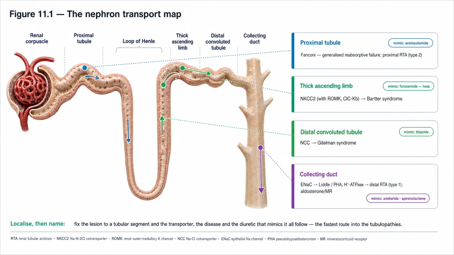
The organising principle of the inherited tubulopathies is the nephron-segment map. Each segment reabsorbs solutes through characteristic transporters, and a genetic defect in one of those transporters produces a syndrome whose features reflect the segment's normal job. The proximal tubule reabsorbs the bulk of filtered solutes (glucose, amino acids, phosphate, bicarbonate), so its disorders cause generalised wasting (Fanconi syndrome) or specific defects (Dent disease, cystinuria). The thick ascending limb reabsorbs sodium chloride via the NKCC2 cotransporter — the target of loop diuretics — so its defect (Bartter syndrome) mimics chronic loop-diuretic use. The distal convoluted tubule reabsorbs sodium chloride via the NCC cotransporter — the target of thiazides — so its defect (Gitelman syndrome) mimics chronic thiazide use. The collecting duct fine-tunes sodium (via the epithelial sodium channel, ENaC, the aldosterone/amiloride target), potassium, and acid, so its disorders cause salt-retaining hypertension (Liddle), the distal renal tubular acidosis, and concentrating defects. Learning the map — which transporter, which segment, which diuretic it resembles — makes every tubulopathy predictable rather than memorised. The single most useful pairing is the loop-versus-thiazide analogy for Bartter and Gitelman, developed next.
— Bartter and Gitelman: loop-like and thiazide-like
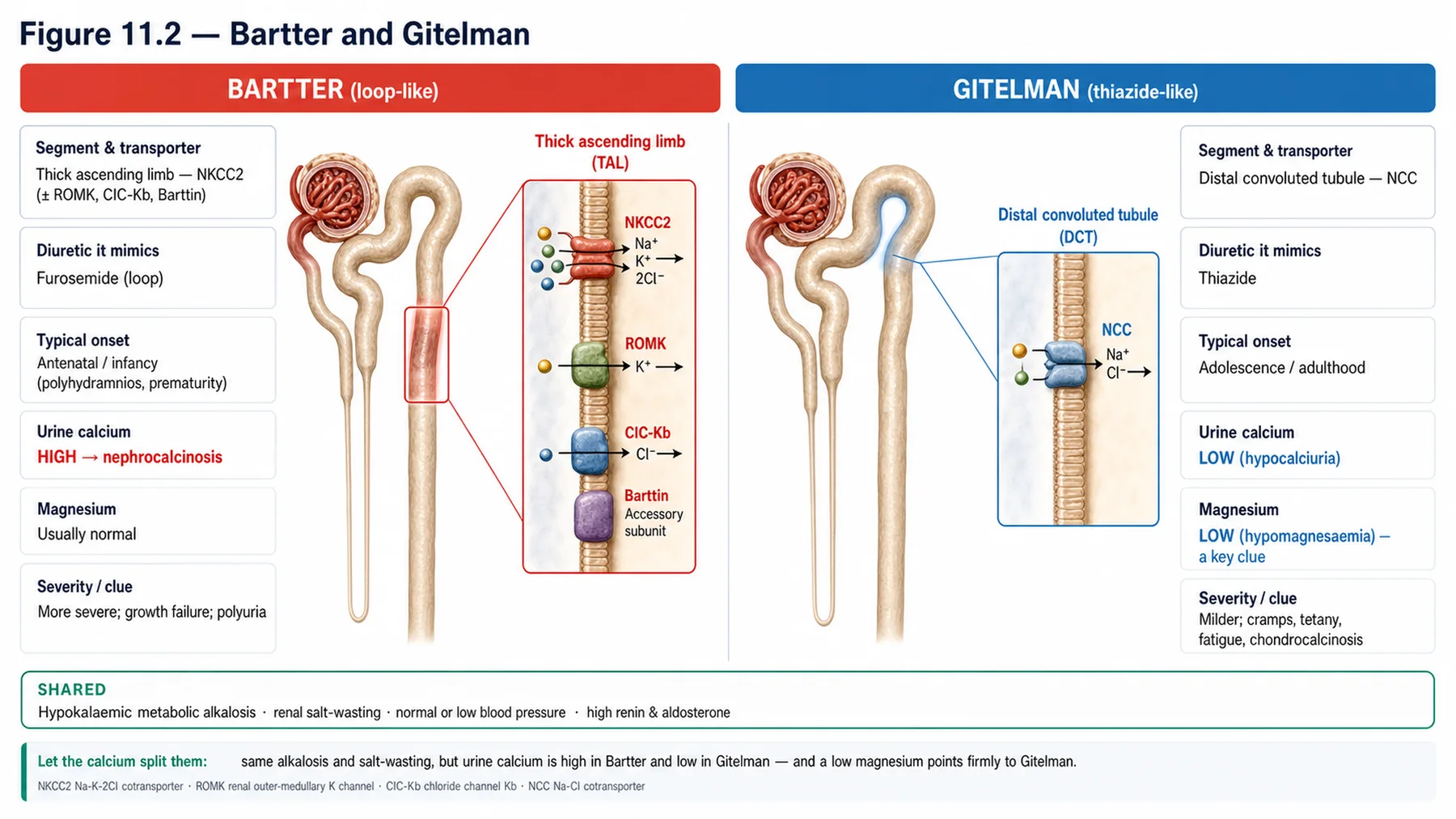
Bartter and Gitelman syndromes are the archetypal salt-wasting tubulopathies, and the diuretic analogy makes them clear. Bartter syndrome is a defect in the thick ascending limb's sodium-chloride reabsorption machinery (NKCC2, ROMK, ClC-Kb, or barttin) — the same machinery loop diuretics block — so it produces a 'furosemide-like' state: renal salt wasting (with volume depletion and an activated renin-angiotensin-aldosterone system), a hypokalaemic metabolic alkalosis, and — because the thick ascending limb's NaCl reabsorption normally drives calcium reabsorption there — hypercalciuria, often with nephrocalcinosis. The blood pressure is normal or low (it is a salt-losing state). Bartter is typically severe and presents early (antenatal and neonatal forms with polyhydramnios, or classic childhood forms). Gitelman syndrome is a defect in the distal convoluted tubule's thiazide-sensitive cotransporter (NCC) — the thiazide target — so it produces a 'thiazide-like' state: salt wasting, a hypokalaemic metabolic alkalosis, and — mirroring thiazides, which lower urinary calcium and waste magnesium — hypocalciuria and hypomagnesaemia, again with a normal-to-low blood pressure. Gitelman is the commonest inherited tubulopathy, milder, and presents later (adolescence or adulthood, often with cramps, tetany, and fatigue from the hypokalaemia and hypomagnesaemia). The crucial distinction between them is the urinary calcium (high in Bartter, low in Gitelman) and the magnesium (low in Gitelman), plus the severity and age of onset — and the loop-versus-thiazide analogy predicts all of it.
— Salt-wasting versus salt-retaining
A second organising axis is whether the tubulopathy wastes or retains salt, because this determines the blood pressure — and divides the syndromes into two camps. The salt-wasting tubulopathies (Bartter and Gitelman) lose sodium chloride, so the patient is volume-contracted with a normal-to-low blood pressure, an activated renin-angiotensin-aldosterone system, and a hypokalaemic metabolic alkalosis. The salt-retaining tubulopathies, by contrast, reabsorb too much sodium, so the patient is volume-expanded and hypertensive, with a suppressed renin. Two of these were met in the monogenic-hypertension discussion of the hypertension volume: Liddle syndrome is a gain-of-function of the epithelial sodium channel (ENaC), causing constitutive sodium reabsorption with hypertension, hypokalaemia, and a low renin and a low aldosterone — and is treated with an ENaC blocker (amiloride), not spironolactone, because there is no aldosterone to antagonise. Gordon syndrome (pseudohypoaldosteronism type 2) increases the activity of the distal sodium-chloride cotransporter (through WNK kinases), causing hypertension with the unusual combination of hyperkalaemia and metabolic acidosis (the mirror image of Gitelman), and is treated with a thiazide that blocks that cotransporter. So the blood pressure sorts the tubulopathies: low-to-normal in the salt-wasters (Bartter, Gitelman), high in the salt-retainers (Liddle, Gordon) — and the potassium and acid-base pattern then narrows it further.
— The renal tubular acidoses
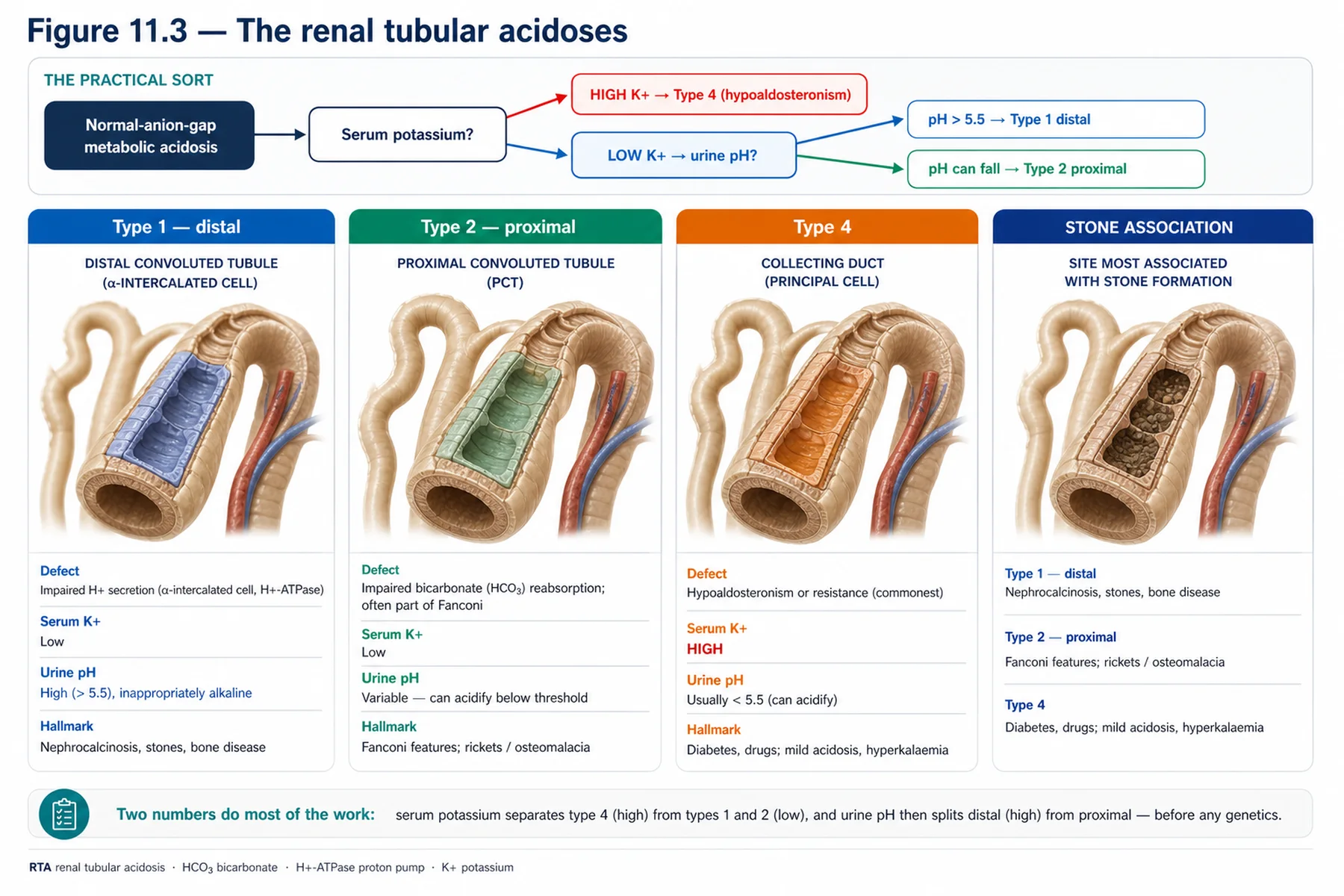
The renal tubular acidoses (RTAs) are a group of tubulopathies defined by a normal-anion-gap (hyperchloraemic) metabolic acidosis from impaired renal acid handling, and distinguishing the three types is a classic exercise. Distal (type 1) RTA results from impaired hydrogen-ion secretion in the collecting duct, so the patient cannot acidify the urine (the urine pH stays inappropriately high despite the acidosis), and develops a hypokalaemic metabolic acidosis with hypercalciuria, nephrocalcinosis, and stones; inherited forms involve transporters such as the anion exchanger AE1 or the proton pump (the latter associated with sensorineural deafness). Proximal (type 2) RTA results from impaired bicarbonate reabsorption in the proximal tubule, so bicarbonate is wasted until the plasma level falls low enough for the distal nephron to reclaim the rest; it is often part of a generalised Fanconi syndrome, and is also hypokalaemic. Type 4 RTA is different — it results from hypoaldosteronism or aldosterone resistance, causing a hyperkalaemic normal-anion-gap acidosis (the hyperkalaemia distinguishing it from the hypokalaemic types 1 and 2). The practical pattern: a normal-anion-gap metabolic acidosis with hypokalaemia and an inability to acidify the urine (with stones) is distal (type 1); with bicarbonate wasting and Fanconi features is proximal (type 2); and with hyperkalaemia is type 4. Treatment is alkali (with potassium in the hypokalaemic types), and the specific defect where relevant.
— The proximal tubulopathies, and treatment
The proximal tubule, reabsorbing the bulk of the filtrate, gives rise to several distinctive disorders. Fanconi syndrome is generalised proximal tubular dysfunction — a global failure of proximal reabsorption causing glucosuria (with normal blood glucose), aminoaciduria, phosphaturia (with hypophosphataemia and rickets/osteomalacia), bicarbonaturia (proximal RTA), uricosuria, and low-molecular-weight proteinuria — with many inherited causes (cystinosis, the metabolic disease of the Fabry chapter, being a classic one). Dent disease is an X-linked proximal tubulopathy (defects in CLCN5 or OCRL) characterised by low-molecular-weight proteinuria, hypercalciuria, nephrocalcinosis, kidney stones, and progressive CKD, often with some Fanconi features. Cystinuria is a defect in the proximal reabsorption of cystine and the dibasic amino acids, causing recurrent cystine kidney stones (the stone chapter). The treatment of the tubulopathies overall follows a simple principle: replace what is lost and target the defect. The salt-wasting syndromes are treated by replacing the losses — potassium and magnesium in Gitelman, potassium and sodium chloride in Bartter (where a prostaglandin inhibitor such as indomethacin can also reduce the prostaglandin-driven losses), often with potassium-sparing diuretics. The salt-retaining syndromes are treated by targeting the overactive transporter — amiloride for Liddle, a thiazide for Gordon. The acidoses are treated with alkali (and potassium where needed), and cystinuria with hydration, urinary alkalinisation, and thiol drugs. The unifying message of the chapter is the segment-by-segment logic: locate the defective transporter in its nephron segment, and the electrolyte pattern, the blood pressure, the urinary calcium, and the treatment all follow — turning a confusing list of syndromes into an intelligible, predictable map.
| 04 | PHASE A · LEVEL 4 · ORIENTATION & KNOWLEDGE Reference Tables |
Table 11.1 — The nephron-segment map
| Segment | Disorders |
| Proximal tubule | Fanconi syndrome; Dent disease; cystinuria; proximal (type 2) RTA |
| Thick ascending limb (NKCC2 — loop) | Bartter syndrome ('furosemide-like') |
| Distal convoluted tubule (NCC — thiazide) | Gitelman syndrome ('thiazide-like') |
| Collecting duct (ENaC) | Liddle (salt-retaining HTN); distal (type 1) RTA; nephrogenic DI |
Table 11.2 — Bartter versus Gitelman
| Feature | Bartter (TAL, loop-like) | Gitelman (DCT, thiazide-like) |
| Electrolytes / acid-base | Hypokalaemic metabolic alkalosis | Hypokalaemic metabolic alkalosis |
| Urinary calcium / magnesium | HYPERcalciuria (± nephrocalcinosis) | HYPOcalciuria + HYPOmagnesaemia |
| Onset / severity / BP | Earlier, more severe; normal-to-low BP | Later, milder; normal-to-low BP |
Table 11.3 — Salt-wasting versus salt-retaining
| Category | Detail |
| Salt-wasting (Bartter, Gitelman) | Volume contracted; normal-to-low BP; RAAS activated; hypokalaemic alkalosis |
| Liddle (ENaC gain-of-function) | HTN, hypokalaemia, LOW renin AND aldosterone → amiloride (not spironolactone) |
| Gordon (PHA2; ↑ NCC) | HTN, HYPERkalaemia, acidosis → thiazide |
| Sorting | Blood pressure: low in salt-wasters, high in salt-retainers |
Table 11.4 — The renal tubular acidoses
| Type | Defect | Pattern |
| Distal (type 1) | Impaired distal H+ secretion | HypoK acidosis; urine can't acidify; stones/nephrocalcinosis |
| Proximal (type 2) | Impaired HCO3 reabsorption | HypoK; bicarbonate wasting; often Fanconi |
| Type 4 | Hypoaldosteronism / resistance | HYPERkalaemic normal-anion-gap acidosis |
Table 11.5 — The proximal tubulopathies
| Disorder | Detail |
| Fanconi syndrome | Generalised proximal dysfunction — glucosuria, aminoaciduria, phosphaturia, type 2 RTA, LMW proteinuria |
| Dent disease | X-linked (CLCN5/OCRL) — LMW proteinuria, hypercalciuria, nephrocalcinosis, stones, CKD |
| Cystinuria | Defective cystine/dibasic amino acid reabsorption → cystine stones |
| Causes of Fanconi | Cystinosis, inherited and acquired (drugs, paraproteins) |
Table 11.6 — Treatment principle: replace and target
| Disorder | Treatment |
| Gitelman | Replace potassium and magnesium |
| Bartter | Replace potassium and sodium chloride; indomethacin (prostaglandin); K-sparing diuretics |
| Liddle / Gordon | Amiloride (Liddle); thiazide (Gordon) — target the transporter |
| RTA / cystinuria | Alkali (± potassium); hydration/alkalinisation/thiol drugs for cystine stones |
| 05 | PHASE B · LEVEL 5 · VISUALISE & MAP Imaging & Flowchart Specifications |
| 06 | PHASE B · LEVEL 6 · VISUALISE & MAP Concept Maps |
Each chain runs from mechanism to a named clinical insight; read the arrows as “leads to.”
Nephron-segment map. Defect in a transporter → features reflect the segment's normal job → proximal (Fanconi), TAL (Bartter), DCT (Gitelman), collecting duct (Liddle/RTA) → ACTION: locate the defect to predict the syndrome.
Bartter (loop-like). Thick-ascending-limb NaCl defect (NKCC2) → furosemide-like → salt wasting, hypokalaemic alkalosis, HYPERcalciuria → ACTION: recognise the loop-like, hypercalciuric, severe/early pattern.
Gitelman (thiazide-like). Distal-convoluted-tubule NCC defect → thiazide-like → salt wasting, hypokalaemic alkalosis, HYPOcalciuria + HYPOmagnesaemia → ACTION: recognise the thiazide-like, hypocalciuric, hypomagnesaemic, mild/late pattern.
Salt-wasting vs salt-retaining. Salt-wasting (Bartter/Gitelman) → low/normal BP; salt-retaining (Liddle/Gordon) → HTN → ACTION: use the blood pressure to split the tubulopathies, then potassium/acid-base to narrow.
The RTAs. Normal-anion-gap acidosis → distal (type 1, hypoK, can't acidify, stones), proximal (type 2, HCO3 wasting/Fanconi), type 4 (hyperK) → ACTION: use potassium and urine pH to type the RTA.
| 07 | PHASE B · LEVEL 7 · VISUALISE & MAP Decision Pathways |
| R1 | IF facing a confusing tubulopathy, THEN locate the defect on the nephron-segment map — the segment's normal job predicts the syndrome. |
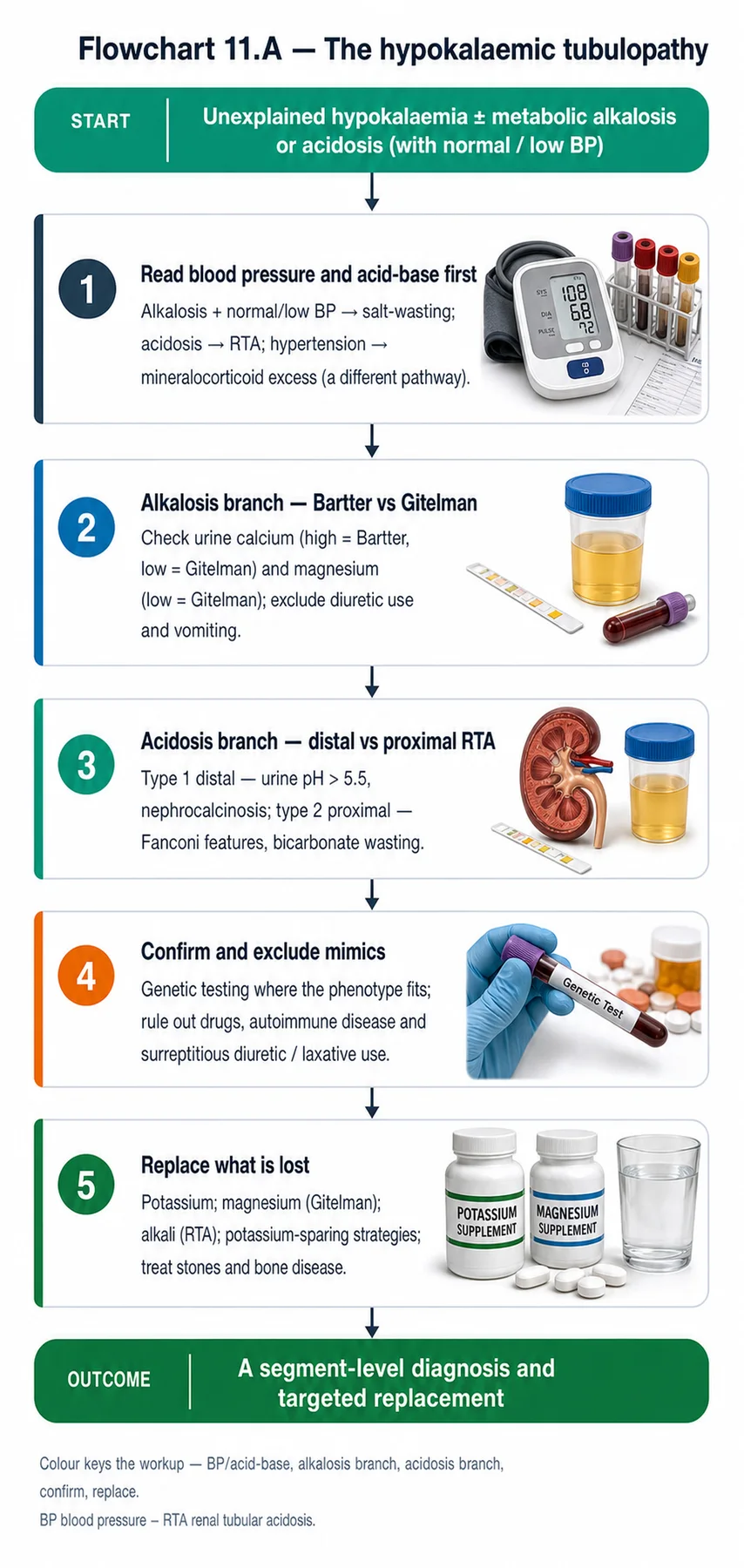
| R2 | IF there is a salt-wasting hypokalaemic metabolic alkalosis with normal/low BP, THEN it is Bartter or Gitelman — check the urinary calcium and magnesium. |
| R3 | IF the urinary calcium is HIGH (± nephrocalcinosis), THEN it is Bartter (loop-like, severe/early). |
| R4 | IF the urinary calcium is LOW with hypomagnesaemia, THEN it is Gitelman (thiazide-like, mild/late). |
| R5 | IF a tubulopathy causes hypertension, THEN it is salt-retaining — Liddle (hypokalaemia, low aldosterone → amiloride) or Gordon (hyperkalaemia, acidosis → thiazide). |
| R6 | IF there is a normal-anion-gap metabolic acidosis, THEN type the RTA by the potassium (hypoK = type 1/2; hyperK = type 4) and the urine pH/Fanconi features. |
| R7 | IF there are proximal features (glucosuria, aminoaciduria, phosphaturia, LMW proteinuria), THEN consider Fanconi syndrome, Dent disease, or cystinuria. |
| R8 | IF treating a tubulopathy, THEN replace what is lost (K, Mg, salt) and target the defect (amiloride, thiazide, alkali). |
| 08 | PHASE C · LEVEL 8 · CLINICAL REASONING Clinical Cases |
| CASE 1 | LOOP OR THIAZIDE? Calcium distinguishes them Bartter versus Gitelman |
Presentation
Two patients have a hypokalaemic metabolic alkalosis with salt wasting and a normal-to-low blood pressure. One — a neonate — has hypercalciuria with nephrocalcinosis; the other — an adult with cramps — has a low urinary calcium and a low magnesium. A trainee finds them indistinguishable.
❖ Pause and reflect What distinguishes these two salt-wasting tubulopathies? |
Analysis
The urinary calcium (and magnesium, and the age/severity) distinguishes them: the first is Bartter, the second Gitelman. Both share the salt-wasting hypokalaemic metabolic alkalosis with a normal-to-low blood pressure, which is why they seem indistinguishable on those features alone. But the diuretic analogy sorts them. Bartter is a thick-ascending-limb defect ('furosemide-like'): like a loop diuretic, it causes hypercalciuria (often with nephrocalcinosis) and tends to be severe and early (the neonate with nephrocalcinosis fits). Gitelman is a distal-convoluted-tubule defect ('thiazide-like'): like a thiazide, it causes hypocalciuria and magnesium wasting (hypomagnesaemia), and is milder and later (the adult with cramps and a low calcium and magnesium fits). So the urinary calcium is the key — high in Bartter, low in Gitelman — with the hypomagnesaemia and the milder, later course confirming Gitelman. The loop-versus-thiazide framing predicts all of it.
Plan
Diagnose Bartter in the neonate (hypercalciuria, nephrocalcinosis, loop-like, severe) and Gitelman in the adult (hypocalciuria, hypomagnesaemia, thiazide-like, mild), using the urinary calcium and magnesium to distinguish them, and treat by replacing the losses. Distinguish Bartter from Gitelman by the urinary calcium.
Teaching point
Bartter (loop-like) causes hypercalciuria and is severe/early; Gitelman (thiazide-like) causes hypocalciuria and hypomagnesaemia and is mild/late — the urinary calcium distinguishes them.
Cross-reference
Exercises rules R2, R3, and R4; the nephron-map and Bartter/Gitelman concept maps; Figures 11.1, 11.2; Table 11.2.
| CASE 2 | HIGH PRESSURE, LOW POTASSIUM Salt-retaining tubulopathy Liddle versus Gordon |
Presentation
A young patient with hypertension and hypokalaemia has a low renin and a low aldosterone. A second young hypertensive patient has hyperkalaemia and a metabolic acidosis. Both are being investigated for endocrine causes.
❖ Pause and reflect What salt-retaining tubulopathies do these two patterns suggest? |
Analysis
Liddle syndrome and Gordon syndrome — two salt-retaining (hypertensive) tubulopathies with opposite potassium patterns. The first patient, with hypertension, hypokalaemia, and a low renin AND a low aldosterone, has Liddle syndrome: a gain-of-function of the epithelial sodium channel (ENaC) causing constitutive sodium reabsorption (hence hypertension and a low renin), with potassium wasting (hypokalaemia) but a low aldosterone (the channel is active independent of aldosterone) — which is why it is treated with the ENaC blocker amiloride, not spironolactone (there is no aldosterone to antagonise). The second patient, with hypertension, hyperkalaemia, and a metabolic acidosis, has Gordon syndrome (pseudohypoaldosteronism type 2): increased activity of the distal sodium-chloride cotransporter causes sodium retention (hypertension) with the distinctive hyperkalaemia and acidosis (the mirror of Gitelman), treated with a thiazide that blocks that cotransporter. The blood pressure marks both as salt-retaining; the potassium (low in Liddle, high in Gordon) and the aldosterone distinguish them. These are tubulopathies, not primary endocrine disease.
Plan
Diagnose Liddle in the hypokalaemic, low-aldosterone hypertensive (treat with amiloride) and Gordon in the hyperkalaemic, acidotic hypertensive (treat with a thiazide), recognising both as salt-retaining tubulopathies. Use the potassium and aldosterone to distinguish the salt-retaining syndromes.
Teaching point
Salt-retaining tubulopathies cause hypertension — Liddle (hypokalaemia, low aldosterone → amiloride) and Gordon (hyperkalaemia, acidosis → thiazide).
Cross-reference
Exercises rule R5; the salt-wasting-versus-retaining concept map; Figure 11.3; Table 11.3; monogenic hypertension in Volume 8.
| CASE 3 | ACID AND THE GAP Typing the RTA The renal tubular acidoses |
Presentation
Three patients have a normal-anion-gap metabolic acidosis: one with hypokalaemia, an inappropriately high urine pH, and kidney stones; one with hypokalaemia, bicarbonate wasting, and glucosuria/aminoaciduria; and one with hyperkalaemia. A trainee labels them all 'RTA' without distinguishing.
❖ Pause and reflect How are these three renal tubular acidoses distinguished? |
Analysis
By the potassium, the urine pH, and the associated features — they are distal (type 1), proximal (type 2), and type 4 RTA respectively. All three share a normal-anion-gap (hyperchloraemic) metabolic acidosis from impaired renal acid handling, but they are distinct. The first — hypokalaemia, an inappropriately high urine pH (inability to acidify the urine), and stones/nephrocalcinosis — is distal (type 1) RTA, from impaired collecting-duct hydrogen-ion secretion. The second — hypokalaemia, bicarbonate wasting, and proximal features (glucosuria, aminoaciduria) — is proximal (type 2) RTA, from impaired proximal bicarbonate reabsorption, often as part of a Fanconi syndrome. The third — hyperkalaemia — is type 4 RTA, from hypoaldosteronism or aldosterone resistance, the hyperkalaemia distinguishing it from the hypokalaemic types 1 and 2. So the potassium first (hypo = type 1 or 2; hyper = type 4), then the urine pH and proximal features (can't acidify + stones = type 1; bicarbonate wasting + Fanconi = type 2). Lumping them as 'RTA' misses the type-specific cause and treatment.
Plan
Type the RTAs — distal (type 1: hypoK, high urine pH, stones), proximal (type 2: hypoK, bicarbonate wasting, Fanconi), type 4 (hyperK) — using the potassium, urine pH, and associated features, and treat with alkali (and potassium where needed). Type the RTA rather than lumping them.
Teaching point
Type the RTAs by potassium and urine pH: type 1 (distal, hypoK, can't acidify, stones), type 2 (proximal, hypoK, bicarbonate wasting/Fanconi), type 4 (hyperK).
Cross-reference
Exercises rule R6; the RTA concept map; Figure 11.3; Table 11.4; acid-base in Volume 7.
| CASE 4 | PROTEIN, CALCIUM, STONES An X-linked proximal tubulopathy Dent disease |
Presentation
A young man has low-molecular-weight proteinuria, hypercalciuria, nephrocalcinosis, recurrent kidney stones, and slowly progressive CKD, with a similar picture in a maternal uncle. He has been worked up for a glomerular disease without success.
❖ Pause and reflect What proximal tubulopathy fits this picture? |
Analysis
Dent disease — an X-linked proximal tubulopathy. The picture is characteristic: low-molecular-weight proteinuria (a proximal tubular marker, not glomerular), hypercalciuria, nephrocalcinosis, recurrent kidney stones, and slowly progressive CKD, with an X-linked family pattern (the affected maternal uncle). Dent disease is caused by defects in CLCN5 (or OCRL) affecting proximal tubular function, often with some Fanconi features. The low-molecular-weight proteinuria is the key clue that this is a proximal tubular, not a glomerular, disease — which is why the glomerular work-up was fruitless. Recognising the combination — low-molecular-weight proteinuria, hypercalciuria, stones, and an X-linked pattern — points to Dent disease, confirmed by genetics. The broader lesson is that proximal tubulopathies (Dent, Fanconi, cystinuria) present with proximal markers (low-molecular-weight proteinuria, glucosuria, aminoaciduria, hypercalciuria, stones), distinct from glomerular disease.
Plan
Diagnose Dent disease (X-linked proximal tubulopathy) from the low-molecular-weight proteinuria, hypercalciuria, nephrocalcinosis, stones, and X-linked pattern, confirm genetically, and manage the hypercalciuria/stones and CKD — rather than pursuing a glomerular work-up. Recognise the proximal tubulopathy by its proximal markers.
Teaching point
Dent disease (X-linked proximal tubulopathy) gives low-molecular-weight proteinuria, hypercalciuria, nephrocalcinosis, and stones — the proximal markers distinguish it from glomerular disease.
Cross-reference
Exercises rule R7; the nephron-map concept map; Figure 11.1; Table 11.5; stones in Chapter 16; Fanconi/cystinosis in Chapter 9.
| 09 | PHASE C · LEVEL 9 · CLINICAL REASONING Clinical Implications |
One triad per mechanism the narrative exposed: the physiology, why it matters, and the clinical move.
MECHANISM Each tubulopathy is a defect in a specific transporter in a specific nephron segment. |
WHY IT MATTERS The segment's normal job predicts the syndrome's features. |
ACTION Locate the defect on the nephron-segment map. |
MECHANISM Bartter impairs thick-ascending-limb NaCl reabsorption (the loop-diuretic target). |
WHY IT MATTERS It is 'furosemide-like' — salt wasting, hypokalaemic alkalosis, hypercalciuria. |
ACTION Recognise the loop-like, hypercalciuric, severe pattern as Bartter. |
MECHANISM Gitelman impairs the distal thiazide-sensitive cotransporter. |
WHY IT MATTERS It is 'thiazide-like' — salt wasting, hypokalaemic alkalosis, hypocalciuria, hypomagnesaemia. |
ACTION Recognise the thiazide-like, hypocalciuric, hypomagnesaemic pattern as Gitelman. |
MECHANISM Salt-retaining tubulopathies reabsorb too much sodium. |
WHY IT MATTERS They cause hypertension (Liddle, Gordon), unlike the salt-wasters. |
ACTION Use the blood pressure to split the tubulopathies, then potassium to narrow. |
MECHANISM The renal tubular acidoses impair renal acid handling at different sites. |
WHY IT MATTERS Type 1 (distal), type 2 (proximal), and type 4 differ in potassium and urine pH. |
ACTION Type the RTA by the potassium and urine pH. |
| 10 | PHASE C · LEVEL 10 · CLINICAL REASONING Clinical Pearls |
| Tubulopathies = transporter defects, each mapping to a nephron segment. | Locate the defect → the segment's job predicts the syndrome. |
| Bartter = thick ascending limb (NKCC2), 'furosemide-like'. | Bartter: salt wasting, hypokalaemic alkalosis, HYPERcalciuria ± nephrocalcinosis; severe/early; normal/low BP. |
| Gitelman = distal convoluted tubule (NCC), 'thiazide-like'. | Gitelman: salt wasting, hypokalaemic alkalosis, HYPOcalciuria + HYPOmagnesaemia; mild/late; commonest tubulopathy. |
| Bartter vs Gitelman: urinary calcium (high vs low) is the key. | Salt-wasting (Bartter/Gitelman) → low/normal BP; salt-retaining → HTN. |
| Liddle (ENaC gain-of-function): HTN, hypokalaemia, low renin AND aldosterone → amiloride. | Gordon (PHA2, ↑NCC): HTN, HYPERkalaemia, acidosis → thiazide. |
| Distal (type 1) RTA: hypoK, can't acidify urine, stones/nephrocalcinosis. | Proximal (type 2) RTA: hypoK, bicarbonate wasting, often Fanconi. |
| Type 4 RTA: HYPERkalaemic normal-anion-gap acidosis (hypoaldosteronism). | Fanconi: generalised proximal dysfunction (glucosuria, aminoaciduria, phosphaturia, LMW proteinuria). |
| Dent disease: X-linked proximal — LMW proteinuria, hypercalciuria, stones, CKD. | Treat: replace losses (K, Mg, salt) and target the defect (amiloride/thiazide/alkali). |
| 11 | PHASE D · LEVEL 11 · SAFETY & EVIDENCE Red Flags & Never-Do |
Panel A — Red flags
| ▲ | Hypokalaemic metabolic alkalosis with salt wasting and normal/low BP — Bartter or Gitelman; check urinary calcium/magnesium. |
| ▲ | Hypertension with hypokalaemia and a low aldosterone — consider Liddle (treat with amiloride, not spironolactone). |
| ▲ | Hypertension with hyperkalaemia and acidosis — consider Gordon (treat with a thiazide). |
| ▲ | Normal-anion-gap acidosis — type the RTA (potassium, urine pH) rather than lumping them. |
| ▲ | Low-molecular-weight proteinuria with hypercalciuria and stones — a proximal tubulopathy (Dent), not glomerular. |
Panel B — Never do
| ✖ NEVER — treat Liddle syndrome with spironolactone — use amiloride (no aldosterone to block). |
| ✖ NEVER — lump the renal tubular acidoses as one — type them by potassium and urine pH. |
| ✖ NEVER — confuse Bartter and Gitelman — the urinary calcium distinguishes them. |
| ✖ NEVER — pursue a glomerular work-up for a proximal-tubular picture (LMW proteinuria). |
| 12 | PHASE D · LEVEL 12 · SAFETY & EVIDENCE Common Pitfalls |
Pitfall 1 — Confusing Bartter and Gitelman
| ✖ | WRONG Treating Bartter and Gitelman as indistinguishable. |
| ✓ | RIGHT Distinguishing by urinary calcium (and magnesium, severity). |
| ✉ | WHY Bartter is hypercalciuric (loop-like); Gitelman hypocalciuric (thiazide-like). |
Pitfall 2 — Spironolactone for Liddle
| ✖ | WRONG Treating Liddle syndrome with spironolactone. |
| ✓ | RIGHT Treating it with amiloride (an ENaC blocker). |
| ✉ | WHY Liddle has a low aldosterone — there is nothing for spironolactone to block. |
Pitfall 3 — Lumping the RTAs
| ✖ | WRONG Calling every normal-anion-gap acidosis 'RTA' without typing. |
| ✓ | RIGHT Typing by potassium and urine pH (type 1/2/4). |
| ✉ | WHY The types differ in cause, potassium, and treatment. |
Pitfall 4 — Missing the salt-retaining cause
| ✖ | WRONG Working up hypertension with hyperkalaemia as endocrine disease. |
| ✓ | RIGHT Recognising Gordon syndrome (treat with a thiazide). |
| ✉ | WHY Increased NCC activity gives this distinctive picture. |
Pitfall 5 — Glomerular work-up for a proximal disease
| ✖ | WRONG Pursuing a glomerular cause for low-molecular-weight proteinuria. |
| ✓ | RIGHT Recognising the proximal tubulopathy (Dent disease). |
| ✉ | WHY LMW proteinuria is a proximal tubular, not glomerular, marker. |
| 13 | PHASE D · LEVEL 13 · SAFETY & EVIDENCE Evidence Grading |
GRADE A HIGH CONFIDENCE The effect is real and the estimate is stable. RCTs at low risk of bias; multiple concordant prospective cohorts; meta-analyses. |
GRADE B MODERATE CONFIDENCE The effect is likely real but may shift with new data. Observational studies, registries, mechanistic human studies. |
GRADE C LOW CONFIDENCE Rests on physiology, reasoning, or consensus rather than outcomes. Pathophysiological reasoning; extrapolation; consensus without outcomes. |
Graded statements (by evidence type)
| Statement | Grade | Basis (evidence type) |
| Each inherited tubulopathy maps to a specific transporter and nephron segment. | A | Molecular physiology |
| Bartter is a thick-ascending-limb (loop-like) salt-wasting defect with hypercalciuria. | A | Physiology and clinical data |
| Gitelman is a distal (thiazide-like) defect with hypocalciuria and hypomagnesaemia. | A | Physiology and clinical data |
| Liddle syndrome is treated with amiloride, not spironolactone. | A | Established physiology |
| Gordon syndrome (↑ NCC) is treated with a thiazide. | A | Established physiology |
| The renal tubular acidoses differ by potassium and urine pH. | A | Established physiology |
| Proximal tubulopathies present with proximal markers (LMW proteinuria, etc.). | A | Clinical data |
| 18 | PHASE F · LEVEL 18 · APPLY & TEST Cheat Sheet |
| Tubulopathies map to nephron segments (transporter defects). | Proximal: Fanconi, Dent, cystinuria, type 2 RTA. |
| Thick ascending limb (NKCC2): Bartter ('furosemide-like'). | Distal convoluted tubule (NCC): Gitelman ('thiazide-like'). |
| Collecting duct (ENaC): Liddle, distal (type 1) RTA, nephrogenic DI. | Bartter: hypoK alkalosis, HYPERcalciuria, severe/early, normal/low BP. |
| Gitelman: hypoK alkalosis, HYPOcalciuria + HYPOmagnesaemia, mild/late. | Bartter vs Gitelman = urinary calcium (high vs low). |
| Salt-wasting (Bartter/Gitelman) → low/normal BP. | Liddle: HTN, hypoK, low renin AND aldosterone → amiloride. |
| Gordon: HTN, HYPERkalaemia, acidosis → thiazide. | Distal (type 1) RTA: hypoK, can't acidify, stones. |
| Proximal (type 2) RTA: hypoK, HCO3 wasting, Fanconi. | Type 4 RTA: HYPERkalaemia (hypoaldosteronism). |
| Dent: X-linked proximal — LMW proteinuria, hypercalciuria, stones. | Treat: replace losses + target the defect. |
| 19 | PHASE F · LEVEL 19 · APPLY & TEST Flashcards |
| CARD 1 | Q. How are the inherited tubulopathies best organised? A. By the nephron segment and transporter affected: a genetic defect in a specific transporter produces a syndrome whose features reflect that segment's normal job — proximal (Fanconi, Dent, cystinuria), thick ascending limb (Bartter), distal convoluted tubule (Gitelman), and collecting duct (Liddle, distal RTA). DETAILED. Locating the defect predicts the syndrome. CLINICAL. Use the nephron-segment map to make sense of them. |
| CARD 2 | Q. What is Bartter syndrome? A. A defect in the thick ascending limb's sodium-chloride reabsorption machinery (the loop-diuretic target) — 'furosemide-like' — causing renal salt wasting, a hypokalaemic metabolic alkalosis, hypercalciuria (often with nephrocalcinosis), and a normal-to-low blood pressure; it is typically severe and presents early. DETAILED. It mimics chronic loop-diuretic use. CLINICAL. Recognise the loop-like, hypercalciuric, severe pattern. |
| CARD 3 | Q. What is Gitelman syndrome? A. A defect in the distal convoluted tubule's thiazide-sensitive cotransporter — 'thiazide-like' — causing salt wasting, a hypokalaemic metabolic alkalosis, hypocalciuria, hypomagnesaemia, and a normal-to-low blood pressure; it is the commonest inherited tubulopathy, milder, and presents later (often with cramps and tetany). DETAILED. It mimics chronic thiazide use. CLINICAL. Recognise the thiazide-like, hypocalciuric, hypomagnesaemic pattern. |
| CARD 4 | Q. How are Bartter and Gitelman distinguished? A. Both cause a salt-wasting hypokalaemic metabolic alkalosis with a normal-to-low blood pressure, but Bartter is hypercalciuric (loop-like) and more severe/earlier, whereas Gitelman is hypocalciuric with hypomagnesaemia (thiazide-like) and milder/later. DETAILED. The urinary calcium is the key distinguisher. CLINICAL. Check the urinary calcium and magnesium to tell them apart. |
| CARD 5 | Q. How do the salt-retaining tubulopathies present, and how are they treated? A. They cause hypertension: Liddle syndrome (an overactive epithelial sodium channel) gives hypertension with hypokalaemia and a low renin and aldosterone, treated with amiloride (not spironolactone); Gordon syndrome (increased NCC activity) gives hypertension with hyperkalaemia and acidosis, treated with a thiazide. DETAILED. The blood pressure marks them as salt-retaining. CLINICAL. Distinguish by potassium; treat Liddle with amiloride and Gordon with a thiazide. |
| CARD 6 | Q. How are the renal tubular acidoses distinguished? A. All cause a normal-anion-gap metabolic acidosis: distal (type 1) has hypokalaemia, an inability to acidify the urine, and stones; proximal (type 2) has hypokalaemia, bicarbonate wasting, and often Fanconi syndrome; type 4 has hyperkalaemia from hypoaldosteronism. DETAILED. The potassium and urine pH type them. CLINICAL. Type the RTA by potassium and urine pH. |
| CARD 7 | Q. What are the proximal tubulopathies? A. Fanconi syndrome (generalised proximal dysfunction — glucosuria, aminoaciduria, phosphaturia, type 2 RTA, low-molecular-weight proteinuria), Dent disease (X-linked — low-molecular-weight proteinuria, hypercalciuria, nephrocalcinosis, stones, CKD), and cystinuria (cystine stones). DETAILED. Proximal markers distinguish them from glomerular disease. CLINICAL. Recognise the proximal markers (LMW proteinuria, glucosuria, aminoaciduria). |
| CARD 8 | Q. What is the principle of treating the tubulopathies? A. Replace what is lost and target the defect: replace potassium and magnesium in Gitelman, potassium and salt in Bartter (with indomethacin to reduce prostaglandin-driven losses); use amiloride for Liddle and a thiazide for Gordon; give alkali for the acidoses; and hydration, alkalinisation, and thiol drugs for cystine stones. DETAILED. The treatment follows the mechanism. CLINICAL. Replace losses and target the specific transporter defect. |
| 20 | PHASE F · LEVEL 20 · APPLY & TEST One-Minute Preceptor |
| SCENE 1 | The intern confusing Bartter and Gitelman |
GET A COMMITMENT. “You can't tell these two salt-wasting, hypokalaemic-alkalosis patients apart — what would distinguish them?”
PROBE FOR EVIDENCE. “They look identical” — ask: “What does each resemble — a loop diuretic or a thiazide — and what does that predict for the urinary calcium?”
TEACH A GENERAL RULE. Bartter is loop-like (hypercalciuria, severe/early); Gitelman is thiazide-like (hypocalciuria, hypomagnesaemia, mild/late) — the urinary calcium distinguishes them.
REINFORCE WHAT WAS RIGHT. Recognising both as salt-wasting tubulopathies was correct.
CORRECT A MISTAKE. Check the urinary calcium and magnesium to tell them apart.
| SCENE 2 | The resident giving spironolactone for Liddle |
GET A COMMITMENT. “You've prescribed spironolactone for this hypertensive, hypokalaemic, low-aldosterone patient — why?”
PROBE FOR EVIDENCE. “It's mineralocorticoid hypertension” — ask: “If the aldosterone is low, what is spironolactone meant to block?”
TEACH A GENERAL RULE. Liddle syndrome is a gain-of-function of the sodium channel, independent of aldosterone — so the aldosterone is low and spironolactone fails; amiloride, an ENaC blocker, is the treatment.
REINFORCE WHAT WAS RIGHT. Recognising a salt-retaining picture was reasonable.
CORRECT A MISTAKE. Treat Liddle with amiloride, not spironolactone.
| 22 | PHASE F · LEVEL 22 · APPLY & TEST Board-Style Questions |
| Q 01 | Bartter syndrome is a defect of which nephron segment, and resembles which diuretic? |
| A | Distal convoluted tubule; thiazide |
| B | Thick ascending limb; loop diuretic (furosemide) |
| C | Collecting duct; amiloride |
| D | Proximal tubule; acetazolamide |
Rationale Bartter affects the thick-ascending-limb NaCl machinery, mimicking a loop diuretic (Figure 11.1, Table 11.2). A is Gitelman; C and D are wrong. |
| Q 02 | Which distinguishes Gitelman from Bartter syndrome? |
| A | Both have hypercalciuria |
| B | Gitelman has hypocalciuria and hypomagnesaemia; Bartter has hypercalciuria |
| C | Both have hypertension |
| D | Neither affects potassium |
Rationale The urinary calcium (and magnesium) distinguishes them (case 1, Table 11.2). A, C, and D are incorrect. |
| Q 03 | A salt-wasting hypokalaemic metabolic alkalosis with a normal-to-low blood pressure suggests: |
| A | Liddle or Gordon syndrome |
| B | Bartter or Gitelman syndrome |
| C | Primary aldosteronism |
| D | Type 4 RTA |
Rationale Salt-wasting with low/normal BP is Bartter/Gitelman (Figure 11.3, Table 11.3). A is salt-retaining/HTN; C is HTN; D is hyperkalaemic. |
| Q 04 | Liddle syndrome (hypertension, hypokalaemia, low renin and aldosterone) is treated with: |
| A | Spironolactone |
| B | Amiloride (an ENaC blocker) |
| C | A thiazide |
| D | A loop diuretic |
Rationale Liddle's channel is active independent of aldosterone, so amiloride works and spironolactone does not (case 2, Table 11.3, rule R5). A fails; C is for Gordon; D is wrong. |
| Q 05 | Hypertension with hyperkalaemia and a metabolic acidosis suggests: |
| A | Gitelman syndrome |
| B | Gordon syndrome (treated with a thiazide) |
| C | Bartter syndrome |
| D | Liddle syndrome |
Rationale Increased NCC activity in Gordon syndrome gives this picture, treated with a thiazide (case 2, Table 11.3). A, C, and D cause hypokalaemia/low BP. |
| Q 06 | A normal-anion-gap acidosis with hypokalaemia, an inappropriately high urine pH, and stones is: |
| A | Proximal (type 2) RTA |
| B | Distal (type 1) RTA |
| C | Type 4 RTA |
| D | A high-anion-gap acidosis |
Rationale Inability to acidify the urine with stones is distal (type 1) RTA (case 3, Table 11.4). A wastes bicarbonate; C is hyperkalaemic; D is wrong. |
| Q 07 | Type 4 RTA is distinguished by: |
| A | Hypokalaemia |
| B | Hyperkalaemia (from hypoaldosteronism) |
| C | A high anion gap |
| D | An inability to acidify the urine |
Rationale Type 4 RTA is a hyperkalaemic normal-anion-gap acidosis (case 3, Table 11.4). A describes types 1/2; C and D are wrong. |
| Q 08 | Low-molecular-weight proteinuria with hypercalciuria, nephrocalcinosis, and stones in an X-linked pattern suggests: |
| A | Glomerular disease |
| B | Dent disease (a proximal tubulopathy) |
| C | Gitelman syndrome |
| D | Liddle syndrome |
Rationale This is the proximal tubulopathy Dent disease; LMW proteinuria is a proximal, not glomerular, marker (case 4, Table 11.5). A, C, and D do not fit. |